Introduction
Cowpea Vigna unguiculata (L.) Walp. (Fabaceae) is the most important grain legume crop in sub-Saharan Africa (Adati et al., Reference Adati, Tamò, Yusuf, Downham, Singh and Hammond2008; Dugje et al., Reference Dugje, Omoigui, Ekeleme, Kamara and Ajeigbe2009). It is highly proteinaceous, well adapted to different soil types and intercropping systems, and resistant to drought with proven ability to improve soil fertility and prevent erosion (Adati et al., Reference Adati, Tamò, Yusuf, Downham, Singh and Hammond2008; Dugje et al., Reference Dugje, Omoigui, Ekeleme, Kamara and Ajeigbe2009). The sale of stems and leaves as animal feed during the dry season is a major source of income for smallholder farmers across Africa (Langyintuo et al., Reference Langyintuo, Lowenberg-DeBoer, Faye, Lambert, Ibro, Moussa, Kergna, Kushwaha, Musa and Ntoukam2003; Adati et al., Reference Adati, Tamò, Yusuf, Downham, Singh and Hammond2008; Dugje et al., Reference Dugje, Omoigui, Ekeleme, Kamara and Ajeigbe2009). Despite its importance in improving food security and alleviating poverty, growers consider cowpea production risky due to the plethora of insect pests that attack the crop (Singh and Allen, Reference Singh and Allen1979; Singh and Emden, Reference Singh and Emden1979; Jackai and Daoust, Reference Jackai and Daoust1986; Adati et al., Reference Adati, Tamò, Yusuf, Downham, Singh and Hammond2008). Moreover, infestation can cause up to 100% yield loss if not effectively controlled (Dugje et al., Reference Dugje, Omoigui, Ekeleme, Kamara and Ajeigbe2009). Among these different insect pests, the legume pod borer Maruca vitrata (Fabricius) (Lepidoptera: Crambidae) is regarded as one of the most important pests, causing yield losses of 20–100% (Karel, Reference Karel1985; Sharma, Reference Sharma1998; Ranga Rao and Shanower, Reference Ranga Rao and Shanower1999; Sharma et al., Reference Sharma, Saxena and Bhagwat1999; Srinivasan, Reference Srinivasan2012). For example, in Kenya, losses of up to 80% are common on different varieties of cowpea (Okeyo-Owuor et al., Reference Okeyo-Owuor, Agwaro and Simbi1983). Infestation starts in the terminal shoots, later spreading to the reproductive structures with the larvae feeding on floral buds, flowers and pods (Jackai, Reference Jackai1981; Sharma, Reference Sharma1998). Currently, the majority of smallholder farmers who produce the bulk of cowpea across Africa rely exclusively on chemical insecticides to combat damage due to M. vitrata with little guidance on their use and application frequency. The direct consequences of inappropriate application and overuse are destruction of indigenous natural enemies, pesticide resistance and human and environmental hazards (Okeyo-Owuor et al., Reference Okeyo-Owuor, Agwaro and Simbi1983; Ekesi, Reference Ekesi1999; Ton, Reference Ton2000; Ulrichs et al., Reference Ulrichs, Mewis and Schnitzler2001; Chopra et al., Reference Chopra, Leemans, Kumar and Simons2005). Resistance by M. vitrata to commonly used insecticides, including organochlorines, organophosphates, carbamates and synthetic pyrethroid compounds has been documented in Kenya (Okeyo-Owuor et al., Reference Okeyo-Owuor, Agwaro and Simbi1983), Benin (Atachi and Sourokou, Reference Atachi and Sourokou1989), Nigeria (Ekesi et al., 1995) and Southeast Asia (Ulrichs et al., Reference Ulrichs, Mewis and Schnitzler2001).
There is therefore a need to identify alternative management methods. A recent advance involves engineering cowpea-encoding genes that express the Cry1Ab delta endotoxin of Bacillus thuringiensis (Berliner) subsp. kurstaki (Popelka et al., Reference Popelka, Gollasch, Moore, Molvig and Higgins2006; Chaudhury et al., Reference Chaudhury, Madanpotra, Jaiwal, Saini, Ananda Kumar and Jaiwal2007; Adesoye et al., Reference Adesoye, Machuka and Togun2008; Huesing et al., Reference Huesing, Romeis, Ellstrand, Raybould, Hellmich, Wolt, Ehlers, Dabiré-Binso, Fatokun, Hokanson, Ishiyaku, Margam, Obokoh, Mignouna, Nang'ayo, Ouedraogo, Pasquet, Pittendrigh, Schaal, Tamò and Murdock2011), but this technique carries the risk of resistance development as noted for the stem borer, Busseola fusca (Fuller) (Campagne et al., Reference Campagne, Kruger, Pasquet, Le Ru and Van den Berg2013).
Several entomopathogens have been reported to attack Ma. vitrata in nature (Otieno et al., Reference Otieno, Odindo, Okeyo-Owuor and Sabwa1983; Odindo et al., Reference Odindo, Otieno, Oloo, Kilori and Odhiambo1989; Okeyo-Owuor et al., Reference Okeyo-Owuor, Oloo and Agwaro1991; Huang et al., Reference Huang, Peng and Talekar2003). Consequently, entomopathogenic fungi (EPF) used as a biopesticide has been proposed as an important component of an IPM strategy (Tamò et al., Reference Tamò, Maniania, Cherry, Neuenschwander, Borgemeister and Langewald2003). In earlier attempts to exploit these pathogens, Ekesi et al. (Reference Ekesi, Adamu and Maniania2002a, Reference Ekesi, Maniania and Luxb) reported ovicidal effects of Nigerian isolates of Metarhizium anisopliae (Metchnikoff) Sorokin and Beauveria bassiana (Balsamo) Vuillemin (Hypocreales: Clavicipitaceae) against Nigerian populations of M. vitrata causing 71–100% egg mortality and 61–100% deferred mortality in larvae that hatched from the treated eggs. Additional studies using the same isolates against East African populations yielded lower than 45% mortality on eggs (S. Ekesi et al., unpublished data), suggesting the existence of host specificity of EPF to varying populations of the insect. Indeed, there is documented evidence to suggest the existence of various populations and host races of M. vitrata, and variations have been reported in the responses of populations in West Africa, Southeast Asia and South Asia to synthetic sex pheromone lures (Downham et al., Reference Downham, Tamo, Hall, Datinon, Adetonah and Farman2004; Hassan, Reference Hassan2007; Schläger et al., Reference Schläger, Ulrichs, Srinivasan, Beran, Bhanu, Mewis and Schreiner2012; Srinivasan, Reference Srinivasan2012). The difference in populations may be responsible for the lack of efficacy of Nigerian isolates of EPF to M. vitrata in East Africa. Therefore, there is a need to identify potent isolates of EPF that are efficacious against different populations or races of the pest in this region.
The biology and behaviour of M. vitrata (Taylor, Reference Taylor1967; Usua and Singh, Reference Usua and Singh1979; Jackai, Reference Jackai1981; Sharma, Reference Sharma1998) have been considered the reasons why this pest is difficult to manage. There is usually a very short period of exposure on leaves and flowers mostly by the first instar larvae immediately after hatching and before they burrow into flowers and pods. However, a potentially important window of opportunity is to target the first instar larvae with an evening spray of EPF when they emerge from eggs. Although this application strategy would appear to be optimal, it may not always be possible to fully cover this larval stage with biopesticidal spray. There is also a need to test for the virulence of selected isolates against other developmental stages of the pest, because they also periodically wander around on plant structures and could benefit from secondary uptake of biopesticidal spores. Furthermore, variations in virulence across life stages by EPF have been reported for other pests (Ekesi et al., Reference Ekesi, Adamu and Maniania2002a, Reference Ekesi, Maniania and Luxb; Angel-Sahagún et al., Reference Angel-Sahagún, Lezama-Gutiérrez, Molina-Ochoa, Galindo-Velasco, López-Edwards, Rebolledo-Dominguez, Cruz-Vázquez, Reyes-Velázquez, Skoda and Foster2005; Kirubakaran et al., Reference Kirubakaran, Sathish-Narayanan, Revathi, Chandrasekaran and Senthil-Nathan2014), but such information is lacking for M. vitrata. If applications are appropriately timed in the evenings, it would also protect the EPF from the vagaries of UV radiation. Further protection of EPF and enhancement of their efficacy can be achieved through effective formulation (Daoust et al., Reference Daoust, Ward and Roberts1983; Jackson et al., Reference Jackson, Dunlap and Jaronski2009), and oil-formulated mycoinsecticides are generally reported to be more effective than aqueous formulations (Prior et al., Reference Prior, Jollands and le Patourel1988; Bateman et al., Reference Bateman, Carey, Moore and Prior1993; Lomer et al., Reference Lomer, Bateman, Godonou, Kpindou, Shah, Paraiso and Prior1993; Inglis et al., Reference Inglis, Johnson and Goettel1996).
Mass production is another important factor to consider in the selection of candidate EPF for biopesticide development (Goettel and Inglis, Reference Goettel, Inglis and Lacey1997). On solid media, hyphomycetes produce differentiated asexual spores that are genetically stable (Mangenot and Reisinger, Reference Mangenot, Reisinger, Weber and Hess1976); in submerged culture, they usually form single cells called blastospores through schizolytic separation at septa or by mechanical fragmentation of hyphae by yeast-like budding (Rombach, Reference Rombach1989). For different isolates, the formation of these structures, in solid and/or liquid media, and the quantity and quality of the propagules that are produced for use against different insects, can be affected by the composition of the medium and culture conditions (Fargues et al., Reference Fargues, Maniania, Delmas and Smits1992; Jackson et al., Reference Jackson, Mcguire, Lacey and Wraight1997; Vidal et al., Reference Vidal, Fargues, Lacey and Jackson1998; Bae et al., Reference Bae, Sinha, Park, Song and Yun2000).
This study was, therefore, undertaken to: (1) identify potent isolates of B. bassiana and/or Me. anisopliae for the management of East African populations of Ma. vitrata through screening; (2) evaluate the effect of nutrition in liquid media on propagule concentration and biomass production by the two most virulent isolates and (3) assess the effect of different formulations of the most virulent isolate against various developmental stages of M. vitrata.
Materials and methods
Insects
A colony of M. vitrata was started from a batch of larvae obtained from infested cowpea plants in a field at the International Centre of Insect Physiology and Ecology, Thomas Odhiambo Campus (icipe-TOC), Mbita, on the shores of Lake Victoria, Kenya (0°26′ 06.19″S, 34°12′53.13″ E; altitude 1137 masl). The larvae from the field were introduced to a semi-synthetic diet prepared following the procedure described by Onyango and Ochieng'-Odero (Reference Onyango and Ochieng'-Odero1993), and the culture was maintained in the rearing room at 26–30°C, 60–75% relative humidity (RH) and a 12 hr of light:12 hr of dark photoperiod. When larvae pupated, the pupae were carefully harvested from the diet using a pair of soft forceps onto Petri plates (14 cm diameter) and later introduced into a sleeved Perspex glass cage (30 × 30 × 30 cm). Emerging adults were collected and introduced into another sleeved cage (40 × 40 × 40 cm) made of an iron rod frame with a mosquito netting enclosure. They were fed with 10% sugar solution on soaked balls of cotton wool placed at the base of each cage. The sucrose solution was replenished after every 2 days. Potted cowpea seedlings (approximately 2 weeks old) were introduced into the cages where the adults were placed to serve as oviposition sites for female moths. The seedlings were destructively harvested and placed in well-secured ventilated clear plastic cups and incubated at 26–30°C for eggs to hatch (2–3 days). The hatched first instar larvae were then introduced to a freshly prepared diet as described earlier, and the cycle was repeated. The insects were used for the various experiments after 10 generations. Field collected populations of M. vitrata were introduced into the laboratory colony after every 6 months to enhance genetic vigour.
Fungi
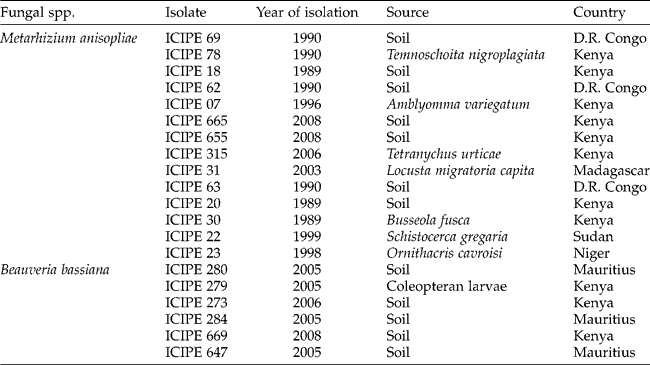
Fungal isolates used in this study were obtained from the Arthropod Germplasm Centre, icipe, and their geographical areas of origin are given in Table 1. The isolates were first passaged through M. vitrata larvae (third instar) to control against attenuation (Butt and Goettel, Reference Butt, Goettel, Navon and Ascher2000), and spores from mycosed cadavers were propagated on Sabouraud dextrose agar (SDA) medium to obtain cultures for bioassays. Fungal suspensions were prepared by harvesting conidia from 2- to 3-week-old sporulating cultures and suspending them in sterile distilled water containing 0.05% Triton X-100 in 10-ml glass bottles with 10 glass beads (3 mm). Bottle contents were vortexed on a mechanical shaker (5 min, 10 cm of vertical travel, and 700 oscillations/min) to produce a homogeneous conidial suspension. The conidia were then quantified using the improved Neubauer haemocytometer (Hausser, Scientific Horsham, USA) (Goettel and Inglis, Reference Goettel, Inglis and Lacey1997; Inglis et al., Reference Inglis, Enkerli, Goettel and Lacey2012).
Fungal isolates tested against first instar larvae of Maruca vitrata

Conidial viability was assessed by spread-plating 0.1 ml of a suspension at a concentration of 3 × 10⁶ conidia/ml on SDA medium. Inoculated plates were sealed with parafilm membrane and incubated at 25 ± 2°C under complete darkness. At 18 hr post-inoculation, lactophenol cotton blue was added to terminate germination and also stain the spores for ease of counting. Two sterile microscope coverslips were placed on the surface of the 18-hr-old culture in each plate. The percentage of germination was determined by counting 100 spores, categorized as germinated and non-germinated, under each coverslip on each Petri plate under a light microscope ( × 400 magnification). A conidium was considered to have germinated if the germ tube was at least twice the size of the spore (Goettel and Inglis, Reference Goettel, Inglis and Lacey1997; Inglis et al., Reference Inglis, Enkerli, Goettel and Lacey2012). Each plate served as a replicate with four replications per fungal isolate originating from different plates.
Screening of EPF isolates against first instar larvae of M. vitrata
Screening for pathogenicity
Each fungal isolate was assayed by dipping five freshly harvested cowpea flowers into 10 ml of a standard concentration of 1 × 10⁸conidia/ml for 30 s. The flowers were placed on a paper towel for c. 30 min to drain off excess suspension and thereafter transferred into clear plastic dishes (11 × 4 cm). A group of 30 first instar larvae were then transferred onto the treated flowers and the dishes were covered with a screened/ventilated lid. Flowers in the control lots were dipped in sterile distilled water containing 0.05% Triton X-100. Insects were fed ad libitum on treated flowers and were provided with fresh flowers (surface-sterilized and untreated) daily as required. Larval mortality was recorded daily for 7 days. Dead insects were transferred to Petri plates lined with moist filter paper to facilitate the development of mycosis. Mortality due to fungal infection was confirmed by microscopic examination of hyphae and conidia on the surface of the cadaver. The screening bioassay was carried out in five batches, with each batch consisting of four fungal isolates and a control; each isolate was replicated four times.
Dose–response bioassay
A dose–response bioassay was performed for the best two fungal isolates, M. anisopliae ICIPE 18 and ICIPE 69, which had been selected based on cumulative mean mortality and lethal time to 50% mortality (LT50) values from the initial screening. The isolates were tested at five conidial concentrations as follows: 1 × 10⁴, 1 × 10⁵, 1 × 10⁶, 1 × 10⁷ and 1 × 10⁸conidia/ml. Five freshly harvested cowpea flowers were placed on Petri plates and sprayed with 10 ml of fungal suspension using the Burgerjon spray tower (Burgerjon, Reference Burgerjon1956). Before flowers were loaded, the Petri plates were lined with filter paper to absorb excess suspension. Flowers in the control lots were sprayed with 10 ml of sterile distilled water containing 0.05% Triton X-100. For each isolate and dosage, 20 first instar larvae of M. vitrata were transferred onto the treated flowers in a plastic dish, and the dish was covered with a ventilated lid. Insects also fed on treated flowers ad libitum within the first 24 hr of treatment, and further feeding protocol and monitoring of treated insects for mortality and handling of cadavers were carried out as described earlier. Each isolate and dosage under test was replicated four times.
Assessment of conidia production on cadavers of first instar larvae
The two best isolates, M. anisopliae ICIPE 18 and ICIPE 69, were further evaluated for conidial production on first instar larval cadavers. The larvae were exposed for 24 hr to cowpea flowers treated with suspensions of ICIPE 18 and ICIPE 69 at a concentration of 1 × 10⁸conidia/ml. On day 1 post-inoculation, fresh untreated cowpea flowers were added as food. Dead insects were collected on day 2 post-inoculation and incubated as described earlier. On day 6 post-incubation, five mycosed insects were collected and dried in an oven at 35°C for 30 min, and transferred individually into 2-ml cryogenic tubes containing 1 ml of sterile 0.05% Triton X-100. The tube was then vortexed for 5 min to dislodge conidia from the insect. Concentration of the resultant conidial suspension was determined as described earlier, providing an estimate of conidial yield by the cadaver. Each insect served as a replicate with five replications per fungal isolate.
Performance of the selected M. anisopliae isolates ICIPE 18 and ICIPE 69 in four liquid media
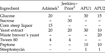
We evaluated the impact of different four media, Adámek, Jenkins–Prior, Arthropod Pathology Unit 1 (APU1) and APU2 (Table 2) on propagule concentration and biomass production of the two selected isolates. The Adámek medium was originally formulated for producing submerged conidia of M. anisopliae (Adámek, Reference Adámek1963). The Jenkins–Prior medium was developed for liquid production of submerged conidia of M. flavoviride (Jenkins and Prior, Reference Jenkins and Prior1993). The APU1 medium is a standard medium used in the APU of icipe and the APU2 medium is a slight modification of APU1. For each medium, 100ml suspension was dispensed into 250-ml shake flasks and then sterilized and cooled. The flasks were inoculated with a suspension of aerial conidia of the two M. anisopliae isolates to yield a final concentration of 1 × 10⁶conidia/ml culture. The flasks were incubated on a reciprocal shaker (Innova® 44, New Brunswick Scientific, NJ, USA; 100 ± 2 travels/min) at 28 ± 0.5°C.
Composition of four liquid media (g/l distilled water) used for evaluating propagule and biomass yield of two Metarhizium anisopliae isolates, ICIPE 18 and ICIPE 69
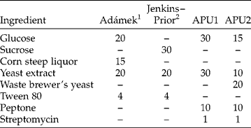
1 Adámek (Reference Adámek1963).
2 Jenkins and Prior (Reference Jenkins and Prior1993).
To estimate the concentration of propagules, culture flasks were sampled at 72hr by removing 0.5ml aliquots from each flask and counting at × 400 magnification using a Neubauer haemocytometer. The dry weight was determined by weighing 1ml sample of the fresh weight matter and centrifuging at 3500rpm at 5°C (Medifuge centrifuge, Heraeus Sepatech GmbH, Osterode, Germany). The resulting precipitate was washed twice with deionized water, weighed and dried in an oven at 95°C for 24hr. Biomass was estimated by determination of dry weight. Five replicated flasks were maintained for each medium.
Efficacy of different formulations of ICIPE 69 against eggs, and larval and adult stages of M. vitrata
Preparation of fungal formulations
The conidia used for formulation were prepared using the same method as described by Ekesi et al. (Reference Ekesi, Maniania and Lux2005). Briefly, the conidia were produced on a substrate of maize and pumice in the ratio of 1:2. The substrate mixture was autoclaved for 1 hr at 121°C, transferred to plastic bowls (33 × 25 × 13 cm), inoculated with a 3-day-old culture of blastospores (50 ml) and covered with a sterile polyethylene bag. The culture was incubated for 21 days at ambient conditions (20–26°C, 40–70% RH) and allowed to dry for 5 days at room temperature. The conidia were harvested by sifting the substrate through a sieve (295-μm mesh size) and then stored in plastic bags in a refrigerator (4–6°C, 40–50% RH) until required for the various experiments. In a viability test, between 84 and 90% of the conidia germinated after 24 hr on SDA plates.
Three formulations were tested, aqueous, oil and dry conidia, for the various assays. The aqueous and oil formulations were tested against eggs and the larval stage and the dry conidia formulation was tested against the adult moths. The aqueous formulation of ICIPE 69 was prepared by suspending the harvested conidia in water as described earlier. The oil formulation was prepared by emulsifying 1% (v/v) corn oil (Bidco Oil Refineries Ltd., Thika, Kenya) in sterile distilled water containing 0.05% Triton X-100 and 0.1 g of harvested conidia. For both formulations, the concentration was adjusted to 1 × 10⁸conidia/ml. Control for the oil formulation was prepared by emulsifying 1% oil in sterile distilled water (v/v) containing 0.05% Triton X-100; the control for the aqueous formulation consisted of only sterile distilled water containing 0.05% Triton X-100. The dry conidial formulations for the two isolates, ICIPE 18 and ICIPE 69, utilized for the adult bioassay were applied directly as harvested from the production substrate.
Ovicidal activity assay
Eggs oviposited by female M. vitrata on potted cowpea seedlings within a period of 12 hr were collected using destructive harvesting of the leaves and counted under a dissecting microscope ( × 16). Pieces of leaves (ca. 15 × 15 cm) carrying 20 eggs were cut off from the whole leaf, placed in sterile Petri plates and sprayed with 10 ml of either oil- or aqueous-formulated fungal suspension of ICIPE 69 using the Burgerjon spray tower. Eggs for the control groups were sprayed with 10 ml of oil or aqueous control suspensions, prepared as described earlier. The treated eggs were thereafter separately transferred into ventilated clear plastic cups (6 cm diametre × 7 cm high), and incubated at 25 ± 2°C. On day 4 post-treatment, the neonates that hatched from the eggs were counted and the percentage of egg mortality was computed. Four replicates were maintained for each formulation.
Larvicidal activity assay
Larval instars were treated by spraying cowpea flowers with 10 ml of fungal suspensions, prepared as described earlier. Flowers for the control groups were treated with 10 ml of oil or aqueous control suspensions. Insects were then released onto the treated flowers as described previously. Twenty insects were used for the first, second and third larval instars; 10 insects were used for the fourth instar as supplies were limited. Larval mortality was recorded daily for 7 days for all larval instars, starting on day 2 post-treatment. Dead insects were transferred to Petri plates lined with moist filter paper to facilitate the development of mycosis. Mortality due to fungal infection was confirmed by microscopic examination of hyphae and conidia on the surface of the cadaver. Three replications were used per formulation and for each larval stage.
Adulticidal activity assays
Two bioassays involving the adult stage of M. vitrata were performed using isolate ICIPE 69. The first experiment assessed fungal effects as a result of direct uptake of spores by male or female moths from an autoinoculative device. The second experiment assessed virulence on adult moths that could result from the transfer of inoculum from contaminated males (as donors) to non-contaminated females (as recipients).
In the first bioassay, individual adult males and females were contaminated with dry fungal conidia following the procedure described by Maniania et al. (Reference Maniania, Oketch, Adino, Opere and Ekesi2013) with a slight modification. Briefly, one-day-old virgin adult males and females of M. vitrata were contaminated individually using small velvet-coated plastic vials (1.5 cm diameter × 6 cm high) as autoinoculative devices. The devices were contaminated with 0.3 g of dry conidia. Thereafter, moths were introduced into the device for 3 min to pick up spores. In the controls, dry spores were used, inactivated by autoclaving for 1 hr at 121°C and dried at ambient conditions. After 3 min of exposure, contaminated insects were transferred in groups of 10 males or females into sleeved Perspex cages (30 × 30 × 30 cm) and provided with 10% sucrose solution as food every 2 days. Adult mortality was monitored daily for 7 days. Dead insects were transferred to Petri plates lined with moist filter paper to facilitate mycosis. Mortality due to fungal infection was confirmed by the presence of hyphae and conidia on the surface of the cadaver. Three replications were used per fungal isolate for male and female moths.
In the second bioassay, 1-day-old males were individually treated as described earlier. Each treated male was then transferred into a sleeved Perspex cage (12 × 12 × 12 cm) containing an uninfected 1-day-old virgin female. A fresh cowpea leaf mounted in a glass vial containing water and plugged with moist cotton wool was introduced into the cage to serve as an oviposition substrate. Cotton wool soaked with 10% sucrose solution that was replaced after every 2 days was also provided for feeding. The cowpea leaf was removed from each cage after 24 hr and eggs laid by each female were counted and a new leaf was introduced. Walls of the cage were also inspected and any oviposited eggs were counted and removed to avoid repeated counting. Egg counting continued until the female died, and longevity and fecundity were recorded. Each treatment had five couples with each couple serving as a replicate.
Statistical analyses
Mortality data were analysed using Abbott's formula (Abbott, Reference Abbott1925) to correct for natural mortality before analysis of variance (ANOVA). In the ANOVA, trial replicates carried out over time were included in the model as a random effect to control for trial-to-trial differences. Data on the concentration of propagules and biomass were Log10 transformed before ANOVA. The dose–mortality and time–mortality data were analysed by Generalized Linear Model (GLM), using the function ‘dose.p’ from the MASS library, to generate LC50 and LT50 estimates, along with slopes of the regression curves. Using the Standard error (SE) values, the 95% confidence intervals (CIs) [LC50± (1.96 × SE)] were calculated. In the case of LT50 estimation, the GLM analysis was run for each replication and the resultant LT50 values and their respective slopes were subjected to ANOVA to generate means. Whenever treatment effects were found to be significant (P= 0.05), means were separated using the Tukey (HSD) test (P= 0.05). For the primary screening bioassay, isolates that caused greater than 45% mortality were considered for LT50 estimation. On the other hand, LC50 estimates were subjected to pairwise comparison and considered to be significant at the 5% level if their respective 95% CIs did not overlap. All analyses were performed using R v2.15.1 (R Development Core Team, 2008).
Results
Screening of EPF isolates against first instar larvae of M. vitrata
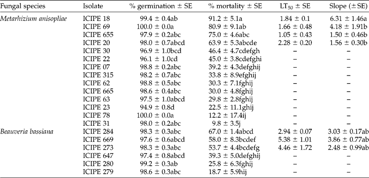
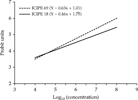
Conidial viability of the test isolates ranged from 95 to 100%, an indication that the bioassay measured true differences in virulence between the fungal isolates (Table 3). There were significant differences in mortality caused by the fungal isolates tested at the concentration of 1 × 10⁸conidia/ml (F= 10.6; df = 19, 60; P< 0.0001). Among the 20 isolates tested, two isolates of M. anisopliae – ICIPE 18 and ICIPE 69 – caused significantly higher mortality (81 and 91%) than other isolates (10–75%) at day 7 post-inoculation (Table 3). The same isolates recorded the shortest LT50 values of 1.7 and 1.8 days. Slopes of mortality curves differed significantly among isolates with ICIPE 18 and ICIPE 69 having the steepest slopes (6.31 and 4.18) compared with the other isolates (Table 3). In terms of lethal concentration to 50% mortality (LC50), ICIPE 69 had a lower LC50 value (3.01 × 10⁶conidia/ml) than isolate ICIPE 18 (1.07 × 10⁷conidia/ml) (Table 4). The plot of Log-probit regressions for the two isolates reveals a steeper slope of the regression line for ICIPE 69 than ICIPE 18, but both were significantly different from zero (P= 0.05) (Fig. 1). ICIPE 69 produced relatively more conidia (1 × 10⁶/cadaver) than ICIPE 18 (0.8 × 10⁶), but the two were not significantly different (F= 0.23; df = 1; P= 0.6418).
Pathogenicity of entomopathogenic fungi applied at a concentration of 1×10⁸conidia/ml to first instar larvae of Maruca vitrata at 7 days post-inoculation+
Means within a column followed by different letters are significantly different by the Tukey (HSD) test (P= 0.05).
+ LT50 estimates for fungal isolates that never attained 50% larval mortality were higher than 7 days (i.e. the experimentation period) and were omitted from the table.
LC50 values for selected isolates of Metarhizium anisopliae against Maruca vitrata

Significance codes at P= 0.05: 0 ‘***’; 0.001 ‘**’, 0.01 ‘*’; 0.05 ‘.’; 0.1; ‘ ’ 1.
Log-probit regressions of mortality caused by Metarhizium anisopliae isolates ICIPE 18 and ICIPE 69 on first instar larvae of Maruca vitrata.

Performance of isolates ICIPE 18 and ICIPE 69 in liquid media
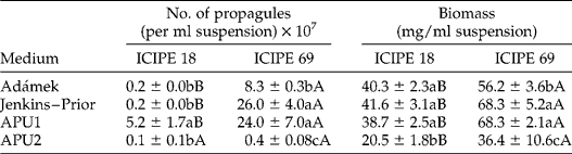
There was a significant difference among the four media evaluated with respect to propagule production (F= 22.4; df = 3, 32; P= 0.0001) and isolates tested (F= 41.3; df = 1, 32; P= 0.0010). The interaction between media and isolate was also significant (F= 9.54; df = 3, 32; P= 0.0011). Among the four media tested, isolate ICIPE 18 produced a significantly higher propagule concentration (5.2 ± 1.7 × 10⁷/ml) on APU1 than on the other media. Jenkins–Prior (2.6 ± 0.4 × 10⁸/ml) and APU1 (2.4 ± 0.7 × 10⁸/ml) were the best media for ICIPE 69 (Table 5). Comparison between the two isolates revealed a significantly higher concentration of propagules produced by ICIPE 69 on the two most effective media (APU1 and Jenkins–Prior), compared with ICIPE 18 (Table 5).
Propagule and biomass production of two isolates of Metarhizium anisopliae ICIPE 18 and ICIPE 69 after 72 hr in four submerged culture media
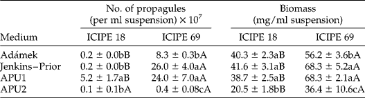
For each parameter, means within a column followed by different lower case letters and within a row followed by different upper case letters differ significantly by the Tukey (HSD) test (P= 0.05).
The amount of biomass produced by the two isolates after 72 hr of oscillation significantly differed among the four media evaluated (F= 19.8; df = 3, 32; P= 0.0001) and the two isolates tested (F= 12.6; df = 1, 32; P= 0.0006) (Table 5). The interaction between media and isolate was also significant (F= 7.14; df = 3, 32; P= 0.00 231). Among the four media, ICIPE 18 produced significantly higher biomass (38.7 ± 2.5 to 41.6 ± 3.1 mg/ml) on Adámek, Jenkins–Prior and APU1 than on APU2. Biomass production by ICIPE 69 was significantly higher on Jenkins–Prior (68.3 ± 5.2) and APU1 (68.3 ± 2.1 mg/ml) than on the other media. Biomass produced by ICIPE 69 on the best two media was significantly higher than that of ICIPE 18 (Table 5).
Efficacy of different formulations of M. anisopliae against eggs, and larval and adult stages of M. vitrata
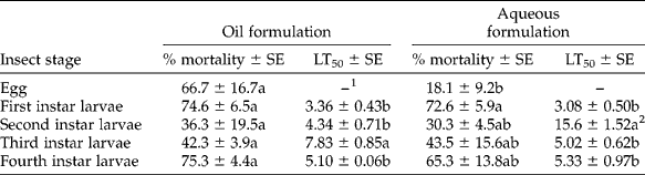
Formulation as a factor had no significant effect on mortality (F= 1.96; df = 1, 28; P= 0.1725), but significant effects were observed due to developmental stage (F= 4.28; df = 4, 25; P= 0.0089) and the interaction (i.e. developmental stage × formulation) (F= 3.31; df = 9, 20; P= 0.0123). In the oil-formulated conidia, mortality across developmental stages was highest on eggs and first and fourth instar larvae (68–75%), although this was not significantly different from the other instars (F= 2.32; df = 4, 10; P= 0.1283) perhaps due to high levels of standard errors in eggs and at the second instar stage. The rapidity of kill in terms of LT50 was lower on first, second and fourth instars (3.36–5.10 days) than on the third instar (7.83 days) (Table 6). Applications of aqueous formulation of isolate ICIPE 69 also caused the highest mortality to first and fourth instar larvae (65–73%) compared with the other stages (F= 4.60; df = 4, 10; P= 0.0229). LT50 was lower at first, third and fourth instars (3.08–5.33 days) compared with the second instar (15.6 days) (Table 6).
Pathogenicity of oil and aqueous formulations of Metarhizium anisopliae isolate ICIPE 69 against the egg and larval stages of Maruca vitrata
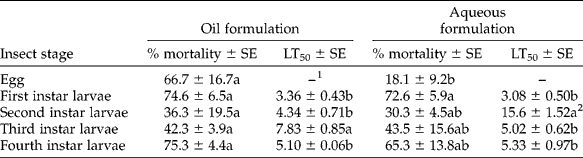
Means within a column followed by different letters are significantly different by the Tukey (HSD) test (P= 0.05).
1 Eggs were excluded from LT50 analysis as egg mortality was assessed at once (i.e. 4 days post-treatment) as opposed to the 7-day period for the larval mortality assessment.
2 LT50 for the aqueous formulation against the second instar larvae is an estimate beyond the 7-day mortality assessment period.
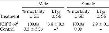
Significant differences were observed in mortality incurred by male (F= 198.61; df = 1, 4; P= 0.0001) and female (F= ∞; df = 1, 4; P< 0.0001) M. vitrata adults (Table 7) treated with dry conidia of isolate ICIPE 69 compared with those treated with autoclaved conidia (controls). Isolate ICIPE 69 caused 100% mortality in male and female M. vitrata with LT50 of 3.4 and 2.9 days. Mortality in the controls did not exceed 3%.
Mortality of male and female Maruca vitrata adults as a result of exposure to dry conidia of isolate ICIPE 69 using an autoinoculative device
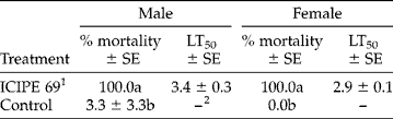
Means within a column followed by different letters under each parameter, are significantly different by the Tukey (HSD) test (P= 0.05).
1 Metarhizium anisopliae isolate ICIPE 69.
2 LT50 analysis for the control was not performed, as mortality values for most replications in this group were zero.
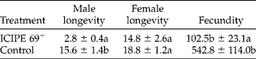
There was a significant difference in the longevity of adult males directly contaminated with isolate ICIPE 69 and the control group (F= 81.92; df = 1, 8; P< 0.0001), with the untreated males living longer (16.0 days) than the treated males (2.8 days). However, no significant difference (F= 1.89; df = 1, 8; P= 0.2068) was observed among adult females maintained with males contaminated with ICIPE 69 and the control males. Fecundity differed significantly (F= 14.33; df = 1, 6; P= 0.0091) between female M. vitrata maintained with contaminated males (103 eggs) and the control group (543 eggs) (Table 8).
Horizontal transmission of fungal infection from male to female Maruca vitrata moths and impact on longevity and fecundity
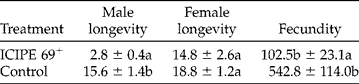
Means within a column followed by different letters under each parameter, are significantly different by the Tukey (HSD) test (P= 0.05).
+ Metarhizium anisopliae isolate ICIPE 69.
Discussion
Results from the primary screening bioassay indicated that all the fungal isolates tested against M. vitrata were pathogenic to the insects, but their virulence varied greatly among the isolates, perhaps not surprisingly because inter- and intra-specific variations in virulence among fungal species and isolates within fungal species are well documented for different species of insects (Poprawski et al., Reference Poprawski, Marchal and Robert1985; Feng and Johnson Reference Feng and Johnson1990; Maniania, Reference Maniania1992; Ekesi et al., Reference Ekesi, Maniania, Onu and Löhr1998).
Among the 20 isolates screened, results indicated that M. anisopliae isolates ICIPE 18 and ICIPE 69 caused significantly higher mortality than the others and isolates of M. anisopliae induced a higher mortality than those of B. bassiana. A pathogen that infects several host species in an ecosystem has a better chance of contacting susceptible hosts, thereby increasing pathogen population density and disease prevalence (Fuxa, Reference Fuxa1987). Both M. anisopliae and B. bassiana are among the hyphomycetes reported to have a wide range of hosts among invertebrates (Butt and Goettel, Reference Butt, Goettel, Navon and Ascher2000), although some isolates can be host specific. Indeed, the two isolates of M. anisopliae, ICIPE 18 and ICIPE 69, identified in this study as highly pathogenic to M. vitrata, have also been reported to cause high mortality in other tropical insects. For example, from among 22 isolates screened, Ekesi et al. (Reference Ekesi, Maniania, Onu and Löhr1998) identified ICIPE 69 as one of the isolates most pathogenic to the legume flower thrips Megalurothrips sjostedti (Trybom). It has also been found to be effective against the adult pea leafminer, Liriomyza huidobrensis (Blanchard) (Migiro et al., Reference Migiro, Maniania, Chabi-Olaye and Vandenberg2010). Similarly, ICIPE 18 has been found to be highly pathogenic to species of African tephritid fruit flies (Ekesi et al., Reference Ekesi, Adamu and Maniania2002a, b; Dimbi et al., Reference Dimbi, Maniania, Lux, Ekesi and Mueke2003), L. huidobrensis (Migiro et al., Reference Migiro, Maniania, Chabi-Olaye and Vandenberg2010) and sandfly Phlebotomus duboscqi (Neveu-Lemaire) (Ngumbi et al., Reference Ngumbi, Irungu, Ndegwa and Maniania2011). This suggests that although widely distributed fungi such as B. bassiana and M. anisopliae could be relatively host specific as pathotypes, some isolates, including those selected in this study, can be effective against a broad range of insects and could benefit from the extension of labels to other crops upon registration and commercialization.
In our study, LT50 values for the two most pathogenic isolates, ICIPE 18 and ICIPE 69, were shorter (1.7 and 1.8 days), than for other isolates. Host death by EPF is considered to be due to a combination of actions including production of toxins, physical obstruction of blood circulation, nutrient depletion and invasion of organs (Goettel and Inglis, Reference Goettel, Inglis and Lacey1997; Butt and Goettel, Reference Butt, Goettel, Navon and Ascher2000; Inglis et al., Reference Inglis, Enkerli, Goettel and Lacey2012). On SDA, these isolates have been observed to exhibit more rapid growth than the others (S. Ekesi, unpublished data), and this action probably assures faster penetration of the insect cuticle by the fungus. It is also possible that these isolates produce toxins that are responsible for the rapid death of the host but this requires further investigation using chemical and/or molecular techniques.
Susceptibility of most insects is dependent on spore dosage, and it is presumed that a threshold exists whereby a certain number of propagules are necessary to overcome the host (Butt and Goettel, Reference Butt, Goettel, Navon and Ascher2000). The slope of regression line for isolate ICIPE 69 was steeper than that of ICIPE 18, suggesting that the target insect will be more vulnerable over a given time with increasing doses of conidia (Ekesi et al., Reference Ekesi, Adamu and Maniania2002a, b) for isolate ICIPE 69 than for ICIPE 18.
This study further reveals that nutritional conditions of the four media tested strongly affected propagule and biomass production by the two isolates. In terms of propagule production, there was the tendency for isolate ICIPE 69 to produce more propagule and biomass than ICIPE 18. Our results concur with previous studies. For instance, Kleespies and Zimmermann (Reference Kleespies and Zimmermann1992) reported that the concentration of propagules produced by three strains of M. anisopliae varied significantly (in the range of 2.08 × 10⁷–2.93 × 10⁸blastospores/ml) according to the media used for their cultivation. Fargues et al. (Reference Fargues, Smits, Vidal, Vey, Vega, Mercadier and Quimby2002) also reported that concentrations of propagules produced by two strains of M. anisopliae var. acridum ranged from 1.4 to 2.4 × 10⁸propagules/ml in four different liquid media: Adámek, Catroux, Jackson and Jenkins–Prior. By and large, the level of propagule production in our experiment is within the range reported by these authors working with Metarhizium spp.
In our experiments, biomass production was highest in Jenkins–Prior, APU1 and Adámek compared with APU2, and ICIPE 69 outperformed ICIPE 18 in terms of the quantity produced. Other researchers have shown that biomass production in hyphomycetes including M. anisopliae on favourable media could range from 14 mg/ml (Humphreys et al., Reference Humphreys, Matewele, Trinci and Gillespie1989) to as much as 60.3 mg/ml (Vidal et al., Reference Vidal, Fargues, Lacey and Jackson1998). Clearly, between the two isolates evaluated in our study, ICIPE 69 produced higher propagule and biomass yield than ICIPE 18 on both Jenkins–Prior and APU1 media. The reason is unclear for the high performance of Jenkins–Prior and APU1 media compared with the others in our experiment but could be attributed to the higher sugar source, as suggested by Vidal et al. (Reference Vidal, Fargues, Lacey and Jackson1998). Kleespies and Zimmermann (Reference Kleespies and Zimmermann1992) also demonstrated that not only the amount but also the nature of sugars, whereby glucose and fructose were better than lactose for blastospore production in M. anisopliae.
Based on dose–response studies and the performance of ICIPE 18 and ICIPE 69 on liquid media, we tested the efficacy of different formulations of ICIPE 69 on different developmental stages of M. vitrata. There was a variation in the susceptibility of the various stages to the fungal formulations tested. Butt and Goettel (Reference Butt, Goettel, Navon and Ascher2000) also noted that not all stages in the life cycle of an insect are equally susceptible to fungal infection, with pupal stages often being indicated to be the most resistant stage and adults the most susceptible. Most recently, Kirubakaran et al. (Reference Kirubakaran, Sathish-Narayanan, Revathi, Chandrasekaran and Senthil-Nathan2014) reported variations in susceptibility of the adults and the second, third and fourth instar larvae of the rice leaf folder Cnaphalocrocis medinalis (Guenée) to oil-formulated M. anisopliae and B. bassiana.
Our results further indicate that, among the larvae, the first instar larvae were highly susceptible to fungal infection incurring greater than 70% mortality and had the shortest LT50 values of approximately 3 days. By targeting early developmental stages of M. vitrata, damage by subsequent, highly mobile and destructive mature fourth and fifth instar larvae will be minimized. Upon egg hatching, first instar larvae of M. vitrata wander around on the plants (Usua and Singh, Reference Usua and Singh1979; Sharma, Reference Sharma1998), and, by so doing, pick up lethal doses of inoculums before they bore into the reproductive structures. We also noted that fourth instar larvae were equally highly susceptible to infection compared with the second and third instar larvae. Although the reason for this variation is unclear, fungal infection in early instar larvae boring into flower buds and flowers and that in fourth instar larvae hiding in flowers and pods will evidently benefit from the increased micro-climatic humidity within the reproductive structures and pods. A high RH is an essential factor in the development of fungal infection (Benz, Reference Benz, Fuxa and Tanada1987).
The oil-formulated M. anisopliae had significantly higher ovicidal activity than the aqueous formulation. Although no significant differences were observed generally in the mortalities among the larval instars treated with both these formulations, the oil formulation was relatively more efficacious at the stage when they were eggs. Oil-formulated mycoinsecticides are reported to show a greater efficiency than aqueous formulations (Prior et al., Reference Prior, Jollands and le Patourel1988; Bateman et al., Reference Bateman, Carey, Moore and Prior1993; Lomer et al., Reference Lomer, Bateman, Godonou, Kpindou, Shah, Paraiso and Prior1993; Inglis et al., Reference Inglis, Johnson and Goettel1996). This is attributed to the ability of oil to protect fungi against adverse environmental conditions, especially UV radiation, thus prolonging field persistence (Daoust et al., Reference Daoust, Ward and Roberts1983; Jackson et al., Reference Jackson, Dunlap and Jaronski2009). Oil also improves the adhesion of conidia to the insect cuticle and in conveying the spores to the intersegmental membrane where infection is maximized (Jackson et al., Reference Jackson, Dunlap and Jaronski2009).
As dry conidia of M. anisopliae ICIPE 69 tested in this study were highly effective against adult M. vitrata inducing 100% mortality with LT50 values of 3–4 days, this presents a highly promising management option for this pest. With this option, dry conidia can be delivered through an autodissemination device combined with a pheromone lure. Autodissemination of EPF presents a cost-effective method for reducing insect pest populations, whereby insects are used as vectors to transmit the inoculum among conspecifics in the environment after they have been attracted and have acquired the pathogen (Vega et al., Reference Vega, Meyling, Luangsa-ard and Blackwell2012). Fungal autodissemination has been described for other adult insect pests such as fruit flies (Dimbi et al., Reference Dimbi, Maniania, Lux, Ekesi and Mueke2003; Dimbi et al., Reference Dimbi, Maniania and Ekesi2013) and leafminers (Migiro et al., Reference Migiro, Maniania, Chabi-Olaye and Vandenberg2010). There is mounting evidence that M. vitrata is attracted to synthetic sex pheromone lures (Downham et al., Reference Downham, Tamo, Hall, Datinon, Adetonah and Farman2004; Hassan, Reference Hassan2007; Schläger et al., Reference Schläger, Ulrichs, Srinivasan, Beran, Bhanu, Mewis and Schreiner2012; Srinivasan, Reference Srinivasan2012). Thus, delivering the fungal conidia along with these lures using an appropriate autoinoculation device that ensures maximum spore uptake by the insects and prolonged spore viability should have a highly effective impact on M. vitrata populations. Although the longevity of untreated female ‘recipient’ moths was not significantly different from fungus-treated male ‘donor’ moths when they were maintained together, results obtained from 15-day fecundity data showed that egg laying was significantly lower (103 eggs/female) in contaminated moths compared with the uncontaminated control (543 eggs/female). Dimbi et al. (Reference Dimbi, Maniania and Ekesi2013) achieved 82% reduction in fecundity in Ceratitis capitata Wied., 73% in C. fasciventris (Bezzi) and 37% in C. cosyra (Walker), following contamination of these African fruit flies with dry conidia of M. anisopliae. Reduced egg laying has also been reported in the onion maggot Delia antiqua (Meigen), the carrot fly Psila rosae Fabricius (Eilenberg, Reference Eilenberg1987) and the house-fly Musca domestica Linn. infected by Entomophthora muscae (Cohn) Fres. (Watson and Petersen, Reference Watson and Petersen1993). This phenomenon could be attributed to the invasive effects of fungal mycelia or the toxic effects of fungal metabolites or a combination of both (Castillo et al., Reference Castillo, Moya, Hernandez and Primo-Yufera2000), although, according to Meadow et al. (Reference Meadow, Vandenberg and Shelton2000), the female flies may have been weakened by the infection. The observed reduction in oviposition is one of the benefits of fungal pathogens infecting pest insects, as it would result in further reduction in the potential host populations and offset the slow speed of kill associated with EPF.
This study has demonstrated that EPF are a promising alternative to the use of synthetic insecticides in the management of M. vitrata. Although two isolates of M. anisopliae (ICIPE 18 and ICIPE 69) were found to outperform other EPF screened in this study, Jenkins–Prior and APU1 media were found to be most suitable for production of ICIPE 69, and the propagule and biomass production of this isolate was superior to that of ICIPE 18. Isolate ICIPE 69 was also found to be highly pathogenic to first and fourth instar larval stages as well as eggs and adult stage of M. vitrata. We have been able to demonstrate that conidia in oil enhance the efficacy of this isolate against eggs and larval instars, with the effect being more pronounced on eggs. We conclude that M. anisopliae isolate ICIPE 69 therefore holds great potential as a biopesticide for managing the East African population of M. vitrata. Two liquid media, Jenkins–Prior and APU1, which are based on locally available materials, should facilitate production of a low-cost biopesticide of ICIPE 69 that can be used by smallholder cowpea producers, especially in East Africa.
Acknowledgements
The authors are grateful to the Federal Ministry for Economic Cooperation and Development (BMZ) for funding to the Maruca IPM project through the World Vegetable Center, Taiwan, and the German Academic Exchange Service (DAAD) and the International Centre of Insect Physiology and Ecology (icipe) for the fellowship awarded to the first author through the African Regional Postgraduate Programme in Insect Science (ARPPIS).