

Previous Brain Bytes(Reference Sæther, Das and Lagopoulos1) have provided an introduction and overview of the technical aspects of proton magnetic resonance spectroscopy (1H-MRS) and have discussed the limitations of this imaging modality with respect to the hardware, software and acquisition techniques involved. Building on this discussion, this issue of Brain Bytes briefly considers the clinical difficulties of 1H-MRS and some of the inherent challenges of analysis.
A recent Pictures & Prose that described the putative link between oxidative stress and mood disorders (Reference Malhi, Tanious, Cashman and Das2) posited that depression arises because of both genetic and environmental factors and in particular their interplay. Further, it suggested that stress is perhaps a key driver of the illness both at the time of initial onset and during the many episodic recurrences that typically occur lifelong. In this brief article, glutathione (GSH) was highlighted because of its anti-oxidant properties; however, it was noted that reliable, in vivo proton spectroscopy of GSH is difficult. By contrast, glutamate (Glu) is easier to detect and quantify and possibly of equal, if not greater, interest in relation to understanding depression.
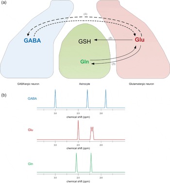
Functionally, Glu is possibly one of the most exciting neurotransmitters implicated in depression. It is the major excitatory neurotransmitter in the mammalian brain and the direct precursor for the main inhibitory neurotransmitter, γ-aminobutyric acid (GABA) (see Fig. 1a). It therefore acts as a neuromodulator and is additionally important in the synthesis of metabolites such as GSH and other peptides. Being an amino acid, Glu is unable to pass through the blood brain barrier and therefore has to be produced centrally within neurons and glia. Derived mainly from glucose and α-ketoglutarate, along with a smaller component from glutamine (Gln), the largest concentration of Glu is in glutamatergic neurons with smaller amounts in GABAergic neurons and astroglia. By contrast, Gln that is synthesised from Glu by the action of Gln synthase is found predominantly in astrocytes. Like its precursor, Gln is an amino acid and in conjunction with Glu forms the Glu–Gln neurotransmitter cycle (see Fig. 1a).
(a) Schematic depiction of the relationship between glutamatergic neurons, GABAergic neurons and astrocytes. Glu is the main excitatory neurotransmitter in the mammalian brain, and is a precursor for the production of the inhibitory neurotransmitter GABA. Gln is an amino acid synthesised in astrocytes that serves as an intermediary in neurotransmitter metabolism. GSH is an anti-oxidant synthesised in astrocytes that plays an important role in amino acid transport and has been implicated in mood disorders. (1) Represents the enzymatic pathways linking Glu and GABA; (2) Glu when released into the synaptic cleft is taken up by astrocytes and is used to synthesise GSH and (3) is the Glu–Gln neurotransmitter cycle. (b) Schematic representation of high-field 1H-MRS spectra for GABA, Glu and Gln. Note that the peaks are not shown to scale and that the signals from all three metabolites overlap considerably. It is this that makes partitioning and quantification of individual metabolites difficult.

Technological advances in 1H-MRS imaging and the development of specialised analysis techniques have made it possible to quantify metabolites such as Glu, GABA and Gln in vivo. However, issues remain with respect to the reproducibility and relative accuracy of the techniques that are used to make these measurements (Reference Henry, Lauriat, Shanahan, Renshaw and Jensen3). For example, to detect Glu, GABA and Gln in vivo a number of techniques (e.g. GABA-edited MEGA-PRESS, 2D J-resolved MRS and single-echo 30 ms PRESS) have been developed that permit separate and simultaneous measurement of these metabolites but even with high-field magnets and optimal 1H-MRS sequences reliable separation is not always possible (see Fig. 1b).
These technical difficulties have the potential to limit the salience of results from clinical studies. For instance, early research that relied on magnets of low-field strength generally measured composites of Glu and Glu-related metabolites termed Glx and not surprisingly, the findings from these studies were somewhat equivocal. However, more recent 1H-MRS studies that have been able to better discriminate between the various metabolites have achieved greater consistency, and as a consequence some valuable insights are gradually emerging. For example, studies of major depressive disorder have found that in depression the neural concentrations of Glx are significantly reduced but normalise with successful treatment (Reference Yüksel and ÖNgür4). By contrast, Glx is elevated in bipolar disorder and the change is seemingly independent of mood state.
At this point it is important to emphasise that these findings are at best preliminary and that there are exceptions to these results across both disorders. Further, only a handful of brain regions have been investigated thus far, and the effects of age, medication, gender, duration of illness, severity of symptoms, all of which are no doubt important have yet to be investigated. Nevertheless, the possibility that two subtypes of mood disorder can perhaps be distinguished using spectroscopy is an exciting prospect and the likelihood that the effects of medications may be measurable in the near future is of great potential value.
Current indications are that Glu, Glu-related metabolites and others such as myo-inositol will feature increasingly in depression and bipolar disorder pathophysiology. Thus in mood disorders, 1H-MRS is proving to be a valuable tool. Future research that combines spectroscopy findings with those from clinical investigations, neurocognitive studies and other imaging modalities is likely to provide novel insights into many neuropsychiatric disorders.