Implications
Resumption of ovulation and oestrous cycles are required to facilitate rebreeding of postpartum cows. Beef cows have a prolonged interval to resumption of ovulation compared with dairy cows because of suckling and maternal bond-inhibiting ovulation. The time to ovulation is limited by the lack of ovulation stimulus and not lack of follicle growth. In dairy cows, the key inhibitors of resumption of ovulation include energy balance changes, body condition score at calving, dry matter intake and health disorders. First, ovulation postpartum is generally silent (no expression of oestrous behaviour) and is generally followed by a short cycle.
Introduction
Reproductive efficiency in dairy and beef cows is dependent on achieving high submission rates and high conception rates per service. However, to achieve good submission and conception rates, cows must resume ovarian cycles, have normal uterine involution, be detected in oestrus and be inseminated at an optimum time. In seasonally calving herds, the aim is to achieve conception by 75 to 85 days, depending on breed, following parturition so that calving-to-calving intervals are maintained at 365 days. Reproductive performance of cows affects the efficiency of milk and beef production because of its influence on the calving to first service interval, subsequent calving pattern, length of lactation and culling rate, and culling rates for failure to conceive.
The pattern of resumption of ovarian function in both dairy and beef cows has been previously reviewed (Roche et al., Reference Roche, Crowe and Boland1992; Crowe, 2008). Resumption of ovarian cyclicity is largely dependent on LH pulse frequency. Both dairy and beef cows have early resumption of follicular growth within 7 to 10 days postpartum. The fate of the dominant follicle within the first follicular wave is dependent on LH pulse pattern. This paper updates these earlier reviews and will focus on the factors contributing to resumption of ovulation and affecting uterine health in postpartum dairy and beef cows.
Follicle growth
Ovarian follicle growth takes a period of 3 to 4 months and can be categorized into gonadotrophin-independent and gonadotrophin-dependent stages (Webb et al., Reference Webb, Garnsworthy, Gong and Armstrong2004). Gonadotrophin-dependent follicle growth in cattle occurs in waves (Rajakoski, Reference Rajakoski1960; Matton et al., Reference Matton, Adelakoun, Couture and Dufour1981; Ireland and Roche, Reference Ireland and Roche1987; Savio et al., Reference Savio, Keenan, Boland and Roche1988; Sirois and Fortune, Reference Sirois and Fortune1988). Each wave of growth involves emergence, selection and dominance followed by either atresia or ovulation. Emergence of a follicle wave is defined as growth of a cohort of follicles ⩾5 mm in diameter and coincides with a transient increase in FSH secretion (Adams et al., Reference Adams, Matteri, Kastelic, Ko and Ginther1992; Sunderland et al., Reference Sunderland, Crowe, Boland, Roche and Ireland1994). Follicle selection occurs in the face of declining FSH concentrations and is the process by which the growing cohort of follicles is reduced to the ovulatory quota for the species (in cattle it is generally one; Sunderland et al., Reference Sunderland, Crowe, Boland, Roche and Ireland1994). The selected follicle survives in an environment of reduced FSH owing to the development of LH receptors in the granulosa cells (Xu et al., Reference Xu, Garverick, Smith, Smith, Hamilton and Youngquist1995; Bao et al. Reference Bao, Garverick, Smith, Smith, Salfen and Youngquist1997) and increased intrafollicular bioavailable IGF-I (Austin et al., Reference Austin, Mihm, Evans, Knight, Ireland, Ireland and Roche2001; Canty et al., Reference Canty, Boland, Evans and Crowe2006). The increased bioavailable IGF-I is achieved by reduced IGF-I-binding proteins (IGFBP) because of increased IGFBP protease activity. Dominance is the phase during which the single selected follicle actively suppresses FSH concentrations and ensures the suppression of all other follicle growth on the ovaries (Sunderland et al., Reference Sunderland, Crowe, Boland, Roche and Ireland1994). The fate of the dominant follicle is then dependent on the prevailing LH pulse frequency during the dominance phase. In the presence of elevated progesterone (luteal phase of cyclic animals), LH pulse frequency is maintained at 1 pulse every 4 h and the dominant follicle undergoes atresia; in the follicular phase (preovulatory period in cyclic animals), the LH pulse frequency increases to 1 pulse per hour and this stimulates final maturation, increased oestradiol concentrations and positive feedback on gonadotrophin-releasing hormone (GnRH), LH (and FSH), in a surge that induces ovulation (Sunderland et al., Reference Sunderland, Crowe, Boland, Roche and Ireland1994). Normal follicle waves have an inherent lifespan of 7 to 10 days’ duration from the time of emergence of a wave until emergence of the next wave (indicating either ovulation or physiological atresia of the dominant follicle). In cyclic heifers during the normal 21-day oestrous cycle (range 18 to 24 days), there are normally three waves (sometimes two waves and rarely one or four waves; Savio et al., Reference Savio, Keenan, Boland and Roche1988; Murphy et al., Reference Murphy, Enright, Crowe, McConnell, Spicer, Boland and Roche1991; Forde et al., Reference Forde, Beltman, Lonergan, Diskin, Roche and Crowe2011).
Pregnancy
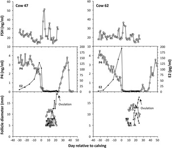
During pregnancy, follicular growth continues during the first two trimesters (Ginther et al., Reference Ginther, Knopf and Kastelic1989 and Reference Ginther, Kot, Kulick, Martin and Wiltbank1996) at regular 7 to 10-day intervals. In late pregnancy (last 22 days), the strong negative feedback of progestagens (mostly from the corpus luteum (CL) of pregnancy and partly of placental origin) and oestrogens (mostly of placental origin) suppresses the recurrent transient FSH rise that stimulates follicle growth (Ginther et al., Reference Ginther, Kot, Kulick, Martin and Wiltbank1996; Crowe et al., Reference Crowe, Padmanabhan, Mihm, Beitins and Roche1998; Figure 1) so that the ovaries are largely quiescent during the last 20 to 25 days of gestation. At parturition, the pituitary stores of LH are low because of the effects of elevated circulating concentrations of oestradiol, of placental origin, in late pregnancy (Nett et al., Reference Nett, Cermak, Braden, Manns and Niswender1987).
FSH, progesterone (P4), oestradiol (E2) and follicular diameter profiles in two representative beef cows from ~30 days prepartum until 50 days postpartum (Crowe et al., Reference Crowe, Padmanabhan, Mihm, Beitins and Roche1998).

Physiology of the postpartum period
At the time of parturition, progesterone and oestradiol concentrations reduce to basal concentrations. Parturition allows the removal of the negative feedback effects of elevated oestradiol and the recommencement of the synthesis of FSH and LH. The synthesized FSH is released into peripheral circulation as evidenced by the almost immediate resumption of recurrent transient increases in blood concentrations of FSH (within 3 to 5 days of parturition) that subsequently occur at 7- to 10-day intervals (Crowe et al., Reference Crowe, Padmanabhan, Mihm, Beitins and Roche1998). The first of these increases stimulates the first postpartum wave of follicle growth that generally produces a dominant follicle by 7 to 10 days postpartum (Murphy et al., Reference Murphy, Boland and Roche1990; Savio et al., Reference Savio, Boland and Roche1990a; Crowe et al., Reference Crowe, Goulding, Baguisi, Boland and Roche1993). The reaccumulation of the anterior pituitary stores of LH is slower than that of FSH and takes 2 to 3 weeks to complete. During this period, circulating concentrations of LH and LH pulse frequency are both low primarily owing to lack of releasable pools of LH in the gonadoptroph cells of the anterior pituitary. This is the case in all cows irrespective of whether they are milked or suckled (Silveira et al., Reference Silveira, Spoon, Ryan and Williams1993; Griffith and Williams, Reference Griffith and Williams1996). The synthesis and sequestration of LH requires only a low level of GnRH pulsatility. Between days 10 and 20 postpartum, the pulsatile release of LH increases in dairy cows (or in beef cows that are weaned). The concurrent LH pulse frequency determines the fate of the first follicular wave-dominant follicle that is dependent on its ability to secrete sufficient oestradiol to induce a gonadotrophin surge. The capacity for oestradiol secretion is in turn dependent on the prevailing LH pulse frequency during the dominant phase of the follicle wave, the size of the dominant follicle and IGF-I bioavailability (Austin et al., Reference Austin, Mihm, Evans, Knight, Ireland, Ireland and Roche2001; Canty et al., Reference Canty, Boland, Evans and Crowe2006). Therefore, the major driver for ovulation of a dominant follicle during the postpartum period is the GnRH/LH pulse frequency. In the beef suckler cows, the suppressive effect of suckling and maternal–offspring bonding on hypothalamic GnRH secretion prevents the establishment of the requisite LH pulse frequency that is required for oestradiol synthesis, induction of a pre-ovulatory LH surge and ultimately ovulation (Murphy et al., Reference Murphy, Boland and Roche1990; Crowe et al., Reference Crowe, Goulding, Baguisi, Boland and Roche1993; Duffy et al., Reference Duffy, Crowe, Boland and Roche2000). Eventually, the beef suckler cow will escape the effect of suckling and maternal–offspring bonding, resulting in an increased frequency of LH pulses and ovulation (Stagg et al., Reference Stagg, Spicer, Sreenan, Roche and Diskin1998). The major physiological difference between beef suckler and dairy cows at 15 to 20 days postpartum is the lower frequency of pulsatile release of LH in beef cows nursing their own calves (Silveira et al. Reference Silveira, Spoon, Ryan and Williams1993; Griffith and Williams, Reference Griffith and Williams1996) compared with the dairy cows.
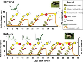
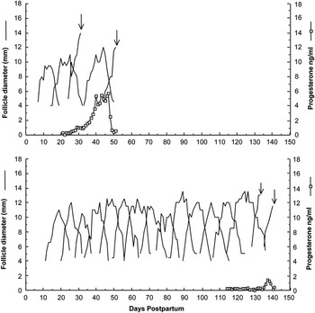
It is well established from studies using sheep as a model (and in cattle to a lesser extent) that LH pulsatile secretion reflects the GnRH pulsatile secretion pattern (Moenter et al., Reference Moenter, Caraty, Locatelli and Karsch1991; Skinner et al., Reference Skinner, Malpaux, Delaleu and Caraty1995; Skinner et al., Reference Skinner, Caraty, Malpaux and Evans1997; Gazal et al., Reference Gazal, Leshin, Stanko, Thomas, Keisler, Anderson and Williams1998). This has been tested and validated by the LH pulsatile infusion studies of Duffy et al. (Reference Duffy, Crowe, Boland and Roche2000) in early postpartum anoestrous beef cows. The LH pulse frequency required to stimulate a dominant follicle towards ovulation is one LH pulse per hour. Figure 2 depicts the likely fate of the early postpartum dominant follicles in beef and dairy cows. In beef cows, the first dominant follicle, and frequently a successive number of dominant follicles, generally fail(s) to ovulate (Murphy et al. Reference Murphy, Boland and Roche1990, Stagg et al. Reference Stagg, Diskin, Sreenan and Roche1995), and rather undergo atresia. With beef cows in good body condition, the first postpartum dominant follicle to ovulate is generally from wave 3.2±0.2 (~30 days; Murphy et al., Reference Murphy, Boland and Roche1990), whereas for beef cows in poor body condition there are typically 10.6±1.2 waves of follicular growth before ovulation occurs (~70 to 100 days; Stagg et al., Reference Stagg, Diskin, Sreenan and Roche1995; Figure 3). In the case of dairy cows’ ovulation of the first postpartum dominant follicle typically occurs in 30% to 80% of cows, whereas it undergoes atresia in 15% to 60% of cows or becomes cystic in 1% to 5% of cows (Savio et al., Reference Savio, Boland, Hynes and Roche1990b; Beam and Butler Reference Beam and Butler1997; Sartori et al., Reference Sartori, Haughian, Shaver, Rosa and Wiltbank2004; Sakaguchi et al., Reference Sakaguchi, Sasamoto, Suzuki, Takahashi and Yamada2004). There is no evidence that lack of FSH or a delayed resumption of ovarian follicle waves are causes of prolonged postpartum anoestrous intervals in either beef (Crowe et al., Reference Crowe, Padmanabhan, Mihm, Beitins and Roche1998; Stagg et al., Reference Stagg, Spicer, Sreenan, Roche and Diskin1998) or dairy cows (Beam and Butler, Reference Beam and Butler1999).
Diagrammatic scheme of resumption of dominant follicles and ovarian cycles during the postpartum period in dairy and beef suckler cows not nutritionally stressed. LH pulse frequency is that occurring during an 8-h window where cows are blood sampled every 15 min. Short cycles occur in most (70%), but not all cows after first ovulation (adapted from Roche et al., Reference Roche, Crowe and Boland1992).

Pattern of growth and regression of dominant follicles from calving to second ovulation in (a) a beef suckler cow with two non-ovulatory follicle waves before the first ovulation, and (b) a beef suckler cow with 14 non-ovulatory waves before the first ovulation. Arrows indicate ovulation. Taken from Stagg et al. (Reference Stagg, Diskin, Sreenan and Roche1995).

First ovulation in both dairy and beef cows is generally silent (i.e. no behavioural oestrus; Kyle et al., Reference Kyle, Callahan and Allrich1992) and is generally (>70%) followed by a short cycle, usually containing just one follicle wave. This first luteal phase is of short duration because of the premature release of prostaglandin F2α (PGF2α; Peter et al., Reference Peter, Bosu, Liptrap and Cummings1989), thought to be induced by increased oestradiol produced from the formation of the postovulatory dominant follicle on days 5 to 8 of the cycle, inducing premature oestradiol and oxytocin (Zollers et al., Reference Zollers, Garverick, Smith, Moffatt, Salfen and Youngquist1993) receptors. Thus, the CL, which also secretes lower quantities of progesterone, regresses prematurely around days 8 to 10 of the cycle, with the second ovulation (of this postovulatory dominant follicle) occurring around days 9 to 11 after the first ovulation. This second ovulation is generally associated with the expression of oestrous behaviour and followed by a luteal phase of normal duration generating normal concentrations of progesterone (Crowe et al., Reference Crowe, Padmanabhan, Mihm, Beitins and Roche1998).
Cyclic cows may have two, three or, very occasionally, four follicle waves during the oestrous cycles that occur in the postpartum period (Savio et al., Reference Savio, Boland and Roche1990a; Sartori et al., Reference Sartori, Haughian, Shaver, Rosa and Wiltbank2004). Unlike non-lactating heifers, lactating Holstein postpartum dairy cows tend to have two follicle waves per 18- to 23-day cycle (Sartori et al., Reference Sartori, Haughian, Shaver, Rosa and Wiltbank2004). Blood concentration of progesterone is the major factor that affects LH pulse frequency in cyclic animals. Generally, lactating Holstein dairy cows tend to have lower blood concentrations of progesterone during the cycle than cyclic heifers (Sartori et al., Reference Sartori, Haughian, Shaver, Rosa and Wiltbank2004; Wolfenson et al., Reference Wolfenson, Inbar, Roth, Kaim, Bloch and Braw-Tal2004). These lower progesterone concentrations tend to allow a subtle increase in LH pulse frequency and allows for prolonged growth of each dominant follicle rather than the faster atresia that occurs in cyclic heifers. Cows with prolonged luteal phases tend to have a 4th follicle wave (Savio et al., Reference Savio, Boland and Roche1990a). The number of follicle waves or rate of turnover of dominant follicles are directly related to the duration of dominance of each dominant follicle, and cattle with shorter durations of dominance for the ovulatory dominant follicles tend to have higher conception rates (Austin et al., Reference Austin, Mihm, Ryan, Williams and Roche1999). Therefore, nutrition, by altering metabolic clearance of progesterone, can affect the duration of dominance of a dominant follicle, the number of follicle waves per cycle and have an indirect effect on conception rates.
Negative feedback effects of oestradiol
From the foregoing, it is clear that the regulation of LH secretion is the key driver of resumption of ovulation in postpartum cows. A decrease in the concentration of LH and a suppression in the frequency of LH pulses had been reported in nutritionally induced anoestrous beef cows (Richards et al., Reference Richards, Wettemann and Schoenemann1989), heifers (Imakawa et al., Reference Imakawa, Day, Garcia-Winder, Zalesky, Kittok, Schanbacher and Kinder1986), and occurs as a result of reduced GnRH secretion from the hypothalamus. Long-term oestradiol pre-treatment during prolonged undernutrition reduced the magnitude of the induced LH surge to just 20% of that observed in lambs not chronically treated with oestradiol, suggesting that low levels of oestradiol have a strong negative feedback effect on undernourished animals. In chronically underfed cows, Richards et al. (1991) also implicated oestradiol negative feedback in the reduced hypothalamic and pituitary response to an oestradiol challenge. More recent evidence suggests that the pituitary sensitivity to GnRH pulses is decreased during undernutrition. Tatman et al. (Reference Tatman, Judkins, Dunn and Moss1990) found that the pituitary content of LH was lower in thin ewes; Kile et al. (Reference Kile, Alexander, Moss, Hallford and Nett1991) found that undernutrition suppressed pituitary synthesis of LH, as the concentration of mRNA for both α and β subunits of LH were less in nutritionally restricted ovariectomized ewes, although pulsatile administration of GnRH was capable of restoring LH synthesis and secretion. Similarly, cows fed restricted diets released more LH in response to exogenous GnRH than cows fed moderate or high diets (Whisnant et al., Reference Whisnant, Kiser, Thompson and Hall1985; Rasby et al., Reference Rasby, Wettemann, Geisert, Wagner and Lusby1991) and had increased concentrations of GnRH in the median eminence stalk of the hypothalamus (Rasby et al., Reference Rasby, Wettemann, Harms, Lusby and Wagner1992), suggesting that the greater sequestration of LH in the anterior pituitary gland and decreased LH secretion in nutritionally restricted animals is because of reduced GnRH release. Therefore, it appears that the nutritionally induced suppression of LH may be at least partly modulated by factors affecting the GnRH pulse generator and the pituitary response to GnRH, but the fuller mechanism(s) responsible remain to be elucidated.
Anovulatory anoestrus
Factors contributing to GnRH/LH pulse frequency in early postpartum beef cows
The major factors that control the GnRH/LH pulse frequency (and therefore the fate of early postpartum dominant follicles) in postpartum beef cows include maternal bond/calf presence (presumably owing to effects on opioid release), suckling inhibition (Myers et al., Reference Myers, Myers, Gregg and Moss1989; Stagg et al., Reference Stagg, Diskin, Sreenan and Roche1995) and poor body condition (Canfield and Butler, Reference Canfield and Butler1990). Calf presence has a very clear negative effect on the resumption of ovulation in beef cows’ nursing calves. Restricted suckling of beef cows (once per day) from day 30, where calves were in an isolated pen away from the sight of cows, significantly shortened the interval from calving to first ovulation (51 days) compared with cows where the calves had ad libitum access (79 days; Stagg et al., Reference Stagg, Diskin, Sreenan and Roche1995). The effect of calf presence can be further compartmentalized into suckling stimuli (mammary sensory pathways) and maternal behaviour/bonding effects (Silveira et al., Reference Silveira, Spoon, Ryan and Williams1993; Williams et al., Reference Williams, Mcvey and Hunter1993) but requires positive calf identification by either sight or olfaction (Griffith and Williams, Reference Griffith and Williams1996). Cows that calve down in poor BCS (<2.5) are more likely to have a prolonged anoestrous period (Stagg et al., Reference Stagg, Diskin, Sreenan and Roche1995), presumably owing to lower LH pulse frequency (Stagg et al., Reference Stagg, Spicer, Sreenan, Roche and Diskin1998).
As beef cows (with prolonged anovulatory anoestrus) approach their first postpartum ovulation, LH pulse frequency increases (observed during each sequential follicle wave from six waves before ovulation until the ovulatory wave; Stagg et al., Reference Stagg, Spicer, Sreenan, Roche and Diskin1998). Concentrations of IGF-I increased linearly from 75 days before first ovulation until ovulation, which was associated with a linear decrease in growth hormone concentrations during the same period (Stagg et al., Reference Stagg, Spicer, Sreenan, Roche and Diskin1998). In addition to increased circulating concentrations of IGF-I that help to stimulate dominant follicle maturation and growth so that there is sufficient secretion of oestradiol to induce an LH surge and ovulation. Management may be used to encourage earlier ovulation by restricting suckling/access of the cows to the calves from approximately day 30 postpartum (Stagg et al. 1998) or by increased plane of nutrition and body condition. The available evidence would indicate that the majority (85%) of beef cows are capable of ovulating by 35 days postpartum (Murphy et al., Reference Murphy, Boland and Roche1990; Crowe et al., Reference Crowe, Goulding, Baguisi, Boland and Roche1993; Duffy et al., Reference Duffy, Crowe, Boland and Roche2000; Mackey et al., 2000). Removal of the suckling/maternal calf bond results in a doubling of LH pulse frequency within 48 h of calf separation followed by subsequent ovulation of the concurrent or next dominant follicle. Interestingly, in the small proportion of cows that fail to respond to the removal of the suckling/maternal calf bond show no increase in LH pulse frequency, and no evidence of an increase in the circulating concentrations of oestradiol. These non-responders typically had prolonged postpartum anoestrous intervals and could be described as being in “deep anovulatory anoestrus” (Sinclair et al., Reference Sinclair, Revilla, Roche, Quintans, Sanz, Mackey and Diskin2002).
Factors contributing to LH pulse frequency in early postpartum dairy cows
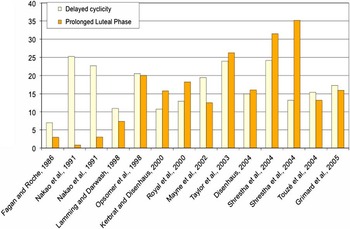
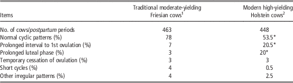
In dairy cows, the major factors affecting resumption of ovulation include BCS and energy balance (yield and dry matter intake), parity, season and disease (Bulman and Lamming, Reference Bulman and Lamming1978; Beam and Butler, Reference Beam and Butler1997; Opsomer et al., Reference Opsomer, Grohn, Hertl, Coryn, Deluyker and de Kruif2000; Wathes et al., Reference Wathes, Bourne, Cheng, Mann, Taylor and Coffey2007). Energy intake, BCS and milk yield interact to affect energy balance in dairy cows. There is evidence to link many of these factors to reduce LH pulse frequency. A number of studies have been conducted on dairy cows of various yield potential that have categorized the pattern of resumption of ovarian function by measuring milk progesterone. These range from a study by Fagan and Roche (Reference Fagan and Roche1986) using what would now be classified as traditional moderate yielding Friesian cows (4000 to 5000 kg milk per lactation) to that of Opsomer et al. (Reference Opsomer, Coryn, Deluyker and de Kruif1998) using modern high-yielding Holstein type cows (6900 to 9800 kg milk per lactation). The data from these two studies are summarized in Table 1. Furthermore, this pattern of resumption of ovarian function has been validated in a series of equivalent papers and the two key problem categories (prolonged interval to first ovulation and prolonged luteal phase) are summarized in Figure 4. Risk factors for these two ovarian abnormalities have been determined in a large epidemiological study by Opsomer et al. (Reference Opsomer, Grohn, Hertl, Coryn, Deluyker and de Kruif2000). The major risk factors for a prolonged interval from calving to first ovulation included (odds ratio in parentheses): acute body condition score (BCS) loss up to 60 days post calving (18.7 within 30 days, 10.9 within 60 days), clinical ketosis (11.3), clinical diseases (5.4), abnormal vaginal discharge (4.5) and difficult calving (3.6).
Percentage of cows defined as having either (i) delayed resumption of ovulation or (ii) prolonged luteal phases based on the evaluation of milk progesterone profiles across a number of studies in dairy cows (compiled by Benedicte Grimard, France, personal communication).

Pattern of resumption of ovarian cyclicity in postpartum dairy cows (traditional moderate-yielding Friesians v. modern high-yielding Holsteins), using milk progesterone profiling (samples collected twice weekly)

*Categories with a major disparity between the two studies.
1 Fagan and Roche (Reference Fagan and Roche1986).
2 Opsomer et al. (Reference Opsomer, Coryn, Deluyker and de Kruif1998).
The greatest of these risk factors is acute BCS loss. Current evidence suggests that dairy cows should calve down in a BCS of 2.75 to 3.0 and not lose more than 0.5 of a BCS unit between calving and first service (Overton and Waldron Reference Overton and Waldron2004; Mulligan et al., Reference Mulligan, O’Grady, Rice and Doherty2006) rather than earlier recommendations of 3.0 to 3.5 (Buckley et al., Reference Buckley, O’Sullivan, Mee, Evans and Dillon2003). Cows that lose excessive body condition (⩾1.0 BCS unit) have a longer postpartum interval to first ovulation. Thus, monitoring BCS from before calving to first service is essential for good reproductive management. BCS changes are good indicators of energy balance and reflect milk yield and dry matter intake. It is necessary to prevent an acute loss of BCS and shorten the duration of severe negative energy balance postpartum. This is best achieved by ensuring that dry matter intake in the early postpartum period is maximized and by having cows in appropriate BCS (2.73 to 3.0) at calving. Cows that are mobilizing tissue at a high rate have increased blood non-esterified fatty acids, and β-hydroxy butyrate, but reduced concentrations of insulin, glucose and IGF-I (Grummer et al., Reference Grummer, Mashek and Hayirli2004). The metabolic status associated with high rates of tissue mobilization increases the risk for hypocalcaemia, acidosis, fatty liver, ketosis and displaced abomasa (Gröhn and Rajala-Schultz Reference Gröhn and Rajala-Schult2000; Overton and Waldron Reference Overton and Waldron2004, Maizon et al., Reference Maizon, Oltenacu, Gröhn, Strawderman and Emanuelson2004). Cows affected by these metabolic disorders are more prone to anoestrus, mastitis, lameness and subsequently reduced conception rate to AI (Fourichon et al., Reference Fourichon, Seegers and Malher1999; Gröhn and Rajala-Schultz Reference Gröhn and Rajala-Schult2000; Lucy Reference Lucy2001; López-Gatius et al., Reference López-Gatius, Santolaria, Yániz, French and López-Béjar2002; Maizon et al., Reference Maizon, Oltenacu, Gröhn, Strawderman and Emanuelson2004). It is hypothesized that serum IGF-I concentrations could be useful as a predictor of nutritional status and hence reproductive efficiency in dairy cows (Zulu et al., Reference Zulu, Nakao and Sawamukai2002a). Plasma IGF-I concentrations before calving and in the first few weeks of lactation have been linked to subsequent cyclicity and conception rate (Diskin et al., Reference Diskin, Murphy and Sreenan2006; Taylor et al., Reference Taylor, Beever, Bryant and Wathes2006). This emphasizes the critical role of correct nutritional management to ensure that the deficit in energy balance post calving is mild rather than severe, and kept to as minimum duration as possible. Current approaches to minimize the energy balance deficit post calving includes: the optimization of BCS at calving (2.75 to 3.0), shorter dry periods and maintenance of normal rumen function (Mulligan et al., Reference Mulligan, O’Grady, Rice and Doherty2006).
In dairy cows, one of the main drivers of negative energy balance is BCS at calving because cows calving above BCS 3.0 have both reduced appetite and mobilize in excess of 1 BCS unit, with subsequent detrimental effects on fertility (Mulligan et al., Reference Mulligan, O’Grady, Rice and Doherty2006). Recently, several laboratories have examined the effects of glucogenic–lipogenic feeding strategies for dairy cows on reproductive function (Garnsworthy et al., Reference Garnsworthy, Fouladi-Nashta, Mann, Sinclair and Webb2009; Friggens et al., Reference Friggens, Disenhaus and Petit2010). There is evidence that the administration of a glucogenic diet, which promotes increased circulating concentrations of insulin and glucose (Van Knegsel et al., Reference Van Knegsel, van den Brand, Dijkstra, Tamminga and Kemp2005), would be expected to improve several reproductive variables. Gong et al. (Reference Gong, Lee, Garnsworthy and Webb2002) found that the administration of a glucogenic diet increased circulating concentrations of insulin and increased the proportion of cows ovulating by 50 days after calving, which is highly desirable. Further support for this concept has come from New Zealand studies (Burke et al., Reference Burke, Kay, Phyn, Meier, Lee and Roche2010) that also recorded a shorter postpartum anoestrous interval in pasture/grass silage-fed dairy cows supplemented with 5 kg/day of corn and barley-based concentrate. However, there is evidence that high circulating concentrations of insulin have negative effects on both oocyte quality (Fouladi-Nashta et al.. Reference Fouladi-Nashta, Gutteriez, Garnsworthy and Webb2005; Garnsworthy et al., Reference Garnsworthy, Lock, Mann, Sinclair and Webb2008a), and in vitro embryo production from overfed, superstimulated heifers (Freret et al., Reference Freret, Grimard, Ponter, Joly, Ponsart and Humblot2006). In contrast, lipogenic diets increased the oestradiol-secreting capacity of preovulatory follicles, thus providing enhanced substrate for progesterone production (Leroy et al., Reference Leroy, Van Soom, Opsomer and Bols2008) and improved blastocyst development rates. Garnsworthy et al. (Reference Garnsworthy, Sinclair and Webb2008b) proposed a strategy of glucogenic diets during the early postpartum period to hasten the onset of regular oestrous cycles followed by more lipogenic diets to lower circulating insulin and improve oocyte quality at breeding. Although this has a strong physiological basis, there is a need for confirmatory studies of the concept.
Disease state may also regulate follicle fate via LH and other mechanisms. Uterine conditions such as retained foetal membranes, endometritis and metritis contribute to reproductive efficiency via various mechanisms. Other diseases such as mastitis Huszenicza et al. (Reference Huszenicza, Janosi, Kulcsar, Korodi, Reiczigel, Katai, Paters and de Rensis2005)and lameness (Petersson et al., Reference Petersson, Strandberg, Gustafsson and Berglund2006) delay resumption of luteal activity by 7 to 17 days, respectively. For these there is considerable evidence that this is mediated due to acute stressors reducing GnRH and hence LH pulse frequency, leading to decreased oestradiol production by dominant follicles and preventing or reducing the gonadotrophin surge, thus delaying ovulation.
Role of insulin
Insulin is primarily involved in glucose homeostasis but also serves as a metabolic signal influencing pituitary release of LH (Monget and Martin, Reference Monget and Martin1997) and ovarian responsiveness to gonadotrophins (Stewart et al., Reference Stewart, Spicer, Hamilton and Keefer1995). Plasma insulin concentrations are influenced by both BCS and level of nutrition (Vizcarra et al., Reference Vizcarra, Wettemann, Spitzer and Morrison1998), and may serve as a more sensitive indicator of nutritional status than BCS. In a transnational study, Sinclair et al. (Reference Sinclair, Revilla, Roche, Quintans, Sanz, Mackey and Diskin2002) showed that postpartum anoestrous beef cows with low (<5 mIU/l) plasma concentrations of insulin were unable to ovulate a dominant follicle in response to restricted suckling, unlike cows with higher (>5 mIU/l) plasma concentrations of insulin, notwithstanding an increase in LH pulse frequency. The results of that study are consistent with those of Gong et al. (2001, 2002) who showed that dairy cows fed a diet, which increased circulating concentrations of insulin during the first 50 days postpartum had shorter postpartum anoestrous intervals, independent of any effects on LH or FSH and without affecting milk yield or energy balance. The latter three studies would argue for a direct effect of insulin at the ovarian level. The inability to respond to increased LH pulse frequency may be owing to lack of granulosa cell LH receptors, which are known to be dependent on the combined actions of FSH and oestradiol 17β (Bao and Garverick, Reference Bao and Garverick1998). Follicular oestradiol-17β is, in turn, dependent on LH-stimulated production of androgens from the thecal cells that are dependent on peripheral concentrations of insulin and IGF-I (Stewart et al., Reference Stewart, Spicer, Hamilton and Keefer1995). Therefore, low plasma concentrations of insulin could reduce androgen and oestradiol production and thus compromise the ability of follicles to acquire LH receptors.
Prolonged luteal phases
After the resumption of ovarian cyclicity, prolonged luteal phases are the main cause of irregular oestrous cycles in cows. The incidence of prolonged luteal phases has increased from 3% (Fagan and Roche, Reference Fagan and Roche1986) to 11% to 22% (Lamming and Darwash, Reference Lamming and Darwash1998; Opsomer et al., Reference Opsomer, Coryn, Deluyker and de Kruif1998; Shrestra et al. 2004a,b; Figure 4). It is generally considered that prolonged luteal phases are associated with an abnormal uterine environment that disrupts endometrial PGF2α production. Interestingly, in the study by Opsomer et al. (Reference Opsomer, Coryn, Deluyker and de Kruif1998), where the incidence of cows with prolonged luteal phases was 20% (89/448 cows), only 43/89 cows had abnormal uterine content, 2/89 had ovarian cysts and 44/89 had no detectable abnormalities. However, in that study, abnormalities were identified only by rectal palpation. The major risk factors for a prolonged luteal phase in cows having resumed ovulation included (odds ratio, in descending order of importance, in parentheses; Opsomer et al., Reference Opsomer, Grohn, Hertl, Coryn, Deluyker and de Kruif2000): metritis (11.0), abnormal vaginal discharge (4.4), retained placenta (3.5), parity (2.5 for parity 4+ v. primiparous) and earlier resumption of ovulation (2.8 for resumption<19 days postpartum, 2.4 for resumption 19 to 24 days postpartum). These data support the concept that prolonged luteal phases are related to uterine problems rather than ovarian problems.
Follicular cysts
Follicular cysts occur where dominant follicles in the early postpartum period (often the first dominant follicle postpartum) fail to ovulate. Cysts typically continue to grow to diameters >20 to 25 mm over a 10- to 40-day period in the absence of a CL (Savio et al., Reference Savio, Boland and Roche1990a; Gümen et al. 2002; Hatler et al. 2003). This continued growth appears to be owing to lack of positive feedback induced by oestradiol and thus failure of induction of the LH/FSH pre-ovulatory surge, despite increased LH pulse frequency (to an intermediate level). At this time, systemic concentrations of progesterone are low, whereas concentrations of oestradiol are elevated above normal pro-oestrus concentrations (Savio et al., Reference Savio, Boland, Hynes and Roche1990b; Hatler et al. 2003), resulting in many cases in strong exhibition of oestrous behaviour by cows in the early phases of a follicular cyst. This is followed by a period of time when there is an absence of oestrous behaviour in the second half of the cysts’ lifespan. The elevated oestradiol in conjunction with elevated inhibin suppresses blood concentrations of FSH, so that no new follicle waves emerge during the early active phase of a follicular cyst. The cyst then becomes oestrogen inactive (despite being morphologically still present) and a new follicle wave emerges. The dominant follicle of this new wave may either ovulate, undergo atresia or also become cystic. Many cows with follicular cysts correct themselves, but some develop sequential follicular cysts. The metabolic risk factors associated with dairy cows developing cysts in the early postpartum period are over body-conditioned cows, a reduction in systemic insulin (Vanholder et al., Reference Vanholder, Leroy, Dewulf, Duchateau, Coryn, deKruif and Opsomer2005) and IGF-I, and increased non-esterified fatty acids (Zulu et al., Reference Zulu, Sawamukai, Nakada, Kida and Moriyoshi2002b). The typical incidence for follicular cysts in dairy cows is between 1% and 5% (Opsomer et al., Reference Opsomer, Coryn, Deluyker and de Kruif1998; Beam and Butler, Reference Beam and Butler1999).
Postpartum uterine health
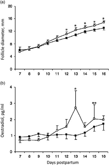
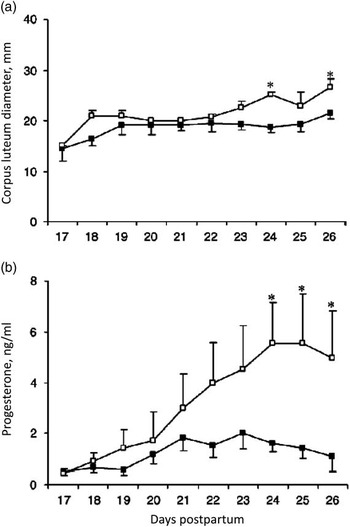
Postpartum uterine infection is a major contributor to reduced fertility in dairy cattle. Following parturition, the uterus becomes contaminated with bacteria, and although many animals can clear this contamination, infection persists in up to 20% of cows as endometritis (Sheldon et al., Reference Sheldon, Cronin, Goetz, Donofrio and Schuberth2009). There is evidence that uterine infections contribute to reduced fertility via a number of mechanisms. Bacterial products or immune mediators produced in response to infection suppress pituitary LH secretion and are associated with the inhibition of folliculogenesis, decreased ovarian steroidogenesis, abnormal luteal phases and a higher incidence of cystic ovarian disease (Peter et al., Reference Peter, Bosu, Liptrap and Cummings1989; Huszenicza et al., Reference Huszenicza, Fodor, Gacs, Kulcsar, Dohmen and Vamos1999, Opsomer et al., Reference Opsomer, Grohn, Hertl, Coryn, Deluyker and de Kruif2000; Mateus et al., Reference Mateus, Lopes da Costa, Bernardo and Silva2002 and Reference Mateus, Lopes da Costa, Diniz and Ziecik2003; Sheldon et al., Reference Sheldon, Noakes, Rycroft, Pfeiffer and Dobson2002; Williams et al., Reference Williams, Fischer, Noakes, England, Rycroft, Dobson and Sheldon2007). In a study on 82 clinically normal postpartum cattle with no risk factors for uterine disease, 75% of the animals had high numbers of uterine pathogens on day 7 postpartum, the predominant isolate being Escherichia coli. These animals also had retarded ovarian follicle growth; the first postpartum dominant follicle grew slower and produced less oestradiol (Figure 5; Williams et al., Reference Williams, Fischer, Noakes, England, Rycroft, Dobson and Sheldon2007 and Reference Williams, Sibley, Miller, Lane, Fishwick, Nash, Herath, England, Dobson and Sheldon2008a). Furthermore, in the animals that ovulated, the CL was smaller and produced less progesterone (Figure 6; Williams et al., Reference Williams, Fischer, Noakes, England, Rycroft, Dobson and Sheldon2007). Within the uterus E. coli may disrupt the mechanisms of PG-induced luteolysis in cyclic cows and therefore contribute to prolonged luteal phases by switching PG synthesis away from PGF2α towards PGE2 (Herath et al., Reference Herath, Lilly, Fischer, Williams, Dobson, Bryant and Sheldon2009; Williams et al., Reference Williams, Herath, England, Dobson, Bryant and Sheldon2008b).
Mean±s.e.m. (a) diameter of the first dominant follicle and (b) peripheral plasma oestradiol concentrations between days 7 and 16 postpartum for cows with a high (■) or low (□) day 7 uterine pathogen growth density. Values differ between groups within day, *P<0.05 and **P<0.01. Adapted from Williams et al. (Reference Williams, Fischer, Noakes, England, Rycroft, Dobson and Sheldon2007).

Mean±s.e.m. (a) diameter of the corpus luteum and (b) peripheral plasma progesterone concentrations between days 17 and 26 postpartum for cows with a high (■) or low (□) day 7 uterine pathogen growth density. At every time point n⩾8. *Values differ between groups within day (P<0.05). Adapted from Williams et al. (Reference Williams, Fischer, Noakes, England, Rycroft, Dobson and Sheldon2007).

These observations show a direct correlation between the presence of uterine pathogens, particularly E. coli, on day 7 postpartum and suboptimal ovarian function for the following 3 weeks. However, the evidence shows that early postpartum uterine disease contributes to infertility in cows by disrupting ovarian function and that these adverse effects may persist after uterine disease has resolved. The specific mechanisms by which uterine infection disrupts ovarian function are many and diverse, but there is substantial evidence that the endotoxin lipopolysaccharide (LPS) is a key disruptor of ovarian function. In addition to being detected in the uterus and peripheral circulation, LPS has been detected in follicular fluid of cattle with uterine disease; unsurprisingly, concentrations of LPS are directly correlated with bacterial load. (Peter et al., Reference Peter, Bosu, Liptrap and Cummings1989; Mateus et al., Reference Mateus, Lopes da Costa, Diniz and Ziecik2003; Herath et al., Reference Herath, Williams, Lilly, Gilbert, Dobson, Bryant and Sheldon2007; Williams et al., Reference Williams, Fischer, Noakes, England, Rycroft, Dobson and Sheldon2007). Administration of intravenous LPS disrupts neuroendocrine activity and results in interference with the oestrous cycle. In heifers, given an intrauterine infusion of LPS, the pre-ovulatory LH surge was blocked, resulting in the formation of cystic follicles (Peter et al., Reference Peter, Bosu, Liptrap and Cummings1989). Intravenous administration of LPS in sheep, suppressed hypothalamic GnRH secretion, inhibited pulsatile LH secretion and reduced pituitary responsiveness to GnRH (Williams et al., 2001). Ovarian function can also be disrupted following LPS administration in the absence of gonadotrophic effects. An inhibition of peripheral plasma oestradiol concentrations was observed, despite normal plasma LH concentrations (Xiao et al., 1998; Battaglia et al., 2000). In heifers, LPS delayed ovulation by interrupting the preovulatory oestradiol rise, thus delaying the LH surge (Suzuki et al., Reference Suzuki, Yoshioka, Iwamura and Hirose2001). Infusion of low concentrations of LPS in utero delayed the time interval over which follicles attained dominance and ovulation, although having no effect on LH or FSH concentrations (Williams et al., Reference Williams, Sibley, Miller, Lane, Fishwick, Nash, Herath, England, Dobson and Sheldon2008a). Furthermore, in cows, uterine infection does not affect peripheral plasma FSH concentrations or the consequent emergence of a wave of growing follicles (Sheldon et al., Reference Sheldon, Noakes, Rycroft, Pfeiffer and Dobson2002; Williams et al., Reference Williams, Fischer, Noakes, England, Rycroft, Dobson and Sheldon2007). These studies provide evidence for localized effects of LPS in the ovary. Indeed, in vitro LPS has been shown to disrupt granulosa cell oestradiol secretion via reduced expression of aromatase enzyme expression (Herath et al., Reference Herath, Williams, Lilly, Gilbert, Dobson, Bryant and Sheldon2007) and acute exposure to LPS increases follicular atresia and reduces the primordial ovarian follicle pool (Bromfield and Sheldon, 2013).
In beef cows, the effect of uterine infection on ovarian function has not been studied as extensively as in the dairy cow. However, it could be expected that the mechanisms by which ovarian functions are disrupted would be very similar. The incidence of uterine inflammation, as determined by neutrophil infiltration was similar, or even higher in beef cows than in dairy cows in the early weeks after calving (Santos et al., Reference Santos, Lamb, Brown and Gilbert2009), although the incidence of clinical endometritis at this time may be lower in beef cows (Williams, unpublished observations). In contrast, the incidence of cytological endometritis is higher in dairy cattle later in the postpartum period and it has been hypothesized that the differences in the timing of the first postpartum ovulation between beef and dairy cattle may influence uterine health during this time (Santos et al., Reference Santos, Lamb, Brown and Gilbert2009).
Induction of oestrus and ovulation in anovulatory anoestrous cows
From the previous sections, it is clear that in many cases (especially with dairy cows) anovulatory anoestrus is associated with management risk factors and other diseases (excessive loss of BCS, severe lameness, uterine disease, displaced abomasum, etc.). Therefore, before embarking on a specific treatment for anoestrus, the underlying factors and diseases should always be first addressed before the commencement of specific treatments for the ovarian problems.
GnRH
The major cause of delayed ovulation in postpartum cows is an infrequent LH pulse frequency (and by inference GnRH pulse frequency). GnRH treatment was used with variable effectiveness in numerous studies on postpartum cows when the follicle status of the animals was unknown. A single injection, two injections 10 days apart, or frequent low-dose injections at 1- to 4-h intervals of GnRH or GnRH analogues failed consistently to induce ovulation in over 90% of treated anoestrous cows (Mawhinney et al., Reference Mawhinney, Roche and Gosling1979; Riley et al., Reference Riley, Peters and Lamming1981; Walters et al., Reference Walters, Short, Convey, Staigmiller, Dunn and Kaltenbach1982; Edwards et al., Reference Edwards, Roche and Niswender1983). However, when a GnRH analogue (20 µg Buserelin) was used at known stages of follicle growth (determined by daily ultrasound scanning) of the first postpartum dominant follicle, all cows ovulated when administered during the growing phase of the dominant follicle (12/12) and the majority (7/10) ovulated when the first postpartum dominant follicle was in its plateau/early declining phase of growth (Crowe et al., Reference Crowe, Goulding, Baguisi, Boland and Roche1993). In a further study conducted by Ryan et al. (Reference Ryan, Mihm and Roche1998), 250 µg GnRH resulted in ovulation in 20 cows when administered at dominance of a follicular wave; this was followed by emergence of a new wave of ovarian follicular growth 1.6±0.3 days later and dominance of the subsequent wave was attained in 5±0.3 days. However, there was no effect of GnRH on follicular dynamics when given at emergence of a follicular wave. The existing cohort of follicles continued to develop unaffected in 17 cows, and dominance occurred 3.6±0.5 days later. Thus, GnRH may cause ovulation or no effect on follicle development depending on the animal’s stage of follicle development at treatment. As a result, when GnRH is used as part of an ovsynch protocol (GnRH-PGF2α-GnRH treatment) in postpartum anoestrous cows the effectiveness of the treatment is wholly dependent on the presence or absence of a dominant follicle at the time the first GnRH injection is administered.
Progesterone
Treatment of anoestrous cows with progesterone (and oestradiol) will induce oestrus and shorten the postpartum interval to conception (Rhodes et al., Reference Rhodes, McDougall, Burke, Verkerk and Macmillan2003). Currently, the use of oestradiol is banned in food producing animals within the EU and elsewhere (Lane et al., Reference Lane, Austin and Crowe2008). Anoestrous cows require progesterone pre-treatment to ensure that the first ovulation is associated with the expression of oestrus and a normally functioning luteal phase. The standard progesterone treatment regime for cows known to be in anoestrus is a 7- to 9-day intravaginal device with the optional use of equine chorionic gonadotrophin (eCG) on the day of removal of the device (Lane et al., Reference Lane, Austin and Crowe2008). The use of eCG may accompany progesterone treatment in cows that are in “deep anovulatory anoestrus” to ensure ovulation (Mulvehill and Sreenan, Reference Mulvehill and Sreenan1977), but care must be taken not to induce too high an ovulation rate by using doses in excess of 500 IU. In a herd situation where there is a mix of anoestrous and cyclic cows, then it is better to treat all cows as if they were cyclic; that is, use a 7- to 9-day progesterone treatment as an intravaginal device that is accompanied with a single GnRH injection at the time of progesterone device insertion, and an injection of PGF2α on the day before device removal (Lane et al., Reference Lane, Austin and Crowe2008).
Restricted suckling (beef cows)
Earlier onset of ovulation in beef cows may be induced by restricting suckling from 30 days postpartum (Stagg et al., Reference Stagg, Spicer, Sreenan, Roche and Diskin1998). Restricted suckling involves once or twice daily access of calves to cows for suckling, and at other times of the day the calves are isolated and out of direct physical contact and possibly sight of the dams (Stagg et al., Reference Stagg, Spicer, Sreenan, Roche and Diskin1998).
Summary and conclusions
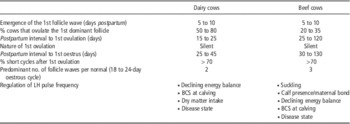
Follicular growth generally resumes within 7 to 10 days postpartum in the majority of both dairy and beef cows and is associated with a transient FSH rise that occurs within 3 to 5 days of parturition. A summary of reproductive parameters for beef and dairy cows is presented in Table 2. Delayed resumption of ovulation is invariably due to a GnRH-mediated lack of LH pulse frequency whether it is primarily because of suckling inhibition in beef cows or metabolic-related stressors in high-yielding dairy cows. First ovulation in both dairy and beef cows is generally silent and followed by a short interovulatory interval (ovarian cycle). The key to optimizing resumption of ovulation in both beef and dairy cows is appropriate pre-calving nutrition and management so that the cows calve down in optimal body condition (BCS 2.75 to 3.0) with postpartum body condition loss restricted to <0.5 BCS units. Genetic selection for increased milk yield in dairy cows increases metabolic and nutritional stressors, and in turn, may affect uterine health and/or uterine immune status that has consequences for clearance of uterine disease.
Reproductive parameters in the early postpartum period of dairy and beef suckler cows

BCS=body condition score.
Acknowledgements
This work was supported by Science Foundation Ireland Strategic Research Cluster grant code 07/SRC/B1156 (the opinions, findings and conclusions, or recommendations expressed in this material are those of the authors and do not necessarily reflect the views of Science Foundation Ireland).