Implications
Previous research has demonstrated an improved nursing ability of sows fed L-arginine (ARG). However, the mechanism underlying changes in the mammary gland tissues of sows under ARG supplementation is not fully understood. The results of this study suggest that supplementing ARG to lactating sows may improve angiogenesis and antioxidant capacity in the mammary gland tissue of lactating sows.
Introduction
As the mammary complex must develop to undertake lactation and support piglet growth, sows commonly undergo metabolic burden during this phase (Berchieri-Ronchi et al., Reference Berchieri-Ronchi, Kim, Zhao, Correa, Yeum and Ferreira2011), resulting in higher reactive oxygen species production mainly of mitochondrial origin. Reactive oxygen species causes oxidative stress when in imbalance with antioxidant defenses, damaging organic constituents and culminating in cell death.
Piglet weight gain is related to milk composition (Farmer et al., Reference Farmer, Palin and Hovey2010) and arginine uptake by mammary glands is higher than its concentration on milk. Therefore, it is possible that arginine may have other functions in mammary tissues beyond milk protein constitution.
Arginine and its metabolic products fulfill numerous functions in the body, such as protein synthesis, ammonia detoxification, cell proliferation, hormone secretion, apoptosis regulation, immune response, tissue repair and blood flow, and it is also considered a conditionally essential amino acid to animals under oxidative stress situations (Tan et al., Reference Tan, Yin, Kong, Li, Li, Gao, Li, Huang and Wu2010). Polyamines and nitric oxide are arginine metabolic compounds obtained from the nitric oxide synthase pathway. They are both related to tissue growth, angiogenesis and antioxidative responses (Flynn et al., Reference Flynn, Meininger, Haynes and Wu2002), which might be the roles of interest in lactating sows.
The available results regarding arginine supplementation for lactating sows are contradictory. Studies evaluating the effects of dietary arginine supplementation to nursing sows have shown performance improvements for suckling piglets and no changes for sows (Zhu et al., Reference Zhu, Guo, Gao, Wang, Chen, Ma and Jiang2017). Opposing outcomes, however, presented no increase in mammary plasma flow, nutrient uptake or milk yield and composition along with no change under arginine supplementation for piglet or sow BW (Krogh et al., Reference Krogh, Oksbjerg, Storm, Feyera and Theil2017). Furthermore, the importance of the various physiological responses promoted by arginine in lactating sows is not completely determined.
As arginine has been shown to be a nitric oxide precursor, here we hypothesized that dietary arginine supplementation promotes angiogenesis and reduces oxidative stress in the mammary gland tissue of lactating sows. Therefore, the present work aimed to investigate the mechanisms modulated by the dietary supplementation of 1.0% ARG in lactating sows, evaluating mammary gland vascular function and antioxidant capacity.
Material and methods
Animals, housing, experimental design and diets
A total of 12 female pigs were used being six gilts and six sows. To avoid possible confounding effects, the number of gilts and sows were equally balanced among treatments. However, during the development of the trial one experimental unit was lost and the experiment remained with a total of six gilts and five sows. Thus, the control treatment had a total of three gilts and two sows, and the supplemented treatment had three gilts and three sows. At 109 days of gestation, sows were transferred to individual farrowing crates equipped with manual feeders and automatic drinker bowls. Farrowing rooms were equipped with datalogger devices (Akso, São Leopoldo, Brazil) to register temperature and humidity.
Sows were fed a gestation diet until farrowing. At farrowing, considered day 0, sows were assigned in a completely randomized design to either a control diet (CON), formulated in order to meet the nutritional requirements of lactating sows (Rostagno et al., Reference Rostagno, Albino, Donzele, Gomes, Oliveira, Lopes, Ferreira, Barreto and Euclides2011), or the CON diet supplemented with 1.0% ARG (Ajinomoto, Saga, Japan) for 21 days.
The level of 1.0% of ARG supplementation in the ARG diet was chosen based on the previous works and on its ability to increase arginine plasma concentration up to 100% in sows (Zhu et al., Reference Zhu, Guo, Gao, Wang, Chen, Ma and Jiang2017). In order to avoid competition for basic amino acid transporters, the ARG diet was designed to not exceed digestible arginine to digestible lysine ratio of 3.0 (Wu et al., Reference Wu, Bazer, Satterfield, Li, Wang, Johnson, Burghardt, Dai, Wang and Wu2013). Experimental diets (Table 1) were provided in individual feeders until day 21. Every day, before the first meal, feed wastage was weighed to calculate daily feed intake. Sows were fed a fixed amount of 6.0 kg/day, subdivided equally in four delivery times (0700 1000, 1300 and 1600 h). The fixed amount of feed supplied daily had the objective of fixing the daily intake of nutrients between the treatments. Although the feed intake of the sows did not reach the maximum established of 6.0 kg/day, it did not vary between experimental groups. Nursing piglets had no access to creep feed. Lactating sows and suckling piglets had free access to water throughout the experimental period.
Ingredients and nutritional composition of experimental diets fed to lactating sows for 21 days

CON=control diet; ARG=1.0% dietary L-arginine supplemented diet.
1 Minimum content per kilogram of product: 80 g of iron, 12 g of copper, 1000 mg of cobalt, 1000 mg of iodine, 40 g of manganese and 100 g of zinc.
2 Minimum content per kilogram of product: 10 000 000 IU of vitamin A, 1 400 000 IU of vitamin D3, 30 000 g of vitamin E, 2800 mg of vitamin K3, 1800 mg of vitamin B1, 5600 mg of vitamin B2, 1600 mg of vitamin B6, 18 000 μg of vitamin B12, 20 g of calcium pantothenate, 30 g of niacin, 1000 mg of folic acid, 240 mg of biotin, 200 mg of selenium and 5000 mg of β-hydroxytoluene.
3 Antioxidant.
4 Values calculated according to Rostagno et al. (Reference Rostagno, Albino, Donzele, Gomes, Oliveira, Lopes, Ferreira, Barreto and Euclides2011).
Data, sample collection and hematological analysis
Sows were weighed with 109 days of gestation, on day 1 after farrowing and at weaning (day 21). Within 24 h post farrowing, litters were cross-fostered to obtain at least 11 contemporary piglets per sow. At weaning, sows were exsanguinated after electro-stunning to obtain blood and mammary tissue samples. Blood was collected from sows’ external pudendal vein using hypodermic needle (40×1.6; Injex, Ourinhos, Brazil) in siliconized tubes of 10 ml (PolyMed, Houston, TX, USA) to obtain blood serum by 1800×g centrifugation (Eppendorf® 5702R) for 12 min at 4°C. Serum samples were stored at −20°C for further hematological analysis. The time elapsed between weaning and slaughter was shorter than 1 h. Whole mammary udder was removed and weighed (Balmak, Santa Bárbara D’Oeste, Brazil), not taking skin load into account. In addition, glandular units were weighted individually. Mammary gland tissue samples were obtained from the three cranial gland pairs, based on their greater uniformity in size and milk production among all glands (Kim et al., Reference Kim, Hurley, Hant and Easter2000). Fragments were sampled from the most central portion of mammary glands 4.0 cm caudal to teat nipples. For gene expression, samples of 0.25 cm3 were disposed in tubes filled with RNAlater (Life Technologies, Carlsbad, CA, USA) and stored at 4°C.
For histological analysis, samples of 1.0 cm3 were obtained from the same glandular region and fixed in 10% buffered formalin solution. Tissue fragments were transferred to a 70% ethanol solution for storage at room temperature 24 h after sampling. Serum samples were used to quantify urea (Ureal Cobas c 311; Roche Diagnostics GmbH, Basel, Switzerland), IGF1 (Immulite® 2000 IGF-1 Kit; Siemens Healthineers, Erlangen, Germany) and insulin (Kit Architect Insulina; Abbott Laboratórios do Brasil Ltda., São Paulo, Brazil).
Gene expression analysis
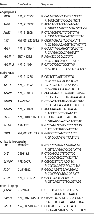
Total RNA extraction was performed using Trizol (Life Technologies) following the manufacturer instructions. The RNA concentration was estimated by NanoVueTM Plus spectrophotometer (GE Healthcare, Freiburg, Germany), and RNA integrity was evaluated through 1% agarose gel electrophoresis. Complementary DNA synthesis was performed according to Goscript Reverse Transcription System protocol (Promega, Madison, WI, USA; Applied Biosystems, Foster City, CA, USA). The primers sequences (Table 2) had their specificity verified in silico, using Nucleotide BLAST tool (https://blast.ncbi.nlm.nih.gov/Blast.cgi).
Oligonucleotides used on gene expression analysis of sows’ mammary gland at 21 days of lactation

ENOS=endothelial nitric oxide synthase; ANG1=angiopoietin1; ANG2=angiopoietin2; TIE2=receptor tyrosine kinase Tie2; VEGF=vascular endothelial growth factor; VEGFR-1=vascular endothelial growth factor receptor 1; VEGFR-2=vascular endothelial growth factor receptor 2; IGF-1R=IGF receptor 1; IGFBP2=IGF-binding protein 2; IGFBP3=IGF-binding protein 3; IGFBP5=IGF-binding protein 5; PRL-R=prolactin receptor; GLU-R=glucocorticoid receptor; COX1=prostaglandin synthase 1; LTF=lactoferrin; CAT=catalase; GSH-PX=glutathione peroxidase; SOD1=superoxide dismutase 1; SOD2=superoxide dismutase 2; GAPDH=glyceraldehyde-3-phosphate dehydrogenase; HPRT1=hypoxanthine–guanine phosphoribosyltransferase.
Primers were used for reverse transcription quantitative PCR with GoTaq qPCR Master Mix (Promega) in ABI Prism 7300 Sequence Detection Systems thermocycler (Applied Biosystems). Geometric means of Ct values of β-actin and Glyceraldehyde-3-Phosphate Dehydrogenase were used to normalize target genes expression (Vandesompele et al., Reference Vandesompele, De Preter, Pattyn, Poppe, Van Roy, De Paepe and Speleman2002). Gene of interest relative expression was calculated by
 $$2^{{{\minus}\Delta \Delta C_{t} }} $$
(Livak and Schmittgen, Reference Livak and Schmittgen2001).
Histological analysis
Samples from the first pair and third right glands were selected for histological analysis, based on the representativeness of samples used for gene expression along with the homogeneity of gland weights (data not shown) and visual conformation among all animals. Tissue samples were embedded in parafin and sectioned at thickness of 5.0 μm using a microtome (RM 2255; Leica Biosystems, Nussloch, Germany). Slides were stained with hematoxylin–eosin solution and qualitatively analyzed using light microscope (BX53; Olympus, Tokyo, Japan). A total of 12 digital images of each glandular unit (n=3) of all animals were obtained randomly on cell Sens Dimensions 1.6 software (Olympus, Tokyo, Japan) using light microscope equipped with digital camera CMOS 1.3 MP BioCAM (Takachiho, Japan) and analyzed with ImageJ software (version 1.50i; National Institutes of Health, Bethesda, MD, USA). The total number of adipocytes and blood vessels were assessed by counting, and the area of each histological image was measured. The volumetric proportion of mammary gland tissue was obtained by counting 4488 points projected onto 12 images per gland captured in histological slides, totalizing 13 464 points per animal. Coincident points were registered in glandular components (parenchyma, blood vessels, connective tissue and adipose cells). The percent of points in each component was calculated using the formula: volumetric proportion (%)=(number of points in each component/(4 488))×100.
Statistical analysis
Data were analyzed in SAS 9.0 (SAS Institute Inc., Cary, NC, USA) considering each sow as an experimental unit in a model including the fixed effect of treatments and post cross-foster litter size as covariate for post cross-foster weight, average daily gain, weaning weight, litter weight gain and litter size at weaning. Differences were considered at P<0.05. P-values between 0.05 and 0.10 were considered a trend. Gene expression data were analyzed using %QPCR_MIXED macro (www.msu.edu/~steibelj/JP_files/QPCR.html), according to methods proposed by Steibel et al. (Reference Steibel, Poletto, Coussens and Rosa2009). BestKeeper was used to obtain the Pearson correlation coefficient between target genes (Pfaffl et al., Reference Pfaffl, Tichopad, Prgomet and Neuvians2004). The angiopoietin2 (ANG2) to angiopoietin1 (ANG1) ratio (ANG2:ANG1) was calculated based on the relative expression of each gene (
 $$2^{{{\minus}\Delta C_{t} }} $$
). Due to a lack of normality verified by the Shapiro–Wilk test (P=0.001) through PROC UNIVARIATE, the data of ANG2:ANG1 ratio were analyzed by using the Wilcoxon test, PROC NPAR1WAY (Pfaffl et al., Reference Pfaffl, Tichopad, Prgomet and Neuvians2004). For histological variables, PROC ANOVA was performed for data with normal distribution (Shapiro–Wilk: P⩾0.05), number of blood vessels per mm2 and percentage of blood vessels. Connective tissue values were submitted to logarithmic transformation for ANOVA. Number of adipocytes per mm2 (Shapiro–Wilk: P<0.001) and percentages of adipocytes (Shapiro–Wilk: P<0.001) and parenchyma (Shapiro–Wilk: P<0.001) did not present normal distribution after logarithmic transformation and their raw data were evaluated by the Wilcoxon test, PROC NPAR1WAY.
Results
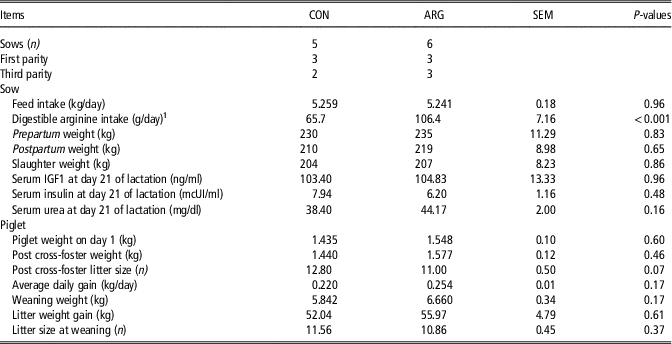
The average of the minimum and maximum temperatures recorded daily in the farrowing room were 19.57±1.79°C and 28.15±1.46°C, respectively, and the average of the daily minimum and maximum relative humidity were 44.73±13.45% and 72.70±7.75%, respectively. Feed intake was similar (P=0.96) between sows receiving CON or ARG diets, whereas arginine intake was higher (P<0.01) in sows fed ARG diet. Sows’ weights did not vary across treatments (P>0.05) at prepartum, postpartum or slaughter (Table 3). No differences on piglets weight were observed after birth (P=0.60) or post cross-foster (P=0.46) (Table 3). Sows fed CON diet tended to have larger (P=0.07) post cross-foster litter size. No differences between treatments were observed for piglets’ average daily gain (P=0.17), piglets’ weight on day 1 (P=0.17), litter weight gain (P=0.61) and litter size at weaning (P=0.37) (Table 3).
Sows’ performance and serological variables during a 21-day lactation period

CON=control diet; ARG=1.0% dietary L-arginine supplemented diet.
1 Values calculated according to Rostagno et al. (Reference Rostagno, Albino, Donzele, Gomes, Oliveira, Lopes, Ferreira, Barreto and Euclides2011).
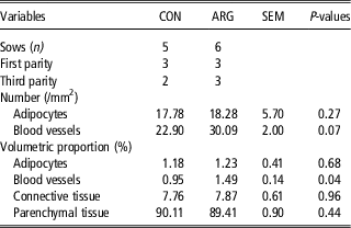
The mammary gland tissues showed no difference (P=0.27) between groups for number of adipocytes per mm2 (Table 4). Sows fed ARG tended (P=0.07) to have a greater number of blood vessels per mm2 and had a greater (P=0.04) percentage of blood vessels in mammary tissue than sows fed CON. Percentage of other mammary gland adipocytes (P=0.68), connective tissue (P=0.96) and parenchyma (P=0.44) did not differ between treatments.
Histomorphometric variables of sows’ mammary glands at 21 days of lactation

CON=control diet, ARG=1.0% dietary L-arginine supplemented diet.
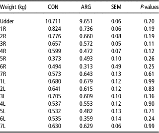
No differences (P>0.05) were observed for udder and individual mammary glands weights at 21 days of lactation (Table 5).
Udder and individual sows’ mammary gland weights at 21 days of lactation

CON=control diet, ARG=1.0% dietary L-arginine supplemented diet.
Numbers followed by ‘R’ or ‘L’ indicate gland order, cranio-caudal direction and right or left sides, respectively.
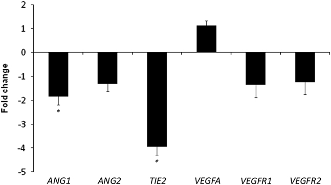
No difference (P=0.71) was observed for expression of endothelial nitric oxide synthase (ENOS) expression between treatments. Expression of ANG1 (P=0.03) and receptor tyrosine kinase (TIE2, P=0.01) was decreased in sows fed ARG diet, whereas expression of ANG2 did not differ (P=0.20) between groups (Figure 1). Similar expression for vascular endothelial growth factor (VEGF; P=0.40), VEGF receptor 1 (P=0.45) and VEGF receptor 2 (P=0.56) (Figure 1) was observed between groups. The ANG2:ANG1 messenger RNA ratio tended (P=0.07) to increase in ARG fed sows.
Fold change between treatments with 1.0% L-arginine (ARG) supplementation (three first parity and three third parity sows) and control (CON; three first parity and two third parity sows) diets regarding the relative expression (positive values mean higher gene expression for ARG fed sows) of respective genes related to angiogenesis in sows’ mammary glands at 21 days of lactation. ANG1=angiopoietin 1; ANG2=angiopoietin 1; TIE2=receptor tyrosine kinase Tie2; VEGFA=vascular endothelial growth factor A; VEGFR1=vascular endothelial growth factor receptor 1; VEGFR2=vascular endothelial growth factor receptor 2. *0.01<P<0.05.

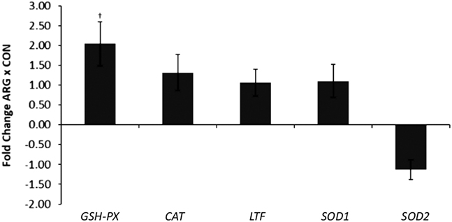
Expression of glutathione peroxidase (GSH-PX) tended to increase in ARG fed sows (P=0.05). Similar expression between groups was observed for catalase (CAT; P=0.27), superoxide dismutase 1 (P=0.57) and superoxide dismutase 2 (SOD2, P=0.47). No changes (P=0.83) were observed for expression of lactoferrin between groups (Figure 2).
Fold change between treatments with 1.0% L-arginine (ARG) supplementation (three first parity and three third parity sows) and control (CON; three first parity and two third parity sows) diets relative to the relative expression (positive values mean higher gene expression for ARG fed sows) of the respective genes related to antioxidant capacity in sows’ mammary glands at 21 days of lactation. GSH-PX=glutathione peroxidase; CAT=catalase; LTF=lactoferrin; SOD1=superoxide dismutase 1; SOD2=superoxide dismutase 2. † P<0.10.

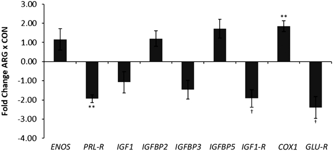
A lower expression of prolactin receptor (PRL-R) (P<0.01) was observed in ARG fed sows. No differences on expressions were observed between CON and ARG groups for IGF1 (P=0.85), IGF-binding protein 2 (P=0.53), IGF-binding protein 3 (P=0.31) and IGF-binding protein 5 (P=0.14). Nevertheless, expression of IGF1 receptor (IGF-1R) tended to be greater (P=0.07) in sows fed ARG (Figure 3). Expression of glucocorticoid receptor (GLU-R) tended to be lower (P=0.06) in ARG fed animals. A greater value of prostaglandin synthase 1 (COX1) was observed in sows fed ARG (P=0.01) (Figure 3). Consistently, the inclusion of arginine in lactation diet had no effect on the levels of IGF (P=0.96), insulin (P=0.48) or urea (P=0.16) (Table 3).
Fold change between 1.0% dietary L-arginine (ARG) supplementation (three first parity and three third parity sows) and control diet (CON; three first parity and two third parity sows) on the relative expression (positive values mean higher gene expression for ARG fed sows) of endothelial nitric oxide synthase (ENOS) and respective genes related to hormonal response in sows’ mammary gland at 21 days of lactation. PRL-R=prolactin receptor; IGFBP2=IGF-binding protein 2; IGFBP3=IGF-binding protein 3; IGFBP5=IGF-binding protein 5; IGF-1R=IGF receptor 1; COX1=prostaglandin synthase 1; GLU-R=glucocorticoid receptor. **P<0.01; †P<0.10.

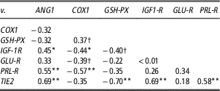
There was correlation between PRL-R/COX1 (r=−0.57, P=0.01), PRL-R/ANG1 (r=0.55, P=0.01), PRL-R/TIE2 (r=0.58, P=0.01), IGF-1R/CAT (r=−0.62, P<0.01), IGF-1R/COX1 (r=−0.44, P=0.04), IGF-1R/ANG1 (r=0.45, P=0.03) and IGF-1R/TIE2 (r=0.69, P<0.01) (Table 6).
The Pearson’s correlation coefficient between differently expressed target genes for sows at 21 days of lactation

ANG1=angiopoietin 1; COX1=prostaglandin synthase 1; GSH-PX=glutathione peroxidase; IGF-1R=IGF receptor 1; GLU-R=glucocorticoid receptor; PRL-R=prolactin receptor; TIE2=receptor tyrosine kinase TIE2.
*0.01<P<0.05; **P<0.01; †P<0.10.
Discussion
Even though mammary gland growth and development to support lactation starts in late gestation (Kensinger et al., Reference Kensinger, Collier, Bazer, Ducsay and Becker1982), we decided to initiate arginine supplementation at parturition because impacts on animal performance were demonstrated during this short period of supplementation previously (Mateo et al., Reference Mateo, Wu, Moon, Carroll and Kim2008). Furthermore, the antioxidant role of arginine could be better assessed during lactation, where energy and amino acid requirement is increasing rapidly, particularly for arginine (Mateo et al., Reference Mateo, Wu, Moon, Carroll and Kim2008).
The pre-determined daily feed intake of 6.0 kg has not been reached. Based on thermoneutral range (16°C to 22°C) for lactating sows (Black et al.,Reference Black, Mullan, Lorschy and Giles1993), it can be inferred that sows in the present study were submitted to periods of heat stress. Heat stress, farrowing stress and the use of primiparous sows, which present low feed consumption, are among factors that may have contributed to observed feed intake values <6.0 kg.
Similar concentrations for serum urea found in this study indicate no excessive nitrogen supply in view of the supplementation with dietary arginine. In addition, this outcome indicates no difference in nitrogen utilization efficiency among diets.
Although the aim of keeping at least 11 contemporary piglets per sow was achieved, there was a trend of greater post cross-foster litter size in the CON group, which led us to use this variable as a covariate in the analyses of piglets performance. Although a numerical increment of 14% in the average daily gain and weaning weight was observed, there was no effect of ARG on piglets’ performance.
We believe that the low number of replicates may have been the cause of the lack of significance as in the present study there was a trend for increased number of blood vessels per mm2 and a higher percentage of blood vessels in the mammary tissue of ARG sows compared with CON sows, which ultimately would cause improved performance of piglets.
As a functional amino acid, arginine supplementation in diets for sows has been recommended to improve health, reproduction and lactation as it has important functions in regulating gene expression, anti-oxidative responses, vasodilation and angiogenesis (Wu et al., Reference Wu, Bazer, Satterfield, Li, Wang, Johnson, Burghardt, Dai, Wang and Wu2013). Our results demonstrate that 1.0% arginine supplementation to lactating sows results in higher number per mm2 and percentage of blood vessels, suggesting an increment in both counting and diameter of blood vessels. Polyamines and nitric oxide stimulate the proliferation of endothelial cells (Flynn et al., Reference Flynn, Meininger, Haynes and Wu2002), indicating that arginine may have played an angiogenic role in mammary tissue through its metabolites in the present study. Possibly, metabolic advantages may have influenced mammary glands, such as mammalian target of rapamycin signaling pathway, leading to greater protein synthesis and cell survival (Kong et al., Reference Kong, Tan, Yin, Gao, Li, Jaeger, Bazer and Wu2012). Greater nutrient yield may have potentiated those arginine effects by enhanced blood flow to the organ, as we observed greater vascularization under histological assessment. Greater tissue vascularization, as a consequence of ARG supplementation, was also found in placental membranes of gestating gilts (Li et al., Reference Li, Bazer, Johnson, Burghardt, Erikson, Frank, Spencer, Shinzato and Wu2010), possibly due to modulation in ENOS gene expression (Liu et al., Reference Liu, Wu, Yin, Liu, Geng, Yang, Blachier and Wu2012). However, the similar expression for ENOS currently found in CON and ARG fed sows was unexpected, particularly considering the increased vascularity in mammary glands.
The result observed can be due to ENOS activation modulated by the enzyme interaction with calmodulin; consequently, ENOS expression is secondary in predicting its metabolites production (Alderton et al., Reference Alderton, Cooper and Knowles2001). In addition, higher ENOS levels evident during first days of arginine supplementation, when tissues are actively proliferating (Nielsen et al., Reference Nielsen, Trottier, Stein, Bellaver and Easter2002), may explain the similar expressions found for ENOS at day 21. It is also important to note that ARG supplementation may increase tetrahydrobiopterin (BH4) availability and thus NO synthesis without change on ENOS expression (Shi et al., Reference Shi, Meininger, Haynes, Hatakeyama and Wu2004).
The relationship between both ANG1 and ANG2 expressions is of great importance as angiopoietins compete for the same receptor, TIE2, to estimate its activation (Hansen et al., Reference Hansen, Singh, Tahir and Brindle2010). This occurs due to ANG1 and ANG2 molecules similarity, and the ability of the second ligand impairing ANG1/TIE2 interaction as well as inhibiting the receptor phosphorylation (Hanahan, Reference Hanahan1997). Increased ANG2 expression is observed at tissue remodeling sites and ANG1 alone does not induce endothelial cell multiplication (Davis et al., Reference Davis, Aldrich, Jones, Acheson, Compton, Jain, Ryan, Bruno, Radziejewski, Maisonpierre and Yancopoulos1996). Instead, ANG1 promotes intercellular adhesion and reduction of vascular permeability, both of which are related to blood vessel stabilization (Hanahan, Reference Hanahan1997). Therefore, the reduction noted for ANG1 and TIE2 expressions in addition to ANG2:ANG1 5.13 unit-increase in ARG fed sows may account for possible destabilization and restructuration of vascular mammary gland network by ARG supplementation. The VEGF-related genes are indispensable to activate angiogenic pathways, including those mediated by angiopoietins (Eklund and Olsen, Reference Eklund and Olsen2006). Then, the absence of difference in expressions for the VEGF reveals that vascular multiplication and modeling was probably diminished at sampling on day 21.
Arginine may increase circulating levels of prolactin, IGF and insulin in sows (Mateo et al., Reference Mateo, Wu, Moon, Carroll and Kim2008; Zhu et al., Reference Zhu, Guo, Gao, Wang, Chen, Ma and Jiang2017). These are important hormones in mammary gland tissue proliferation, differentiation and nutrient uptake (Burnol et al., Reference Burnol, Loizeau and Girard1990). Increased mammary tissue sensitivity to insulin due to lactation might lead to an unnecessary rise in this hormone concentration to provide greater nutrient uptake (Burnol et al., Reference Burnol, Loizeau and Girard1990). Our findings were consistent with this view since similar serum insulin concentrations between sows fed CON or ARG diets on day 21.
Even though indirect effect of GH on mammary tissues occurs through IGFs and IGFBPs, resulting in higher milk production due to acinar cells’ proliferation, they have a minor participation than prolactin on milk yield (Wilde et al., Reference Wilde, Knight and Flint1999). No change for IGF1 and IGFBPs expressions and IGF1 hematological concentrations were observed between experimental groups in the present study. Possibly, the effect of the IGF’s superfamily on mammary gland development was not observed because these genes might be more related to the proliferation of mammary duct cells, which is more significant after 45 days of gestation (Ji et al., Reference Ji, Hurley and Kim2014). Hence, IGF1 gene expression shall be more relevant at the beginning of the glandular development period in comparison to the end of lactation. The IGFBPs present great variability in gene expression levels as well as in their location in the mammary gland of mice females (Wood et al., Reference Wood, Richert, Stull and Allar2000). Supposing analogous observations in sows, it would match with IGFBPs expression patterns observed in the present study. Furthermore, it is possible that both arginine levels used in the current study were high enough to promote the arginine secretagogue effect on insulin and IGF1 (Zhu et al., Reference Zhu, Guo, Gao, Wang, Chen, Ma and Jiang2017), justifying no difference found between CON and ARG fed sows.
The observed decrease in PRL-R gene expression reinforces previous finding on the elevation of circulating prolactin in response to arginine supplementation during lactation (Zhu et al., Reference Zhu, Guo, Gao, Wang, Chen, Ma and Jiang2017), counteracting the described decrease in PRL level during lactation of sows not supplemented with arginine (Farmer et al., Reference Farmer, Palin and Hovey2010). Of note, prolactin has greater importance than IGFs on both milk synthesis and prevention of acinar cell apoptosis in rat mammary glands (Travers et al., Reference Travers, Barber, Tonner, Quarrie, Wilde and Flint1996), being responsible for higher milk yield. Such information sustains the existence of alternative pathways other than those modulated by GH and its mediators for cell survival and metabolism on mammary glands. These pathways can include the possible effects of arginine at the cellular level, for example the activation of proliferative and anabolic mechanisms mediated by nitric oxide, mammalian target of rapamycin and polyamines (Wu et al., Reference Wu, Bazer, Davis, Kim, Li, Rhoads, Satterfield, Smith, Spencer and Yin2009). The Pearson’s correlation coefficient for PRL-R/ANG1 and PRL-R/TIE2 highlights the interactions between proliferative and angiogenic pathways that the authors could not explain.
Another effect of ARG supplementation was the reduction of 2.39-fold for GLU-R gene expression on current research. Glucocorticoids act as lactogenic hormones and on cell differentiation of mammary alveolar epithelium, potentiating protein synthesis stimulated by prolactin for maintenance of lactation (Kobayashi et al., Reference Kobayashi, Oyama, Kuki, Tsugami, Matsunaga, Suzuki and Nishimura2017). Despite these effects of glucocorticoids, elevated GLU-R expression may be undesirable to observe in response to arginine supplementation. Glucocorticoids increase the metabolic rate of arginine in intestinal piglet cells and hinder the amino acid transport decreasing its availability in tissues (Wu et al., Reference Wu, Flynn, Knabe and Jaeger2000), leading to consequent likely restrain of BH4 and NO synthesis and compromising arginine metabolites regulatory functions. To avoid the inconsistent supply of arginine to organs, this amino acid is capable to reduce GLU-R expression and decrease circulating cortisol level in finishing pigs (Ma et al., Reference Ma, Lin, Jiang, Zheng, Zhou, Yu, Cao, Wang and Chen2010), which is a possible analogous outcome for this study. In addition, the secretagogue effect of arginine on prolactin, abovementioned suggested by the reduction in the expression of its PRL-R, may have contributed to the decrease in the expression of the GLU-R as presently found. Considering that both hormone receptors are related to stimulation of milk protein synthesis the increment in one hormone concentration turns unnecessary the other’s presence to ensure lactopoiesis (Kobayashi et al., Reference Kobayashi, Oyama, Kuki, Tsugami, Matsunaga, Suzuki and Nishimura2017).
There is a lack of studies regarding COX1 roles in normal mammary glands, especially during lactation. Nevertheless, it has been shown that COX1 transcripts are more abundant during the initial lactation period compared with other phases of rodents’ reproductive cycle, presenting progressive reduction on its expression during lactation (Chandrasekharan et al., Reference Chandrasekharan, Foley, Jania, Clark, Audoly and Koller2005). The higher relative gene expression of COX1 by 1.85-fold indicates greater synthesis of prostanoid metabolites, stimulating a cascade of intracellular events, through increased calcium influx and promoting cell differentiation, proliferation, angiogenesis and antiapoptotic mechanisms (Chandrasekharan et al., Reference Chandrasekharan, Foley, Jania, Clark, Audoly and Koller2005). Thus, the elevation we observed for COX1 expression, stimulated by ARG supplementation, may prolong growth and differentiation periods of constitutive mammary gland tissues. The negative correlation coefficient noted between PRL-R/COX1 expressions proposes that COX1 expression can be modulated by prolactin levels, indicating that hormonal proliferative effects of prolactin may be mediated by COX1 local activity. Likewise, the aforementioned increased intracellular calcium influx may potentiate nitric oxide-mediated arginine responses, considering ENOS activation dependence on calmodulin interaction. Despite the results found on previous works, more studies are required to characterize COX1 roles during lactation period, particularly in sows.
The increase of 2.04-fold in transcript expression for GSH-PX, an important intracellular antioxidant enzyme, was observed in ARG fed sows. Comparable outcome was observed by dietary supplementation with arginine for finishing pigs (Ma et al., Reference Ma, Lin, Jiang, Zheng, Zhou, Yu, Cao, Wang and Chen2010), indicating that higher arginine intake can increase antioxidant mechanisms, as proposed on present research. Gene expression of the other antioxidant enzymes evaluated demonstrates no additional significant influence in oxidative stress at day 21 across treatments.
The IGF-1R is related to increased resistance to oxidative stress through challenging situations in rat cell culture (Chen et al., Reference Chen, Xu and Song2014). The decrease of 1.92-fold in expression of IGF-1R in sows fed ARG might indicate enhanced antioxidant capacity of mammary glands, by the increase in antioxidant factors, such as the elevation of 2.04-fold in GSH-PX, or lower activation of pro-oxidant mechanisms modulated by arginine. Arginine provision allows a reduction in reactive oxygen species synthesis enhancing the bioavailability of BH4 and nitric oxide. The BH4 is cofactor of ENOS (Shi et al., Reference Shi, Meininger, Haynes, Hatakeyama and Wu2004) and its deficiency increases superoxide production (Stroes et al., Reference Stroes, Hijmering, van Zandvoort, Wever, Rabelink and van Faassen1998), whereas nitric oxide is capable to interact and inactivate oxidative oxygen species (Gardner et al., Reference Gardner, Powlson and Giussani2001). Deficient vascularity may lead to oxidative stress and enhanced angiogenic pathways (Forsythe et al., Reference Forsythe, Jiang, Iyer, Agani, Leung, Koos and Semenza1996). The IGF-1R lesser expression indicates low oxidative stress, turning angiogenesis dismissible during late lactation, as observed through the positive Pearson’s correlation coefficients found for IGF-1R/ANG1 and IGF-1R/TIE2. The IGF-1-R/COX1 negative correlation reveals that proliferative mechanisms mediated by IGFs were possibly replaced by COX1 responses.
In conclusion, our data show that dietary ARG supplementation to lactating sows promotes increase in blood vessel number and calibre, as well as modulating messenger RNA expression of proliferative (PRL-R, IGF-1R, COX1 and GLU-R), angiogenic (ANG1 and TIE2) and antioxidant (GSH-PX) related genes in mammary gland tissues of lactating sows.
Acknowledgments
The authors would like to thank the Conselho Nacional de Desenvolvimento Científico e Tecnológico (CNPq), Coordenação de Aperfeiçoamento de Pessoal de Nível Superior (CAPES) and Instituto Nacional de Ciência Animal (INCT-CA) for financial support.
Declaration of interest
The authors declare no conflict of interest.
Ethics statement
All procedures involving animals were approved by the Universidade Federal de Viçosa Ethical Committee on Animal Use (no. 053/2015).
Software and data repository resources
None.