Case report
A 68-year-old woman was referred to the ENT department two days after frontal trauma with subarachnoidal bleeding and contusion of the right frontal lobe. She had suddenly fallen in her kitchen, probably due to orthostatic reasons. She did not remember the incident. She presented with bloody discharge from the left ear, which had persisted since the trauma, and complained of left-sided, pulsatile tinnitus and vertigo.
Clinical examination revealed a laceration of the left ear canal and a haemotympanum. Audiometry showed near normal hearing on the right and a mixed hearing loss on the left, with an air–bone gap of 60 dB. A computed tomography (CT) scan of the skull had demonstrated a longitudinal petrosal bone fracture, with blood in the mastoid and middle-ear cavity.
Five days later, a near total (House–Brackmann grade five) facial paralysis developed, which was treated with steroids. A bilateral, posterior, benign, paroxysmal, positional vertigo was also found and was treated with Epley's repositioning manoeuvre, which had to be repeated on several occasions. A control CT scan of the petrosal bone showed a left-sided fracture through the roof of the external ear canal, with posterior displacement of the anterior wall and slight stenosis of the external ear canal. The fracture continued through the roof of the middle-ear cavity and ended near the geniculate ganglion, causing an incudomallear luxation.
Four weeks after the trauma, the facial palsy was reduced (to House–Brackmann grade two). Two months after the trauma, facial motility had returned to normal, the middle ear was well ventilated (on otomicroscopy) and both Hallpike tests were negative for bilateral, benign, paroxysmal, positional vertigo.
However, the patient still complained of left-sided, pulsatile tinnitus. Audiometry showed a persisting air–bone gap of 50 dB. At that time, conjunctival vascular injection and suborbital oedema of the left eye were noticed, and the patient was thus referred to an ophthalmologist.
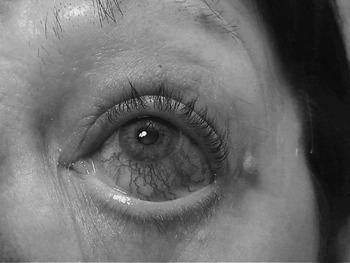
Hyperaemia, chemosis and a small corneal ulcer were seen (Figure 1). Due to the suspicion of a post-traumatic carotico-cavernous fistula, the patient was referred for imaging.
Dilated conjunctival and episcleral vessels of the left eye.

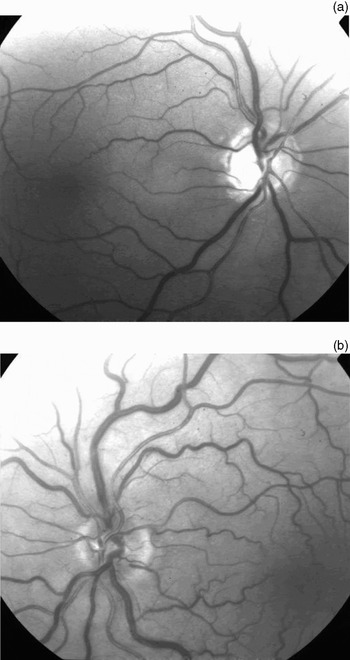
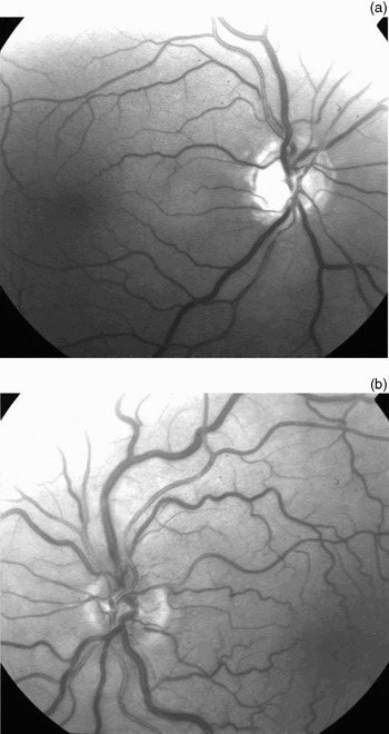
The patient waited one month before presenting again to the ophthalmology department because of diplopia. She had a clear ocular bruit, with normal vision and an ocular pressure of 17 mmHg. Biomicroscopy showed dilatation of the conjunctival and episcleral vessels as well as corneal dehydration caused by orbital oedema in decubitus. Fundoscopy showed greatly dilated retinal vessels on the left (Figure 2).
Pre-operative fundoscopy. (a) Right eye showing normal appearance; (b) left eye showing venous congestion.
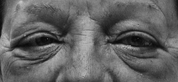
Under a working diagnosis of post-traumatic carotico-cavernous fistula, the patient was readmitted to the neurology department. In the meanwhile, she had developed an abducens palsy, tachycardia and dyspnoea (Figure 3).
Orbital oedema and VIth nerve palsy of the left eye.

The tinnitus disappeared with compression of the left common carotid artery, and the neurological examination was negative.
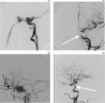
Urgent angiography, 13 weeks after the trauma, showed a massive carotico-cavernous fistula on injection of contrast dye through the left internal carotid artery. There was an early filling of the ophthalmic vein, with a clear varicose appearance, and no filling of the anterior or middle cerebral arteries due to a total steal phenomenon. Angiography of the right carotid artery and the left vertebral artery was normal. The left cerebral hemisphere was supplied by the right internal carotid artery through the anterior communicating artery (Figure 4a and 4b).
Left internal carotid artery, frontal (a, c) and lateral (b, d) view, before (a, b) and after (c, d) endovascular treatment. (a) & (b) Direct shunting of the arterial blood supply to the cavernous sinus, showing early filling of the ipsilateral internal jugular vein, with a steal phenomenon on the arterial blood supply of the left cerebral hemisphere (i.e. no filling of the middle or anterior cerebral artery on the ipsilateral side). On the lateral view, there is shunting of arterial blood in the cavernous sinus, with backflow of arterialised blood into the dilated ophthalmic veins (black arrows) and the sphenoparietal sinus. (c) & (d) A normal blood supply to the left hemisphere after endovascular treatment with a detachable balloon and four Guglielmi detachable coils. The insufflated detached balloon (arrow) in the cavernous sinus can be seen superimposed on the C3 portion of the internal carotid artery.

The patient was subsequently treated with an arterial detachable endovascular balloon (11×19 mm) in combination with Guglielmi detachable coils placed transvenously via the internal jugular vein and superior petrosal sinus. Only by combining a transarterial and transvenous approach could complete occlusion of the carotico-cavernous fistula be achieved (Figure 4c and 4d).
The pulsatile tinnitus completely subsided, but the procedure caused a ptosis and ocular palsy of the left eye. Ophthalmologic follow up revealed resolution of the ocular bruit, but the ocular palsy and vascular dilatation were still present on fundoscopy one week after the procedure.
About nine weeks after the treatment, all ocular symptoms had disappeared. During the final ENT consultation, six months after the initial trauma, a proposal to perform an ossiculoplasty was declined by the patient.
Discussion
Our patient suffered from post-traumatic, bilateral, benign, paroxysmal, positional vertigo and a left-sided, longitudinal petrosal bone fracture, with secondary facial palsy and ossicular luxation. As time passed, other symptoms became more prominent, such as proptosis, chemosis, dilated conjunctival vessels, orbital oedema and, ultimately, diplopia. Angiography diagnosed a direct, high flow, carotico-cavernous fistula (type A). Thirteen weeks passed between the initial trauma and establishment of the diagnosis.
Anatomically, the cavernous sinus is 2 cm long and 1 cm wide, lying on each side of the sphenoid body. The internal carotid artery, the IIIrd, IVth and VIth cranial nerve, and the ophthalmic and maxillary branches of the Vth cranial nerve pass through the cavernous sinus. The most important anterior tributary is the superior ophthalmic vein. Posteriorly, the tributaries are the superior and inferior petrosal sinuses. Carotico-cavernous fistulas are abnormal communications between the internal carotid artery and the cavernous sinus. Contrary to fistulas elsewhere in the body, a tear in the wall of the intracavernous carotid artery or rupture of one of its branches that transverse the sinus produces an arterio-venous fistula without concomitant venous injury.Reference Barrow, Spector, Braun, Landman, Tindall and Tindall1 The venous drainage can be anterior, posterior, inferior, superior or contralateral. Anterior drainage through the superior ophthalmic vein frequently occurs, inducing proptosis and chemosis. The more anterior the fistula, the more prominent the anterior venous drainage.Reference Barrow, Spector, Braun, Landman, Tindall and Tindall1, Reference Debrun, Lacour, Vinuela, Fox, Drake and Caron2
Carotico-cavernous fistulas are most frequently found in middle-aged women and can be divided into four classes, according to pathogenesis, flow characteristics and anatomy (as described by Barrow et al. Reference Barrow, Spector, Braun, Landman, Tindall and Tindall1 and quoted by Robertson et al.).Reference Robertson, Nicolaides and Taylor3 The presenting symptoms are usually diplopia, red eye, proptosis, headache and tinnitus. Signs may include dilated episcleral veins, diminished vision, elevated ocular pressure, chemosis, and IIIrd and VIth nerve palsy (due to compression and ischaemia).Reference Barrow, Spector, Braun, Landman, Tindall and Tindall1, Reference de Keizer4 Carotico-cavernous fistulas may develop spontaneously (class two to four according to Barrow); exophthalmia (96 per cent) and epibulbar loops (97 per cent) are the main signs. Following post-traumatic development (class one according to Barrow), exophthalmia (94 per cent) and tinnitus (88 per cent) are more prominent.Reference de Keizer4
If ophthalmological symptoms occur, a triad of exophthalmia, dilated episcleral veins and elevated intra-ocular pressure is almost pathognomonic for carotico-cavernous fistula. If this condition is suspected, urgent ophthalmological referral is necessary. The ophthalmologist should perform orbital Doppler sonography, ocular pressure reading, fundoscopy and vision testing in order to confirm the diagnosis. Doppler sonography is indicated to differentiate a high flow from a low flow arterio-venous fistula. If high flow is detected, angiography must be performed. When a low flow is detected, magnetic resonance imaging with angiography (MRI/MRA) should be performed.Reference de Keizer4–Reference Duan, Liu, Zhou, Cao, Ruan and Zhao7
The gold standard for diagnosis and endovascular treatment, if possible, is angiography. Angiography has a 100 per cent diagnostic rate for arterio-venous fistulas and, by extension, arterio-venous malformations. However, it is not without risk, as it is an invasive technique which can lead to neurological complications (1.3 per cent), haematomas (0.4 per cent) and allergic cutaneous reactions (0.1 per cent).Reference Barrow, Spector, Braun, Landman, Tindall and Tindall1, Reference Debrun, Lacour, Vinuela, Fox, Drake and Caron2, Reference de Keizer4, Reference Weissman and Hirsch8–Reference Shin, Lalwani and Dowd11
The length of time between onset of symptoms and diagnosis is five months on average. Follow up of low flow carotico-cavernous fistulas usually reveals spontaneous resolution of signs and symptoms. When a low flow arterio-venous malformation measures more than 2.5 cm, the use of gamma-knife radiosurgery leads to progressive obliteration by endothelial cell proliferation, which results in luminal closure. Complete obliteration generally occurs one to three years after radiosurgery. The goal of this technique is to eliminate the risk of future intracranial bleeding, with minimal morbidity.Reference Barrow, Spector, Braun, Landman, Tindall and Tindall1, Reference Brown, Flemming, Meyer, Cloft, Pollock and Link12, Reference Link, Coffey, Nichols and Gorman13
High flow carotico-cavernous fistulas are treated more invasively, using endovascular embolisation, endovascular closure of the fistula, ligation of the internal carotid artery or open surgery with closure of the tear. The indications for treatment are visual deterioration, diplopia, intolerable tinnitus or headaches, proptosis with untreatable corneal exposure, and deterioration of general health.
Post-therapeutic ocular palsy is seen after 20 per cent of all endovascular treatments and usually spontaneously disappears over the following months. The palsy occurs within hours after treatment and is due to excessive compression of the IIIrd cranial nerve by the inflated balloon.Reference Barrow, Spector, Braun, Landman, Tindall and Tindall1, Reference Debrun, Lacour, Vinuela, Fox, Drake and Caron2
Pulsatile tinnitus is often encountered within daily ENT practice. Although it was a presenting complaint in our patient, it is not one of the major symptoms of carotico-cavernous fistula. In our patient, pulsatile tinnitus could initially be explained by the haemotympanum and ossicular luxation. Pulsatile tinnitus of vascular origin results from non-laminar blood flow generated by increased blood flow or luminal stenosis. It can be the first or sole manifestation of a diverse range of pathology, from arterial hypertension with carotid artery disease to potentially life-threatening conditions. However, the diagnosis is not always clear. In cases of pulsatile tinnitus, it is imperative that the ENT specialist undertake a full ear, nose and throat examination, otoscopy, audiometry, tympanometry, and a thorough auscultation including the chest, heart, neck, retroauricular area and orbits.Reference Lockwood, Salvi and Burkard14–Reference Waldvogel, Mattle, Sturzenegger and Schroth16
• This paper describes the case of a patient with a traumatic carotico-cavernous fistula, presenting with pulsatile tinnitus
• When a pulse-synchronised tinnitus is auscultated in the head and neck region, an arterio-venous malformation should be suspected; angiography is the ‘gold standard’ investigation
• When a bruit is not auscultated, Doppler sonography of all the head and neck vessels should be performed
When a pulse-synchronised tinnitus is discovered by auscultation in the head and neck region, an arterio-venous malformation should be suspected, and angiography is the ‘gold standard’ investigation. When a bruit is not evident, Doppler sonography of all the head and neck vessels should be performed. If this shows vascular changes then an angiography is indicated. If no vascular changes are evident on Doppler sonography, then further investigation with MRI/MRA is warranted.Reference Shin, Lalwani and Dowd11
Waldvogel et al. Reference Waldvogel, Mattle, Sturzenegger and Schroth16 and Shin et al. Reference Shin, Lalwani and Dowd11 demonstrated that 54 per cent of patients with pulsatile tinnitus had underlying vascular pathology and that, in 68 per cent of them, pulsatile tinnitus was the presenting complaint. In these patients with underlying vascular pathology, a pulsatile tinnitus was detected by auscultation in 58 per cent, rising to 87 per cent in cases of arterio-venous malformation. Doppler sonography was helpful and had a sensitivity of 68 to 75 per cent. In a group of 33 patients with almost exclusively subjective pulsatile tinnitus, Shin et al. still found an underlying vascular pathology or anatomic abnormalities in 63 per cent using MRI/MRA.Reference Shin, Lalwani and Dowd11, Reference Sismanis15, Reference Waldvogel, Mattle, Sturzenegger and Schroth16
Conclusion
For patients initially presenting with pulsatile tinnitus, a diagnosis of carotico-cavernous fistula alone is uncommon and possibly life threatening. During investigation, cooperation between specialities is vital. When patients complain of pulsatile tinnitus, ENT specialists must remember to ask for a history of trauma over the preceding few months, and they must perform thorough auscultation of the chest, heart, neck, retroauricular area and orbits. This can then guide the differential diagnosis and the ordering of additional investigations.