Introduction
The mumps is induced by a mumps virus infection and may result in various severe symptoms: meningitis, epididymitis and deafness as well as epidemic parotiditis. Although mumps deafness is a rare disorder after mumps virus infection, it is often profound and incurable.Reference Pomeroy and Jordan1, Reference Everberg2 Although most cases of mumps deafness show a unilateral effect, there are also a few reports of bilateral mumps deafness,Reference Kawaguchi, Kawano, Kanebayashi, Oura and Suzuki3–Reference Yamada, Yamasoba and Kaga6 which is said to severely affect patients' verbal communication.Reference Everberg2
There is no effective therapy for bilateral profound sensorineural hearing loss (SNHL) following mumps other than with a cochlear implant. There are a few articles that report good outcomes of cochlear implants.Reference Kawaguchi, Kawano, Kanebayashi, Oura and Suzuki3–Reference Yamada, Yamasoba and Kaga6 Normally a viral invasion is localised in the organ of Corti and the membranous labyrinth, causing viral endolymphatic labyrinthitis. In contrast, mumps infection often causes a disorder of the central nervous system such as meningitis or encephalitis,Reference Honjo7 which would bring about hearing loss with a retrolabyrinthine disorder. We cannot distinguish a labyrinthine disorder from a retrolabyrinthine disorder by the deafness level, and therefore a CNS disorder becomes clear only after cochlear implant surgery is performed. There is no report of a cochlear implant for such a patient in Japan.
We installed a cochlear implant in two children with profound bilateral SNHL following mumps. One child achieved a good result, but the other did not. We describe their cases and discuss the difference in outcomes below.
Case reports
Case 1
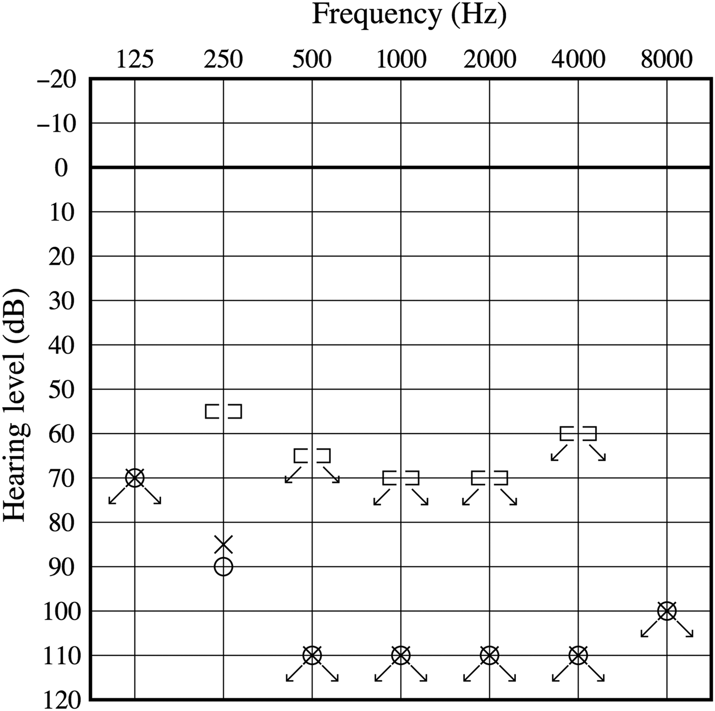
A three-year-old girl had bilateral parotid swelling in March 2001 (day 0). She presented with vomiting, gait disorder and bilateral hearing loss from day 10 and visited a nearby paediatrician's office. After admission to a general hospital on day 12, her mumps antibody titre was found to be rising. Her bilateral severe hearing loss became clear by pure tone audiometry (PTA) (Figure 1) and auditory brainstem response testing on day 20, and she was diagnosed as having mumps deafness. She was given hearing aid by a rehabilitation organisation, but it was not effective. Her parents came to our hospital and requested cochlear implant surgery two months after the deafness onset.
Case 1 audiogram.

We did not detect any abnormal findings in the inspection of the patient's tympanic membrane by computed tomography (CT) and magnetic resonance imaging (MRI). The conditioned orientation reflex audiometric test indicated bilateral severe SNHL. Electronystagmography and a promontory examination could not be performed because she was too young.
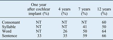
Five months after she lost hearing, we performed a cochlear implantation for the right ear with a Clarion cochlear implant (Advanced Bionics, Valencia, California, USA). Although she had been able to speak normally before mumps, the patient's conversation and reaction to environmental sound were poor. At one year after the cochlear implant surgery, her threshold in the acoustic field was 33 dB, but she achieved only a 33 per cent correct answer rate for sentences on the cochlear implant-2004 (CI-2004) test (ESCOR Ltd., Chiba, Japan).
Although the patient underwent rehabilitation, her language development over the post-operative two years was equivalent to that of a two-year-old, as she became five years old. She began attending a school for deaf persons and grew up learning sign language. As a result of her rehabilitation with ocular vision including sign language, her correct answer rate for sentences on the CI-2004 test gradually rose to 66 per cent, as shown in Table I.
CI-2004 tests for Case 1. A percentage of correct answers in Case 1 for CI-2004 tests. Her speech perception with cochlear implant was not good but gradually developed
NT = not tested
Case 2
An eight-year-old girl developed bilateral parotid swelling in May 2006 (day 0). She complained of bilateral hearing loss from day 2 and had a check up by a nearby otolaryngology doctor. On PTA, the threshold of the right ear was 82 dB, and that of the left ear was off the scale. She was administered a steroid (predonisolone 1 mg/kg, gradual decrease), but her hearing deteriorated and she became deaf on day 6. Since a hearing aid was not effective, she was introduced to our hospital for a cochlear implant consultation on day 35.
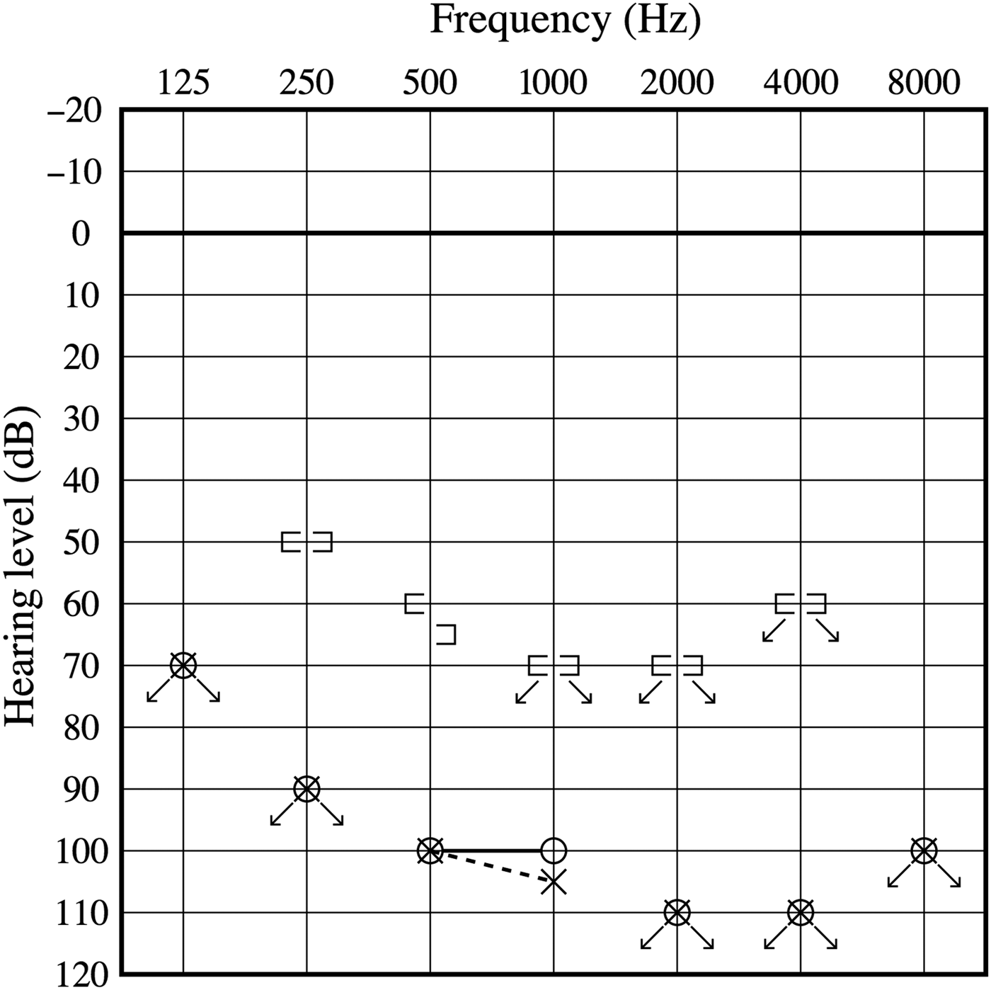
We diagnosed mumps based on her serum immunoglobulin M level. Pure tone audiometry showed that the threshold for both ears was off the scale (Figure 2). The eardrums were both normal. The middle and inner ears did not show abnormality on CT and MRI. Both ears' discrimination was good on promontory examination, and the temporary threshold shift was negative. Electronystagmography showed right moderate canal paralysis.
Case 2 audiogram.
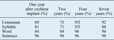
At two months after the onset of hearing loss, we installed a cochlear implant (Conture24, Cochlear, Macquarie Park, NSW, Australia) in the patient's right ear. The intra-operative neural response telemetry indicated a normal evoked compound action potential threshold. She recognised the voice of her mother immediately after the first mapping. On the 24th post-operative day, she caught 100 per cent of vowel sounds and 78 per cent of consonant sounds in different sensitivity tests. She eventually became able to communicate using only her hearing with the cochlear implant, and she returned to the normal-hearing school that she had attended before her hearing loss. Seven years after the cochlear implant operation, her hearing threshold was 33 dB and her correct answer rate for sentences on the cochlear implant-2004 was 96 per cent, as shown in Table II.
CI-2004 tests for Case 2. Each number indicates a percentage of correct answers. The score was initially good
NT = not tested
Discussion
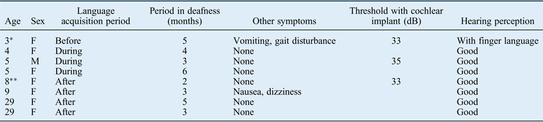
We compared the two cases described above with previous reports of cochlear implantation for patients with mumps deafness (shown in Table III).Reference Kawaguchi, Kawano, Kanebayashi, Oura and Suzuki3–Reference Yamada, Yamasoba and Kaga6 As in most of the reported patients, our Case 2 developed language-hearing ability soon after the cochlear implant surgery. However, Case 1 did not obtain a good outcome. We suggest two causes for these different outcomes. One is the patients' ages, which affect the early stage of language acquisition. The language acquisition period is considered to occur at approx. three to ten years old,Reference Honjo7, Reference Naito8 and thus a hearing loss during this time has a serious adverse effect on speech perception and the development of conversation skills. In addition, because much of the brain's plasticity can deteriorate by four or five years old,Reference Naito8 the influence of a deafened period may be more serious for the language-acquiring child.Reference Geers9 Our Case 1 was just three years old at the onset of hearing loss, an age early in the language acquisition period. In contrast, Case 2, at eight years old, had already acquired significant language skills. Given that the patients who require good hearing perception are older than five years old (in Table III), the timing of hearing loss is important for language acquisition.
Case reports in Japan. Our cases are presented by * (Case 1 as *, Case 2 as **)
F as female and M as male
We suspect that the other cause of the difference in the patients' outcomes is a retrolabyrinthine disorder. Case 1 presented the symptoms of vomiting and gait disturbance 10 days after her mumps. These symptoms indicate that she had vestibular damage or a retrolabyrinthine disorder, such as meningitis. Generally, the mumps virus infects via the spinal fluid as well as blood.Reference Mizushima and Murakami10 According to the results of a temporal bone pathology study reported by Lindsay,Reference Lindsay11 the mumps virus has high affinity to the stria vascularis, cochlear duct, Reissner membrane and phalangeal cells in the organ of Corti. The other experiment using a guinea pig revealed that the mumps virus invaded the stria and affected the outer hair cells as well as the stria vascularis.Reference Tanaka, Fukuda, Terayama, Toriyama, Ishidoya and Ito12 In other words, it is thought that mumps deafness is due to a labyrinthine disorder and is a good candidate for a cochlear implant, the benefits of which have been shown.
• Individuals with bilateral mumps deafness have been considered good candidates for cochlear implants
• We cannot predict the hearing prognosis before a cochlear implant procedure
• A thorough informed consent should be discussed with patients and the parents of minors, especially in cases with risk factors such as vestibular or meningitic symptoms
However, the pathology findings report by LindsayReference Lindsay11, have also suggested the likelihood of a secondary cochlear disorder by postmeningitic inflammation. It is generally thought that meningitis brought about by the mumps virus spreads to the auditory nerve in the columella cochleae, and that the inflammation extends to the peri-lymphatic space through the cochlear aqueduct. Given that 1–10 per cent of mumps-infected children also show aseptic meningitis,Reference Hviid, Rubin and Muhlemann13 there are likely to be patients with post-labyrinthine disorder among children with mumps deafness. The results of a cochlear implant may be not good for individuals with a retrolabyrinthine disorder. In the presented two cases, because of their deafness, we could not determine before the cochlear implant surgery whether the patients also had a retrolabyrinthine disorder. It is thus necessary to carefully consider the appropriateness of a cochlear implant and to obtain full informed consent from the parents of children with mumps deafness accompanied by symptoms suspicious for meningitis such as headache, nausea and vomiting.
Financial support
This research received no specific grant from any funding agency, commercial or not-for-profit sectors.