Introduction
The last decade has witnessed a surge of mortality because of vascular events. Ischaemic heart disease and stroke have been reported as the first and second leading causes of death throughout the world in 2012 (Ref. 1). Atherosclerosis is an inflammatory response that manifests itself as focal thickening of the inner portion of the arterial wall as a consequence of cholesterol deposits in large and medium arteries from macrophages (Ref. Reference Bonomini2). Myocardial infarction (MI) refers to myocardial cell loss from prolonged ischaemia (Ref. Reference Thygesen3). The loss is mainly because of disruption of an atherosclerotic plaque in an epicardial coronary artery, giving rise to a clotting cascade that sometimes leads to total occlusion of the artery (Ref. Reference Tsujita4). Myocardial ischaemia is the primary step in MI in the presence of probable ischaemic symptoms of chest pain and upper extremity, mandibular or epigastric discomfort, or an ischaemic equivalent such as dyspnoea or fatigue (Ref. Reference Thygesen3). MI may lead to ischaemic cardiomyopathy, the myocyte loss and fibrosis in the shape of interstitial collagen deposition.
Stroke is more prevalent among the elderly (Ref. Reference Talebi5). Ischaemic stroke, for which Interleukin-6 is a prognostic factor (Ref. Reference Shaafi6), constitutes more than 80% of strokes (Ref. Reference Markus7). Pathogenetic mechanisms of ischaemic heart disease and stroke are complex and heterogeneous. Genetics plays a vital role as evidenced by a 50–80% greater relative risk of heart attack among siblings and offspring of patients with ischaemic heart disease (Refs Reference Myers8, Reference Murabito9). Likewise, a family history of stroke increases the risk of ischaemic stroke by approximately 75% (Ref. Reference Flobmann, Schulz and Rothwell10). As several nuclear genes are known to be associated with ischaemic heart disease and stroke, it is necessary to distinguish between the roles of nuclear and mitochondrial genes in these disorders. For instance, an association exists between the APOE gene and susceptibility to ischaemic heart disease (Ref. Reference Consortium11) and stroke (Ref. Reference Kessler12). Cerebral autosomal dominant arteriopathy with subcortical infarcts and leuekoencephalopathy (CADASIL), arising from notch3 gene mutations, has increasingly been recognised as a cause of familial subcortical stroke (Ref. Reference Hassan and Markus13).
Normal physiological mitochondrial function is essential for normal vascular cell growth and function (Ref. Reference Madamanchi and Runge14). MtDNA damage and mitochondrial dysfunction also play crucial roles in the pathogenetic mechanisms of cardiovascular disease (Ref. Reference Ballinger15) and stroke (Ref. Reference Niizuma, Endo and Chan16), and recent evidence highlights the involvement of mtDNA variations and mutations in these diseases. The present review summarises the role of mitochondrial dysfunction and mtDNA alterations in susceptibility to cardiovascular disorders vis-à-vis ischaemic heart disease and stroke.
Mitochondria
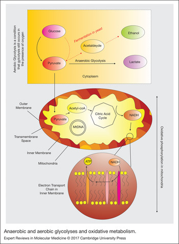
Before venturing into the possible explanations for abovementioned phenomenon, it is important to take a closer look at the mitochondrial functions in general. Mitochondria are cellular engines, responsible for producing adenosine triphosphate (ATP). Mitochondria contribute to cellular signalling and differentiation, cell cycling and growth, and failure of mitochondrial functions contributes to apoptosis. Both Anaerobic and aerobic glycolysis involve the transformation of glucose to pyruvate. The former occurs in the absence of oxygen in cytoplasm, while the latter occurs to a variable extent in all cells when oxygen levels decline below a threshold (Ref. Reference Vaishnavi17), including cancer cells (Refs Reference Lunt and Vander Heiden18, Reference Vander Heiden, Cantley and Thompson19) that use glycolytically generated ATP to proliferate. Aerobic glycolysis accounts for 10–12% of glucose utilised in the adult human brain (Refs Reference Raichle, Posner and Plum20, Reference Gjedde, Marrett and Vafaee21). Oxidative phosphorylation (OXPHOS), the aerobic generation of ATP, takes place in mitochondria, the cellular hosts of the TCA (tricyclic acid) cycle in the mitochondrial matrix that interacts with the electron transport chain complexes in the inner mitochondrial membrane (Refs Reference Robinson and Srere22, Reference Saraste23). The four electron-transporting complexes (I–IV) and the one H+-translocating complex (complex V) drive the ATP synthesis from adenosine diphosphate (Ref. Reference Raha and Robinson24). The TCA operates in the mitochondrial matrix, where it interacts with the electron transport chain complexes in the inner mitochondrial membrane, responsible for the ATP production. Figure 1 depicts the anaerobic and aerobic glycolytic pathways and the oxidative phosphorylation in cells.
Anaerobic and aerobic glycolyses and oxidative metabolism.

Reactive oxygen and nitrogen species
Mitochondria and its electron transport chain complexes are the main sources of reactive oxygen species (ROS) and nitrogen species in cells (Refs Reference Cadenas and Davies25, Reference Muller26), where approximately 90% of cellular ROS production occurs in the mitochondria (Ref. Reference Balaban, Nemoto and Finkel27). Superoxide (O2 −) is the primary ROS (Ref. Reference Murphy28), mostly generated in mitochondria (Ref. Reference Turrens29). O2 −, hydrogen peroxide (H2O2) and hydroxyl radical (OH−) stem from the univalent reduction of dioxygen (Ref. Reference Richter30). Despite the reduction of molecular oxygen to water in the terminal complex IV by a sequential four-electron transfer (Ref. Reference Nicholls and Budd31), roughly 2% oxygen in and around the mitochondria turns into the superoxide radical and ROS (Ref. Reference Inoue32). Complexes I–III are responsible for production of the majority of the superoxide in the mitochondria (Refs Reference Takeshige and Minakami33, Reference Beyer34, Reference Sugioka35, Reference Herrero and Barja36). The influence of ROS results in little damage under normal circumstances, as it enters into a balance with antioxidant systems of enzymes, such as superoxide dismutase (Ref. Reference Fridovich37). The danger lies in excess quantities of ROS: excess mitochondrial ROS injures proteins, mtDNA, and lipids, leading to mitochondrial dysfunction (Refs Reference Irshad and Chaudhuri38, Reference Bulteau, Szweda and Friguet39). The mtDNA rate of mutation is approximately 10 times more frequent than that of nuclear DNA (Ref. Reference Brown, George and Wilson40), owing to the lack of introns and histone patterning, simple mtDNA repair mechanisms, as well as the constant exposure to ROS (Ref. Reference Ito41). The resulting oxidative stress manifests itself as mild or severe injury, depending on the level of metabolic shift.
Oxidative stress ultimately contributes to increased severity of cardiovascular and neurological disorders and malignancy (Ref. Reference Irshad and Chaudhuri38). Moreover, increased oxidative stress plays a pivotal role in the aetiology of psychiatric disorders (Refs Reference Michel, Pulschen and Thome42, Reference Michel43). Interestingly, some of these disorders, such as depression are highly comorbid with both stroke and ischaemic heart disease. The ROS damage mitochondria (Ref. Reference Duchen44) through the liberation of electrons by the coenzymes NADH and FADH that contribute to the electron transport chain (Ref. Reference Puddu45). The enzymes α-ketoglutarate dehydrogenase and monoamine oxidase, present in the outer membrane of mitochondria, also generate considerable amounts of ROS (Refs Reference Andreyev, Kushnareva and Starkov46, Reference Adam-Vizi and Chinopoulos47). NO is both neuroprotective (Ref. Reference Calabrese48) and neurotoxic (Ref. Reference Dawson and Dawson49). Nitrogen dioxide (NO2) is a neurotoxic agent, and NO2 can exert its effect through mitochondrial dysfunction. It alters cortical mitochondrial morphology, diminishes complexes of respiratory chain and ATP production, and augments ROS production (Ref. Reference Yan50).
Mitochondria and ischaemic heart disease
Mitochondrial function protects the myocardium through the mitochondrial Ca2+ transport, the activity of the permeability transition pore, and the improvement of mitochondrial substrate supply (Ref. Reference Suleiman, Halestrap and Griffiths51). However, mitochondria are also involved in heart failure, cardiac dysfunction and myocyte damage through loss of metabolic capacity and release of toxic products (Ref. Reference Lesnefsky52). Additionally, mitochondrial ROS exert a crucial role in the pathogenesis of atherosclerosis because of major involvement in production of superoxide in vascular endothelial cells (Ref. Reference Nishigaki53). Mitochondrial dysfunction is also an important contributor to cardiovascular disease by triggering metabolic perturbation within vascular endothelial and smooth muscle cells, leading to vascular dysfunction (Ref. Reference Takagi54). Mitochondrial energy deficiency contributes to arterial hypertension (Ref. Reference Postnov55) and hypertrophy of myocardium in spontaneously hypertensive rats (Ref. Reference Chen, Tian and Song56). Mitochondrial complex III and IV defects in skin fibroblasts, kidneys and liver emerged in a multi-organ syndrome of severe atherosclerosis at a young age (Ref. Reference Feigenbaum57). In other studies, strains of pigeons prone to atherosclerosis (Ref. Reference Narula58), as well as atherosclerotic swine (Ref. Reference Morrison59), had cardiac mitochondrial dysfunction with deficient OXPHOS.
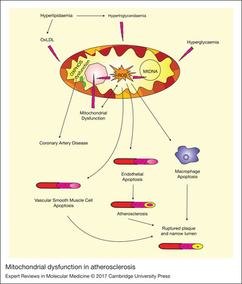
Despite intense investigation into the pathogenesis of atherosclerosis, the underlying factors are poorly understood. Factors associated with vascular pathophysiology include oxidised low-density lipoprotein (oxLDL), triglycerides and free fatty acids, hyperglycaemia, benzo[a]pyrene, and highly active antiretroviral therapy, all of which may result in mitochondrial dysfunction and ROS production (Ref. Reference Madamanchi and Runge14). Mitochondrial dysfunction lowers aerobic capacity with elevation of mitochondrial ROS generation that results in endothelial dysfunction, apoptosis and proliferation of vascular smooth muscle cells, leading to the development of atherosclerosis (Ref. Reference Madamanchi and Runge14). Apoptosis of vascular smooth muscle cells and macrophages from increased ROS generation advances atherosclerotic lesion progression and brings about plaque rupture (Fig. 2).
Mitochondrial dysfunction in atherosclerosis.

Evidence implicates ROS-mediated alterations as important potential contributors of cardiovascular disorder (Refs Reference Massaeli and Pierce60, Reference Dhalla, Temsah and Netticadan61), especially arteriosclerosis (Ref. Reference Victor62). The mitochondrial respiratory chain, in particular complex III, is a major source of ROS under pathological conditions that include myocardial ischaemia and reperfusion (Ref. Reference Chen63). ROS can also be directly involved in atherogenesis through four fundamental mechanisms that include oxidation of LDL, endothelial dysfunction, vascular smooth muscle cell growth and monocyte migration (Ref. Reference Schleicher and Friess64). As mentioned, LDL has an important role in atherosclerosis by virtue of the cytotoxic and atherogenic impacts of oxLDL (Ref. Reference Witztum65), which arises from oxidative and nitrosoxidative stress (Ref. Reference Berliner and Heinecke66). In the complex process of oxidation of LDL, nonenzymatic oxidative alterations of amino acids, proteolysis and cross-links of apoprotein B (apo B) severely change protein composition and structure (Ref. Reference Fong67). The peroxidised lipids decompose with the production of free and core aldehydes and ketones that covalently alter the ε-amino groups of lysine residues in the protein moiety (Ref. Reference Fruebis, Parthasarathy and Steinberg68). The result is Schiff's bases that change the charges on the amino acids, and bring about intra- and intermolecular cross-links between proteolysed apo B; macrophages inevitably scavenge the damaged structures (Ref. Reference Parthasarathy69).
LDL oxidation fails to occur in plasma in the presence of high concentrations of antioxidants and chelators (Ref. Reference Berliner and Heinecke66). Nonetheless, LDL is prone to oxidation in the arterial wall when antioxidants are depleted. In this process, both the protein and the lipids of LDL undergo oxidation. Arteriosclerosis is initiated by transfer of oxLDL to the subendothelial space of the arterial wall at the sites of endothelial damage (Ref. Reference Steinberg70). ROS also contributes to lesion progression and final plaque rupture in arteriosclerosis (Ref. Reference Bonomini2). It is postulated that oxidative stress and disturbed mitochondrial function contribute to arteriosclerosis (Ref. Reference Finsterer71). On the other hand, cholesterol and oxLDL damage the mitochondria (Ref. Reference Ballinger72). OxLDL leads to ROS generation in endothelial mitochondria (Ref. Reference Puddu45).
The process that initiates apoptosis is the activation of mitochondrial complex II (Ref. Reference Cheng73), uncoupled endothelial nitric oxide synthase (eNOS) and the NADPH oxidase activity (Ref. Reference Fleming74). Apoptosis of vascular smooth muscle cells in plaques produced by ApoE at sites of atherogenesis hastens plaque growth and is associated with vulnerability of plaques because of the thin fibrous cap and collagen matrix loss (Ref. Reference Clarke75). In addition, the oxidative energy produced in mitochondria, primarily by fatty acid β-oxidation, mitochondrial respiratory chain and OXPHOS activity, provides the main energy source for cardiac function (Ref. Reference Marin-Garcia, Goldenthal and Moe76) where mitochondrial dysfunction may bring about cardiac failure.
Under normal conditions, high-density lipoprotein (HDL) protects against ischaemia reperfusion injury by inhibition of mitochondrial permeability transition pore opening, an effect mediated by activation of the survivor activating factor enhancement pathway (Ref. Reference Frias77). Subsequent to exacerbation of vascular function and under hyperglycaemic circumstances, additional oxidation of HDL results in apoptosis of endothelium through a mitochondrial defect (Ref. Reference Matsunaga78). Furthermore, ROS signals play a key role in cardiac remodelling, a series of alterations in heart structure and function during recovery from MI (Ref. Reference Grieve79). The ROS activity modulates proliferation of fibroblasts (Ref. Reference Irani80) and synthesis of collagen, and contributes to the activation of MMPs (matrix metalloproteinases) that degrade the cardiac matrix components (Ref. Reference Siwik, Pagano and Colucci81). In the failing myocardium, mitochondrial electron transport complex I has been shown to be a source of ROS signals (Ref. Reference Ide82).
Mitochondria and stroke
Mitochondrial ROS signals also contribute to the pathogenesis of cerebral infarction, as mitochondria are the main source of generation of superoxide in vascular endothelium (Ref. Reference Nishigaki53). Mitochondria are key elements in the development of ischaemic cell death in stroke by failure in ATP production for brain energy metabolism that play important roles in the activation of cell death pathways (Ref. Reference Sims and Muyderman83). Impaired delivery of glucose and oxygen to tissues subject to cerebral ischaemia results in mitochondrial dysfunction. The mitochondrial function is further modified by alterations to fundamental mechanisms during ischaemia and after reperfusion, leading to reduced ATP generation. Impaired ATP production weakens the cellular response to other insults and activates mechanisms of apoptosis, necrosis, or both (Ref. Reference Sims and Muyderman83). The production of ROS relates to the severity of ischaemic injury because ROS damage cellular macromolecules and lead to neuronal loss, which in turn is involved in the breakdown of the blood–brain barrier (BBB) and brain oedema formation (Ref. Reference Moro84).
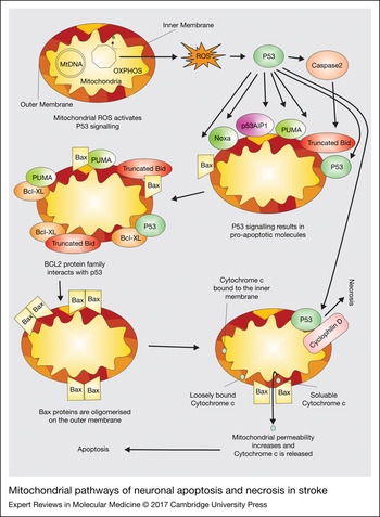
Mitochondria are an important source of ROS in acute brain injuries such as stroke (Ref. Reference Chan85). As the master regulator of cell death, the p53 signalling pathway is involved in the induction of apoptosis (Ref. Reference Schmitt86). Once the p53 signalling pathways are activated, pro-apoptotic molecules (e.g. Bax, Noxa, p53AIP1, PUMA and Bid) act directly on mitochondria (Refs Reference Niizuma, Endo and Chan16, Reference Miyashita and Reed87, Reference Nakano and Vousden88). The interaction of Bcl-2 family proteins and p53 leads to Bax oligomerisation and activation that in turn results in cytochrome c release and neuronal death. Figure 3 illustrates the mitochondrial pathways of apoptosis and necrosis in stroke. Animal studies have suggested that augmented formation of mitochondrial ROS, cytochrome c and Smac liberation, and apoptosis, decline in transgenic (Tg) animals overexpressing cytosolic copper- and zinc-superoxide dismutase subsequent to transient focal or global cerebral ischaemia. This shows that oxidative stress may be an upstream event in the mitochondrial-dependent apoptosis mechanism (Refs Reference Sugawara89, Reference Sugawara90, Reference Fujimura91, Reference Saito92). Apoptotic cell death in animal models of focal ischaemia has been confirmed by DNA fragmentation with analysis of relevant morphological alterations involving a regular pattern of chromatin condensation and apoptotic bodies (Ref. Reference Sims and Anderson93). Following both permanent and temporary middle cerebral artery occlusions, apoptotic cells are present in the entire ischaemic region (Ref. Reference Murakami, Kondo and Chan94). Perifocal tissue, especially the inner border areas next to the ischaemic core, contains most of these apoptotic cells detected as neurons in rats (Ref. Reference Li95). It has also been reported that mitochondrial dysfunction may exist in patients afflicted with CADASIL (Ref. Reference De la Pena96).
Mitochondrial pathways of neuronal apoptosis and necrosis in stroke.

In addition to apoptosis, necrosis is an important mechanism involved in neuronal cell death in ischaemic stroke. In fact, different types of neurons show different tendency to necrosis or apoptosis in ischaemic stroke (Ref. Reference Yuan97). ROS opens the mitochondrial permeability transition pore directly by oxidation of key proteins or activation of p53 (Ref. Reference Manzanero, Santro and Arumugam98). P53 binds to the pore protein cyclophilin D opening the pore. This blocks the mitochondrial respiratory chain and causes mitochondrial swelling leading to necrosis of neuronal cells (Ref. Reference Manzanero, Santro and Arumugam98). Ischaemia after stroke is also a potent initiator of necrosis (Ref. Reference Syntichaki and Tavernarakis99).
Pericytes in MI and stroke
Pericytes are contractile cells that wrap around the endothelial cells in capillaries and venules (Ref. Reference Bergers and Song100). They are important components of the neurovascular unit of endothelial cells, astrocytes and neurons (Ref. Reference del Zoppo101). Novel findings suggest that pericytes play an important role in MI and stroke, as they contribute to crucial and complex cell-specific alterations of signalling pathways and execution cascades (Ref. Reference Moskowitz, Lo and Iadecola102). Failure of ion homeostasis, calcium-ion overload and excitotoxicity, with induction of reactive peroxide, all lead to membrane damage, mitochondrial and DNA injury, inflammation and apoptosis in stroke (Ref. Reference Dirnagl, Iadecola and Moskowitz103), where these events subsequently affect the pericytes (Ref. Reference Wang104). Most recently, the cells interacting and supporting neurons were demonstrated to be equally damaged by ischaemic stroke and the supporting cells also contributed to crucial and complex cell-specific signalling pathways and execution cascades (Ref. Reference Moskowitz, Lo and Iadecola102).
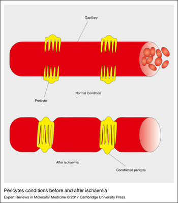
Pericytes regulate BBB impermeability that fails in stroke, giving rise to cerebral oedema (Ref. Reference Armulik105). Capillary blood flow is never fully recovered, even after recanalisation, when cerebral blood flow may be reduced to half (Ref. Reference Andalib106). Figure 4 illustrates pericyte constriction because of cerebral ischaemia, leading to increased resistance to blood flow. Ischaemia constricts CNS capillaries next to pericytes by virtue of the high intracellular calcium concentration in the pericytes when the ATP production declines, inhibiting ion pumping (Refs Reference Bliksøen107, Reference Wang108). Oxidative/nitrative stressors that both follow and lead to mitochondrial dysfunction (Ref. Reference Golomb109) sustain contraction of pericytes on microvessels and impair capillary reperfusion in stroke (Ref. Reference Yemisci110). The same phenomenon is believed to emerge in ischaemic heart disease. Pericytes, the second most-common cell type in the heart, hinder reperfusion of coronary capillaries in a manner similar to that of stroke (Ref. Reference O'Farrell and Attwell111), although their role in ischaemic heart disease is a less divulged topic. Pericyte constriction may be involved in the morbidity of patients by triggering microvascular obstruction (Ref. Reference O'Farrell and Attwell111).
Pericytes conditions before and after ischaemia.

Nuclear DNA alterations in ischaemic heart disease
Family history is an important, nonmodifiable cardiovascular risk factor. Recently, interest in the role of genetics in ischaemic heart disease gave rise to the emerging theory of a genetic basis of arteriosclerosis. Cytogenetic analyses reveal that karyotypes with chromosomal alterations of XXY, +7, +10, +18, −Y and −11, develop human atherosclerotic plaques (Refs Reference Casalone112, Reference Vanni, Cossu and Licheri113, Reference Matturri114). In addition, microsatellite instability is present in human atheromatous plaques (Ref. Reference Spandidos115), for instance at the 3′ region of the H-ras proto-oncogene (Ref. Reference Kiaris116) and the TGF-β1 receptor gene (Ref. Reference McCaffrey117). Loss of heterozygosity also is seen in mismatch repair (Ref. Reference Flouris118) and in NOS (Ref. Reference Grati119) genes. DNA adducts exist in abdominal aorta fragments in patients afflicted with severe atherosclerotic lesions (Ref. Reference Izzotti120). DNA adduct levels in cardiac tissue are correlated with the severity of coronary artery disease (CAD) (Ref. Reference Van Schooten121). Increased oxidative DNA damage occurs with augmented levels of 8-oxo-guanine in lymphocytes associated with atherosclerosis (Ref. Reference Gackowski122). The PON (Ref. Reference Andalib123) and PPAR genes (Ref. Reference Motavallian124) are associated with cardiovascular diseases in different populations. A Glu487Lys of ALDH2 gene contributes to the pathogenesis of MI in elderly Korean men (Ref. Reference Jo125). The C1019T variation in the Connexin 37 gene in men, the 4G-668/5G variation in the plasminogen-activator inhibitor type 1 gene, and the 5A-1171/6A variation in the stromelysin-1 gene in women, all are associated with the risk of MI (Ref. Reference Yamada126). The polymorphic WRN variant, a member of the RecQ family of helicases, is associated with increased risk of MI (Ref. Reference Ye127).
Nuclear DNA alterations in stroke
Recent attention has been paid to the role of nuclear DNA in the pathogenesis of stroke. The risk locus on chromosome 9p21.3 was shown to have one of the strongest associations with ischaemic stroke (Ref. Reference Meschia128), and there are single gene disorders associated with ischaemic stroke. As an example, the autosomal dominant disorder CADASIL stems from a notch3 gene mutation that affects small vessels. Likewise, sickle cell disease affects large arteries and small vessels and arises from a mutation of the HBB gene inherited in an x-linked fashion (Ref. Reference Dichgans129). The autosomal recessive inheritance of the CBS gene mutation is associated with homocystinuria, which affects large arteries and small vessels that result in cardioembolism and arterial dissection.
The mitochondrial, and hence maternally inherited, disorder complex consisting of myopathy, encephalopathy, lactacidosis and stroke (collectively known as MELAS) affects microvascular and neuronal factors. Patients with Marfan syndrome (FBN1 gene mutation), an autosomal dominant disease, present with higher risk of cardioembolic and arterial dissection. Arterial dissection can also be the result of Ehlers–Danlos syndrome type IV that can arise from mutation of the COL3A1 gene with autosomal dominant inheritance. Pseudoxanthoma elasticum with autosomal recessive inheritance that affects large arteries and small vessels stems from an ABCC6 gene alteration (Ref. Reference Dichgans129). Primary and secondary cardiomyopathies, familial atrial myoxoma and familial dysartherimias are Mendelian disorders that trigger ischaemic stroke through a cardioembolic etiology (Ref. Reference Natowicz and Kelley130). Metabolic states (e.g. homocysteinuria (Ref. Reference Mudd131) and dyslipidaemias (Ref. Reference Third132)), prothombotic states such as antithrombin III deficiency, and protein C deficiency, can cause stroke by thrombotic or thromboembolic mechanisms (Refs Reference Vomberg133, Reference Dahlbäck, Carlsson and Svensson134). In addition, multiple studies showed an association between ischaemic stroke and nuclear genes that contribute to the haemostatic system, such as factor V (Ref. Reference De Lucia135), prothrombin (Ref. Reference De Stefano136), fibrinogen (Ref. Reference Carter137), GPIIb/IIIa (Ref. Reference Carter138), GPIb/IX (Ref. Reference Gonzalez-Conejero139), GPIa/IIa (Ref. Reference Carlsson140) and variations in genes involved in renin angiotensin pathway such as ACE (Ref. Reference Sharma141), apo E (Ref. Reference Margaglione142) and apo A1/CIII (Ref. Reference Patsch143).
Mitochondrial DNA
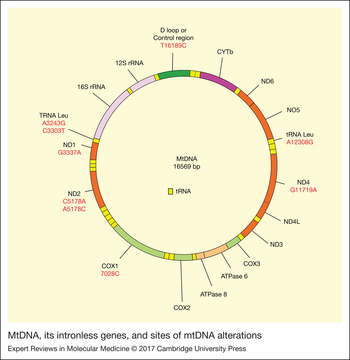
MtDNA is a circular structure with 16 569 base pairs (bp) (Ref. Reference Awadalla, Eyre-Walker and Smith144) and has 37 intronless genes, expressing 13 subunits of the electron-transfer chain, 2 ribosomal RNAs (rRNA) and 22 transfer RNAs (tRNA) (Ref. Reference Chinnery and Schon145). Mitochondrial respiratory chain incorporates five complexes. Of the 46 subunits of complex I, NADH dehydrogenase(ND)1, ND2, ND3, ND4, ND4l, ND5 and ND6 are produced by mtDNA, while nuclear genes express all four subunits of complex II. The only protein of the three subunits of complex III encoded by mtDNA is cytochrome b (cyt b). Among the 13 subunits of complex IV, cytochrome oxidase (COX) I, COX II and COX III are produced by mtDNA, and of 16 subunits of complex V, the A6 and A8 proteins are expressed by mtDNA. MtDNA is only inherited maternally (Ref. Reference Giles146) and complex I genes are the most vulnerable parts (Ref. Reference Gu147). MtDNA and its intronless genes are shown in Figure 5.
MtDNA, its intronless genes, and sites of mtDNA alterations.

MtDNA alterations in ischaemic heart disease and stroke
Several lines of evidence implicate mtDNA in ischaemic heart disease and stroke. Recent studies have suggested that age-dependent accumulation of somatically acquired oxygen damage, as well as deletions in mtDNA, impair cardiac function in relation to age (Ref. Reference Hayakawa148), and mtDNA variations can contribute to respiratory dysfunction (Ref. Reference Lertrit149). ROS resulting from ischaemia/reperfusion and atherosclerosis can damage mtDNA, giving rise to gene deletions on several areas of the genome (Ref. Reference Ramachandran150). Accumulation of damaged mtDNA affects the nuclear OXPHOS gene mRNA (Ref. Reference Ferrari151). ROS can damage endothelial mtDNA, with loss of mitochondrial RNA (mtRNA) transcripts, mitochondrial protein synthesis, and cellular ATP (Ref. Reference Ballinger152). Maternally inherited defects may result in mutations in genes expressing OXPHOS, forming impaired subunits with potential involvement in the pathophysiology of cardiomyopathies (Ref. Reference Luft and Landau153). An animal study also suggested that mitochondrial dysfunction leading to diminished energy metabolism contributes to the myocardial degeneration and changes in mitochondrial membrane system. Structural changes to the inner mitochondrial membrane lead to oxidative stress, which affects mtDNA as a consequence of diminished scavenging activity in the mitochondria (Ref. Reference Tokoro, Ito and Suzuki154). In a study of stroke-prone spontaneously hypertensive rats (SHRSP), myocardial enzymes of the mitochondrial respiratory chain, viz. isocitrate dehydrogenase c (ICDH), COX and the radical scavenger superoxide dismutase (SOD), were shown to have lower activity than those of Wistar–Kyoto rats (Ref. Reference Tokoro, Ito and Suzuki154). Genetic analysis also showed two deletions in the electrophoretic band in the SHRSP myocardium. Another experimental study suggested that mtDNA transcription declines in congestive heart failure, and that PGC-1α is a potential modulator of muscle oxidative capacity (Ref. Reference Garnier155).
Ide et al. (Ref. Reference Ide156) argued that ROS, mtDNA alteration, and the electron transport dysfunction, resulting in additional generation of ROS, are likely to affect the development and progression of left ventricular (LV) remodelling and failure subsequent to MI. Using a murine model of MI and LV remodelling, the authors showed a growth in hydroxyl radicals, stemming from mitochondrial superoxide anion, and lipid peroxide formation in the noninfarcted LV from MI mice. Moreover, Southern blotting revealed that the mtDNA copy number relative to the 18S rRNA nuclear gene preferentially declined by 44% in MI associated with a parallel diminution (30–50% of sham) in the mitochondrial gene transcripts, involving the subunits of complex I (ND1, ND2, ND 3, ND 4, ND 4L and ND 5), complex III (cytochrome b), complex IV (cytochrome c oxidase) and rRNA (12S and 16S) (Ref. Reference Ide156).
Tsutsui et al. (Ref. Reference Tsutsui, Ide and Kinugawa157) implicated ROS in myocyte hypertrophy, apoptosis, and interstitial fibrosis by activating matrix metalloproteinases and mitochondrial oxidative stress. The study suggested an activation of mitochondrial transcription factor A (TFAM) expression that may limit the pathophysiology of myocardial failure. In addition to activation of TFAM, activation of gene expression of peroxiredoxin-3 (Prx-3), which is a mitochondrial antioxidant, facilitates processes involved in mitochondrial dysfunction and myocardial remodelling (Ref. Reference Tsutsui, Kinugawa and Matsushima158). There is also a link between TNF-α, ROS and mtDNA alteration, which contributes to myocardial remodelling and failure (Ref. Reference Suematsu159).
In one study, authors claimed that TFAM, a key regulator of mtDNA copy number encoded by nuclear DNA, may open a new therapeutic approach to cardiac failure, inasmuch as it inhibits LV remodelling subsequent to MI (Ref. Reference Ikeuchi160). In Tg mice overexpressing the human TFAM gene, TFAM overexpression enhanced the activity of mitochondrial complex enzyme in post-MI hearts and reduced the mtDNA copy number, with a significantly higher survival rate post-MI than in wild-type (WT) littermates, despite comparable infarct size, associated with significant attenuation of LV cavity dilatation and dysfunction in the Tg-MI. While LV end-diastolic pressure rose in WT-MI, it declined in Tg-MI. LV function amelioration in Tg-MI coincided with diminution of myocyte hypertrophy, apoptosis and interstitial fibrosis in addition to oxidative stress in the LV without infarct (Ref. Reference Ikeuchi160).
Mitochondrial DNA alterations in the other cardiovascular diseases
The involvement of mitochondria in cardiovascular disorders is evident in an increasing number of observations. MtDNA is pathogenic and gives rise to dilated cardiomyopathy through the mitochondrial apoptotic pathway (Ref. Reference Zhang161). Circulating mtDNA in the plasma recently has been identified as a prognostic marker of MI. The evidence from recent studies suggested that circulating mtDNA is elevated in MI patients (Refs Reference Bliksøen107, Reference Wang108).
In a 6-year-old boy with dilatation cardiomyopathy with abundant ragged red fibres in muscle (20% of total) and severe lactic acidosis (Ref. Reference Vilarinho162), the A3243G mtDNA mutation in the tRNA Leu(UUR) gene constituted 68 and 88% of the total mtDNA in blood and muscle, respectively. Genetic analysis of blood from 16 maternal relatives showed a lower mutation extent exclusively in the oligo-symptomatic mother and brother (Ref. Reference Vilarinho162).
The C3303T mtDNA mutation, altering the aminoacyl stem of the tRNA Leu(UUR), was reported in seven members of an American family with cardiomyopathy and myopathy (Ref. Reference Silvestri163). The proband and two siblings presented with fatal infantile cardiomyopathy, and three maternal relatives suffered the disease in the form of sudden cardiac death or mitochondrial myopathy with cardiomyopathy. Additionally, the proband and her brother were shown to carry the homoplasmic C3303T mtDNA mutation in all tissues, although the mutation was heteroplasmic in the blood of five oligosymptomatic or asymptomatic maternal relatives. In contrast, the mutation was not found in all 70 controls.
Concurrent mutations in tRNA and ND1 mitochondrial genes were demonstrated to act synergistically, influencing the clinical phenotype in patients with cardiomyopathy (Ref. Reference Zifa164) in a Greek investigation. Using a PCR-sequencing, single-stranded conformational polymorphism, and RFLP analyses, a novel homoplasmic G3337A mutation of NADH dehydrogenase 1 gene (ND1) was found in two unrelated cases with cardiomyopathy, although absent in 150 healthy controls. The mutation co-segregated with two known mutations, A12308G (in tRNA Leu[CUN] gene) and C15946T (in tRNA Thr gene). Compared with the controls, subjects with the G3337A mtDNA mutation had decreased mitochondrial respiratory chain complex I activity of platelets (Ref. Reference Zifa164).
An Italian study investigated mtDNA deletion of 4977 bp in 65 CAD subjects, 23 subjects with coronary atherosclerotic plaque, and 22 age-matched healthy controls by means of a nested PCR protocol (Ref. Reference Botto165). There was a significantly higher incidence of mtDNA deletion of 4977 bp in CAD subjects compared with the controls (26.2 versus 4.5%, respectively, P = 0.03), and the relative deletion extent in the CAD subjects was significantly higher than in the controls (P = 0.02). MtDNA deletion of 4977 bp was found in blood samples of 17 of 65 CAD subjects (26.2%), ranging from 0.18 to 0.46% of the total mtDNA (mean ± SD: 0.34 ± 0.02%). Five subjects (21.7%) with coronary atherosclerotic plaque carried the mtDNA deletion of 4977 bp with a range of 0.13–0.45% of the total mtDNA (0.35 ± 0.06%) (Ref. Reference Botto165). It was also shown that the incidence and the relative deletion extent were not significantly affected by atherogenic risk factors and clinical factors in either sample from the subjects.
Corral-Debrinski et al. (Ref. Reference Corral-Debrinski166) investigated the involvement of somatic mtDNA damage in human aging and progressive OXPHOS diseases by assessment of cardiac mtDNA deletions in normal individuals and patients with coronary atherosclerotic disease in the USA. MtDNA deletions were seen in normal individuals after the age of 40 and subsequently increased with age. MtDNA deletion of 4977 bp reached a maximum of 0.007%, with concurrent mtDNA deletions at np 7436 and 10422. There was a 7–220-fold growth of the extent of the mtDNA deletion of 4977 bp over age-matched controls in hearts deprived of mitochondrial substrates because of CAD, with the mtDNA deletions of 7436 and 10422 bps growing in parallel. There was an association between cumulative mtDNA damage and compensatory 3.5-fold induction of nuclear OXPHOS gene mRNA. In addition, areas of ischaemic hearts in the left ventricle showed the highest mtDNA damage accumulation and OXPHOS gene induction (Ref. Reference Corral-Debrinski166).
The mtDNA C5178A variation that changes Leu to Met in the NADH dehydrogenase subunit 2 gene was shown to be antiatherogenic, at least in type 2 diabetic patients (Ref. Reference Matsunaga167). In this study, the mtDNA C5178A variation was assessed by the PCR–RFLP in 1148 Japanese patients with type 2 diabetes, and the findings were compared with clinical characteristics. The association between the variation and early atherosclerotic alterations of the bilateral carotid arteries was also analysed in 412 diabetic cases, randomly chosen from the original cases, with the same frequency of 5178A and 5178C. The frequency of mtDNA C5178A variation in the diabetic cases (454 of 1148; 40%) was shown to not differ from that previously found in healthy controls (114 of 252; 45%) and no significant statistical difference in clinical characteristics of diabetes was found between the mt5178A subjects (n = 454) and the mt5178C subjects (n = 694) (Ref. Reference Matsunaga167). Yet, the mean intima-media thicknesses in the mt5178A group (0.906 ± 0.018 mm) at six regions of the carotid arteries bilaterally were significantly lower than in the mt5178C group (0.995 ± 0.021 mm) (P = 0.022). Moreover, there was a significant correlation between the mt5178 type and both the mean intima-media thickness and the existence of plaques.
A C5178A mtDNA variation of ND2 gene that changes Leu to Met in the peptide chain was shown to be protective against MI in a Japanese population (Ref. Reference Takagi54). Using PCR–RFLP or colorimetry-based allele-specific DNA probe assay, the frequency of the mt5178A genotype was demonstrated to be significantly higher in controls (46.3%) compared with that of subjects with MI (39.3%) (P = 0.0388, OR = 0.73, 95% CI = 0.54–0.98) (Ref. Reference Takagi54). An A5178C mtDNA variation that substituted Met for Leu in the ND subunit was associated with development of MI, especially in younger patients, and represented a risk factor for MI in another Japanese population (Ref. Reference Mukae168). The A5178C variation was analysed in four groups involving 150 cases with MI, 150 cases with essential hypertension, 100 cases with diabetes mellitus (DM), and 150 age/sex-matched controls by using PCR–RFLP test. Compared with the control group, the frequency of the A5178C variation was seen to be higher in the MI group (0.53 versus 0.65, respectively, OR = 1.69, 95% CI = 1.06–2.69), with a considerable difference in younger cases. Further differences were seen in allelic frequencies in the groups of patients with essential hypertension, and in the DM group as well as in the control group, in spite of a higher frequency of the C allele found in the MI patients compared with the essential hypertension and DM groups.
Possibly by virtue of differences in mitochondrial function and ROS between the different haplogroups, the presence of mtDNA variations in atherosclerosis and its ischaemic consequences events, was identified in a study using PCR, single-strand conformation analysis (SSCA), denaturing high-performance liquid chromatography (DHPLC), and sequencing with BigDye chemistry, in an automated ABI310 capillary system (Ref. Reference Palacín169). Five hundred male subjects with early onset MI (<55 years) and at least one atherosclerotic coronary vessel, and 500 controls, were assessed for ten mtDNA variations defining nine common European haplogroups and TFAM gene coding region variations. The study found a strong association between early onset MI and male gender and tobacco smoking. The frequency of mtDNA haplogroup H, defined by allele 7028C, was higher in the early onset MI group of nonsmokers (n = 250) than in healthy controls (n = 300), with an association between the haplogroup and the group of only smokers (250 patients and 200 controls). There was a significant statistical difference between H and other haplogroups for both groups of patients (252/500 [50%]) and for controls (202/500 [40%]) (P = 0.002, OR = 1.50; 95% CI = 1.17–1.92). However, there was no significant difference between the patients and the controls in four common TFAM gene variations (Ref. Reference Palacín169).
In a study of a Central European population, the T16189C variation in the hypervariable D-loop of the control region (CR) of mtDNA was seen to be associated with CAD (Ref. Reference Mueller170). In this study, the authors assessed CR variations and nine main European haplogroups using DNA sequencing and primer extension analysis and found a higher prevalence of T16189C variation in the CAD cases (21.6%), compared with the controls (11.8%) (P = 0, OR = 2.05, 95% CI = 1.6–2.7).
Takeda et al. (Ref. Reference Takeda171) studied the mutation of myocardial mtDNA in human left ventricles from 17 autopsy subjects with DM, MI, cardiomyopathy, cancer and other diseases by means of PCR. In two cases of DM, two cases of MI and one case of pulmonary fibrosis, mtDNA had a 7.4 kb deletion between the D-loop and ATPase 6 genes. Takeda et al. (Ref. Reference Takeda, Singal, Panagia and Pierce172) also reported a case of DM and hypertrophic cardiomyopathy with a point A3243G mutation in tRNA Leu(UUR) mtDNA gene.
Mitochondrial haplogroups in 9254 individuals from the Copenhagen City Heart Study of the general population were assessed by TaqMan assays (Ref. Reference Benn173). The subjects were studied prospectively for the risk of ischaemic cardiovascular disease, morbidity from other causes and mortality, during 25 and 11 years, respectively, but the study found no association of mitochondrial haplogroups (i.e. T, J, K, V, W/I and Z) with risk of ischaemic cardiovascular disease, morbidity from other causes, mortality or longevity.
Association between mitochondrial haplogroups and MI was investigated in a Japanese population (Ref. Reference Nishigaki174). In this study, 2137 unrelated Japanese subjects, involving 1181 subjects with a first MI (920 males and 261 females) and control subjects (522 males and 434 females) were assessed for 28 mtDNA variations of 12 main mitochondrial haplogroups, that is, A, B, D4, D5, F, G1, G2, M7a, M7b, M7c, N9a and N9b, by means of 28-plex PCR and fluorescent beads with sequence-specific oligonucleotide probes. There was a significantly lower prevalence of haplogroup N9b in cases with MI compared with the control group (P = 0.0019). The prevalence of this haplogroup was significantly lower in the male subjects with the disease than that of the male controls subsequent to adjustment for age, sex, body mass index (BMI), and prevalence of smoking, hypertension, hypercholesterolaemia and type 2 diabetes (P = 0.0007). For males, however, there were trends towards higher prevalence of MI in haplogroup G1 (P < 0.05). In females, there was no significant association between the haplogroups and MI; still, haplogroup N9b was held to be protective against MI in males (Ref. Reference Nishigaki174).
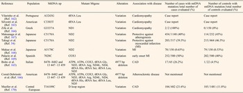
In Central European Caucasian populations, the mtDNA haplogroup T was seen to be associated with CAD and diabetic retinopathy (Ref. Reference Kofler175). The study assessed 487 cases with angiographically confirmed CAD, 149 cases with type 2 DM with or without 78 diabetic retinopathy and 1527 controls without symptoms of atherosclerotic disease. For determination of the major European haplogroups, mtDNA haplotypes were identified by means of multiplex PCR and subsequent multiplex primer extension analysis. There was a high frequency of haplogroup T in subjects with CAD, compared with control subjects (14.8 versus 8.3%, respectively, P = 0.002). Diabetic retinopathy was seen to be significantly associated with a higher prevalence of haplogroup T in type 2 diabetes subjects (12.1 versus 5.1%; P = 0.046) (Ref. Reference Kofler175). MtDNA alterations in cardiovascular diseases are tabulated in Table 1.
MtDNA alterations in ischaemic heart diseases

MtDNA alterations in stroke
MtDNA changes play an important role in the susceptibility to stroke. In addition to spontaneous intracerebral haemorrhage, the levels of plasma nuclear DNA and mtDNA are high in patients with acute ischaemic stroke (Ref. Reference Tsai176), such that these levels may reveal severity of cerebral insult.
MtDNA alterations have been investigated in many neurological disorders such as Leber's Hereditary Optic Neuropathy (Refs Reference Yen177, Reference Johns, Neufeld and Park178), myoclonus epilepsy with ragged-red fibres (Refs Reference Shoffner179, Reference Silvestri180), Kearns–Sayre syndrome (Refs Reference Carod-Artal181, Reference Nelson182), Alzheimer's disease (Refs Reference de la Monte183, Reference Corral-Debrinski184), Parkinson's disease (Refs Reference Andalib, Vafaee and Gjedde185, Reference Autere186), and multiple sclerosis (Refs Reference Andalib106, Reference Andalib187, Reference Andalib188, Reference Andalib189, Reference Andalib190). MtDNA variations have been demonstrated to be associated with susceptibility to stroke in specific populations.
In a study in the UK, mtDNA variation in sub-haplogroup K was demonstrated to be an independent determinant of ischaemic stroke risk (Ref. Reference Chinnery191). This study identified the major European mtDNA haplogroups in two independent sub-populations with 950 individuals in an investigation of ischaemic stroke and transient ischaemic attack (TIA). The haplogroups were compared with those in subjects with acute coronary syndromes in the same populations (n = 340) and with those in an independent control group (n = 2939). The frequency of mtDNA sub-haplogroup K was significantly less in subjects with TIA or stroke than in the control group both in the sub-populations separately and in pooled analysis (P < 0.00001, OR 0.54, 95% CI = 0.39–0.75) (Ref. Reference Chinnery191). However, the association was significant following adjustment for multiple haplogroup comparisons, and the association was significant separately for subjects with TIA and stroke. There was not such association for subjects with acute coronary syndromes. The mtDNA sub-haplogroup K was seen in 8.7% of the total UK population controls and hence decreased by a 4 percentage point (95% CI = 2.2–5.7) in the population attributable risk of TIA and stroke (Ref. Reference Chinnery191).
Haplogroup A increased the risk of atherothrombotic cerebral infarction in Japanese females (Ref. Reference Nishigaki53). In this study, association of mitochondrial haplogroups and atherothrombotic cerebral infarction was investigated in 1081 subjects by using 28-plex PCR and fluorescent beads with sequence-specific oligonucleotide probes. Consequently, 26 mtDNA variations were found in 11 major mitochondrial haplogroups (F [C3970T, G10310A, and G13928 A], B [8272: 9-bp deletion in noncoding region], A [A663G, C8794T], N9a [G5231A, A12358G and G12372A], M7a [C2772T and T4386C], M7b [C4071T, G4048A, T6680C and T12811C], M7c [C4071T and C4850T], G1 [G709A, A4833G, T5108C, T8200C and G15497A], G2 [G709A, A4833G, T5108C and A13563G], D4 [C4883T, C5178A, G3010A] and D5 [C4883T, C5178A and A10397G]). Haplogroup A in females was seen to be associated with atherothrombotic cerebral infarction (P = 0.0294, OR = 2.12, 95% CI = 1.06–4.12). Multivariate logistic regression analysis with adjustment for age, BMI, and prevalence of smoking, type 2 DM, hypertension, and hypercholesterolemia revealed an association between A3759G mtDNA variation, showing subhaplogroup D5b1b and resistance against atherothrombotic cerebral infarction (P = 0.0306, OR = 0.10, 95% CI = 0.01–0.54). For sub-haplogroups A1a1a and D5a2, respectively, C14944T (P = 0.0242, OR = 2.07, 95% CI = 1.10–3.91) and C1310T (P = 0.0360, OR = 4.08, 95% CI = 1.13–16.8) were associated with susceptibility to the disease (Ref. Reference Nishigaki53).
The mitochondrial haplogroup H1 was found to affect the risk of ischaemic stroke. Nineteen mtDNA variations forming the major European haplogroups were assessed in 534 cases with ischaemic stroke and 499 healthy controls (Ref. Reference Rosa192). Comparing each clade against all other haplogroups pooled together, the frequency of haplogroup H1 was seen to be significantly less in cases with stroke than in the control group (P = 0.001, OR = 0.61, 95% CI = 0.45–0.83). However, the pre-HV/HV and U mtDNA lineages was found to be potential genetic factors of stroke risk (P = 0.003, OR = 3.14, 95% CI = 1.41–7.01 and P = 0.021, OR 2.87, 95% CI = 1.13–7.28, respectively). MtDNA variations G3010A, C7028T and G11719A exerted an effect upon risk of ischaemic stroke, their allelic state in haplogroup H1 supporting its protective role (Ref. Reference Rosa192).
In a mitochondrial genome-wide association study (Ref. Reference Anderson193), 2284 ischaemic stroke patients and 1728 healthy control subjects from the International Stroke Genetics Consortium were assessed for 64 mtDNA variations. The study revealed 144 variations, identified in each cohort and in meta-analysis for association with ischaemic stroke. A genetic score of all mtDNA variations in association with ischaemic stroke was assessed, but there was no mtDNA variation reaching adjusted significance in meta-analysis. However, a genetic score that included the summation of contributions from mtDNA variations revealed an association with the disease in meta-analysis (P < 0.0001, OR = 1.13) with minimal heterogeneity (I 2 = 0.00). The ischaemic stroke score was robust to permutation, and associated with the volume of white matter hyperintensity in 792 nested ischaemic stroke subjects (P = 0.037) (Ref. Reference Anderson193).
The T16189C mtDNA variation in the hypervariable D-loop region was shown to be associated with lacunar cerebral infarction in a Taiwanese population (Ref. Reference Liou194), where clinical status, fasting blood sugar (FBS), insulin levels, and insulin resistance index were assessed. Predicated upon the Trial of Org 10172 in Acute Stroke Treatment (TOAST) criteria, individuals with cerebral infarction were subdivided into four subgroups, with a significant difference in occurrence rate of T16189C mtDNA variation between individuals with cerebral infarction (34.2%) and the control group (26.5%). The difference in the occurrence rates between the two groups was statistically significant at P = 0.041. The small vessel subgroup experienced the highest frequency of the variation (41.5%). There was a highly significant difference in occurrence rate between this subgroup and the control group at P = 0.006. These findings correlated well with the increased rates of average fasting blood insulin and the high index of average insulin resistance in the small vessel subgroup with the variation (Ref. Reference Liou194).
The A3243G mtDNA mutation, a common MELAS mutation, was found in young Finnish individuals with occipital stroke (Ref. Reference Majamaa195). In this study, 38 individuals aged 18–45 years with occipital stroke were tested retrospectively for the presence of the mutation in their blood samples. Of four cases (10%) with a clinical or molecular diagnosis of a mitochondrial disorder, two had the mutation, consistent with frequencies of 6 and 14% in cases younger than 45 and 30 years for this mutation, respectively. In addition, two subjects with a clinically definite mitochondrial disorder were found, and the subsequent sequencing of the 22 tRNA mtDNA genes revealed the A12308G mutation in one of them (Ref. Reference Majamaa195).
Another Finnish study demonstrated that the mtDNA haplogroup U defined by the A12308G variation was associated with an occipital stroke in migraine (Ref. Reference Finnilä, Hassinen and Majamaa196). In this study, 14 cases with an occipital stroke carrying the A12308G were identified and by means of confirmation-sensitive gel electrophoresis and direct sequencing, complete mtDNA coding region assessment were conducted for the cases and controls. Sequence data revealed a phylogenetic network of mtDNA haplogroups U and K, involving subclusters U2, U4, U5 and a novel subcluster U7, in addition to cluster K. There were five cases with migrainous stroke in subcluster U5 (P = 0.006). Furthermore, there were multiple unique mutations in cases of occipital stroke involving two tRNA mutations.
Martínez-Fernández et al. (Ref. Reference Martinez-Fernandez197) suggested that ischaemic stroke is likely to be the only or initial manifestation of a mitochondrial disorder. The authors tested 18 subjects with ischaemic stroke of unknown origin and demonstrated that three (17%) subjects had lactic acid hyperproduction subsequent to physical exercise, and six (33%) experienced the mitochondrial respiratory chain dysfunction. The genotyping assessment revealed mtDNA mutations at np 3243 (MELAS mutation), 4216 and 15 928 (Ref. Reference Martinez-Fernandez197). MtDNA alterations in stroke are illustrated in Table 2.
MtDNA alterations in stroke

NM, Not mentioned.
Translation into therapeutic approaches
In stroke and ischaemic heart disease, neuronal and cardiac cells undergo both necrosis and apoptosis. Apoptotic cell loss arises from permeabilisation of mitochondrial membranes, with involvement of ROS and Bcl2 and P53 proteins. Effective restitution of mitochondrial function after ischaemic heart disease or stroke may significantly improve the clinical outcome. Suggested prevention of permeabilisation of mitochondria and inhibition of apoptosis include inhibition of the mitochondrial membrane permeabilisation by ligands of cyclophilin D, openers of mitochondrial ATP-sensitive or Ca2+-activated K+ channels, proteins from the Bcl2 family engineered to cross the cellular membrane, and manipulations modulating the expression or activity of mitochondrial uncoupling proteins (Ref. Reference Fong67). In vivo delivery of a Bcl-xL fusion protein including PTD (protein transduction domain) resulting from the human immunodeficiency TAT protein fused with Bcl-xL limits the effects of ischaemic stroke (Ref. Reference Cao198).
The hydrophilic bile acid, ursodeoxycholic acid (UDCA) and its taurine conjugated derivative (TUDCA) play a unique role in modulating the apoptotic threshold (Ref. Reference Gronbeck199). They can interrupt apoptosis by blocking classic mitochondrial death pathways, while also significantly activating survival pathways. The classic cell death pathway is characterised by mitochondrial membrane permeability transition pore opening, collapse of the transmembrane potential, release of cytochrome c leading to subsequent caspase activation and apoptosis (Ref. Reference Orrenius200). TUDCA significantly inhibits the apoptosis associated with deoxycholic acid, ethanol, transforming growth factor β1, anti-Fas antibody, and okadaic acid, suggesting a basic mechanism common to each of the different apoptotic pathways. At least one mechanism for this ubiquitous antiapoptotic effect involves preservation of the mitochondrial membrane integrity preventing the release of cytochrome c and subsequent caspase activation, as well as the change in transmembrane potential from different apoptotic stimuli (Ref. Reference Rodrigues201).
A variety of conditions can induce oxidative stress of the endoplasmic reticulum and lead to apoptosis, which may have an important role in various diseases, including several neurologic conditions. Preclinical studies have shown the use of TUDCA in ischaemic and haemorrhagic stroke models resulted in improving neurological function (Ref. Reference Rodrigues202). TUDCA has been also shown to be effective in reducing apoptosis and improving cardiac function in a rat model of MI (Ref. Reference Rivard203). Other preclinical studies have shown neuroprotective effect of TUDCA in Huntington's disease, Alzheimer's disease and Parkinson's disease (Ref. Reference Gronbeck199). In clinical settings, TUDCA has been shown to be safe and potentially effective in the treatment of amyotrophic lateral sclerosis (Ref. Reference Elia204). These studies encourage clinical studies for the use of TUDCA in ischaemic heart disease and stroke.
Concluding remarks
Ischaemic heart disease and stroke constitute major global health challenges. Novel tools of biochemistry and molecular genetics have generated a novel paradigm for the investigation of these vascular events. The involvement of mitochondria in ischaemic heart disease and stroke is revealed in an increasing number of observations. Mitochondria produce ROS, which may trigger ischaemic heart disease and stroke through production of oxLDL and induction of apoptosis, respectively. In addition to a number of recently discovered nuclear gene alterations, mtDNA changes have been demonstrated to be associated with ischaemic heart disease and stroke. Yet, there is a need to invest in a substantial effort for the identification of ischaemic heart disease and stroke gene loci. Sophisticated imaging studies are necessary to confirm the influence of mitochondria and their DNA on the complex pathogenesis of ischaemic heart disease and stroke.
Acknowledgements
We thank Kate Lykke Lambertsen for her valuable comments on the paper.
Conflict of interest
None.