Significant outcomes
• Our study revealed that oxidative balance is disrupted in favour of oxidants in patients with somatic symptom disorder.
• Lipid peroxidation seems not to have a role in aetiology of somatic symptom disorder.
• Approaches to reduce oxidative stress may be an alternative option for the prevention and treatment of this disorder.
Limitations
• This research was a cross-sectional study conducted at a single centre with a relatively small group of subjects.
• The oxidative stress parameters were not studied in cerebrospinal fluid (CSF) samples; therefore, serum levels of these oxidative stress parameters may not be fully correlated with their CSF levels.
Introduction
According to fifth edition of the Diagnostic and Statistical Manual of Mental Disorders (DSM-5), somatic symptom disorder is characterised by excessive thoughts, feelings of fear or worry, and anxious behaviours of an individual lasting for 6 months or longer, thinking that the individual has a serious disease as a result of misinterpretations of somatic symptoms (1,Reference Rief and Martin2). Although there are some biopsychosocial theories about its aetiopathogenesis, the aetiologic basis of somatic symptom disorder is not yet known (Reference Sadock, Sadock and Ruiz3).
Oxidative stress is defined as the disruption of the balance between cellular reactive oxygen radicals and antioxidant mechanisms that serve to protect the body against oxidative stress. It is thought that the brain is prone to oxidative damage for various reasons (Reference Poulsen, Specht, Broedbaek, Henriksen, Ellervik, Mandrup-Poulsen, Tonnesen, Nielsen, Andersen and Weimann4). The main reason is that the brain has a high level of oxygen consumption for its metabolism. In addition, the presence of a large amount of redox-active metals such as iron and copper in the brain catalyzes the formation of reactive oxygen radicals. Furthermore, multiple polyunsaturated fatty acids, which exist in large amounts in neuronal cell membranes, are used as substrate in lipid peroxidation (Reference Wang and Michaelis5). The presence of a relatively modest level of important endogenous antioxidant molecules, along with the liability to oxidative reactions, causes the brain’s defense mechanisms against oxidative stress to become vulnerable (Reference Ferreira, de Vasconcelos, da Costa Vilhena, da Silva, da Silva Barbosa, Gomes, Dolabela and Percário6). Hence, there is a great number of studies in the literature evaluating the relationship between psychiatric disorders and oxidative stress. It has been demonstrated that there is a relationship between oxidative stress and various psychiatric disorders such as depression, anxiety disorders, autism, schizophrenia, and bipolar disorder (Reference Salim7,Reference Smaga, Niedzielska, Gawlik, Moniczewski, Krzek, Przegaliński, Pera and Filip8).
Few studies have examined the association between somatisation and pathways related with oxidative stress, including immune-inflammatory pathways and the tryptophan metabolite (TRYCAT) pathway (Reference Rief, Pilger, Ihle, Bosmans, Egyed and Maes9,Reference Rief, Pilger, Ihle, Verkerk, Scharpe and Maes10). These studies showed that there was an increase in inflammatory processes in patients with somatoform disorders (Reference Rief and Auer11). It was reported that activation of inflammatory processes triggered oxidative stress, and that oxidative damage might be related with somatic symptoms (Reference Maes12). However, no studies have shown the status of specific oxidative stress indicators in patients with somatic symptom disorder. Furthermore, it is thought that new studies are needed on this topic due to the manifestation of new probable oxidative stress mechanisms, which may have a role on the aetiology of mental disorders (Reference Maes12).
Aims of the study
In this study, we aimed to investigate oxidative stress parameters including total oxidant status (TOS), total antioxidant status (TAS), paraoxonase (PON), arylesterase (ARE), and thiol-disulphide homeostasis in patients with somatic symptom disorder. Consequently, it was aimed to establish the possible relationship between these oxidative stress parameters and total illness duration and severity of somatic symptoms to contribute to new hypotheses regarding the aetiology of this disorder.
Material and methods
Subjects
The study participants comprised 46 patients who were admitted to the psychiatry outpatient clinic of Ankara Ataturk Research and Training Hospital between January 2016 and September 2017. Patients who were diagnosed as having somatic symptom disorder in accordance with the diagnostic criteria of DSM-5 were screened. Three patients who were suspected of having a rheumatologic disorder and two patients who did not agree to participate in the study were excluded. Accordingly, the study population comprised 41 patients (24 females, 17 males) aged between 18 and 65 years who were medication free for at least 8 weeks. The patients were diagnosed as having somatic symptom disorder by a psychiatrist using a semi-structured psychiatric interview based on the DSM-5.
The exclusion criteria were the presence of any comorbid chronic disease (including fibromyalgia and chronic fatigue syndrome, and any neurologic, endocrinologic or metabolic disorder) or psychiatric disorder (e.g. mood disorders, anxiety disorders, or psychotic disorders), use of any medication, vitamin, or herbal supplement in the last 2 months, use of cigarettes or alcohol, and a history of acute trauma or infection in the last 2 months. To exclude the diagnosis of fibromyalgia, patients with pain symptoms were evaluated according to the ‘ACR 2010 fibromyalgia diagnostic criteria’ (Reference Wolfe, Clauw, Fitzcharles, Goldenberg, Katz, Mease, Russell, Russell, Winfield and Yunus13).
The control group was constituted by 47 healthy individuals (34 females, 13 males) who were matched for age, sex, and other sociodemographic features, and had no medical diseases or psychiatric disorders according to the Symptom Check List 90-Revised test and in the evaluation of the psychiatric interview. Alcohol users, smokers, and those who used any herbal supplements or medication, or had a history of acute infection or trauma in the last 2 months were excluded from the study.
Psychiatric measurements
In the clinical assessment, the Patient Health Questionnaire (PHQ-15) was administered to all patients to evaluate the severity of the somatic symptoms. The PHQ-15 is a 15-item Likert-type self-report scale, which was developed by Spitzer et al (Reference Spitzer, deGruy Ill, Hahn, Brody and Johnson14). The scale asks questions about somatic symptoms and their severity (Reference Kroenke, Spitzer and Williams15). Each item is assessed using scores between 0 and 2.
The Somatic Symptom Amplification Scale (SSAS), which was developed by Barsky et al. (Reference Barsky, Wyshak and Klerman16) to measure the amplification/exaggeration of normal somatic senses, was also administered to the patient group. The Turkish validity and reliability study of the scale was performed by Gulec et al. (Reference Gulec, Sayar and Gulec17). The SSAS is a Likert-type scale comprising 10 questions, which are graded from 1 to 5.
The Beck Depression Inventory (BDI) and Beck Anxiety Inventory (BAI) were conducted to the patient group to assess the severity of their symptoms of depression and anxiety. The BDI is a 17-item Likert-type self-report scale developed by Beck et al. (Reference Beck18) that indicates the emotional, somatic, cognitive, and motivational symptoms of depression. The validity and reliability studies of Turkish version of the scale were conducted by Hisli et al. (Reference Hisli19). The BAI is a 21-item scale that was developed by Beck et al. (Reference Beck, Epstein, Brown and Steer20). The Turkish validity and reliability study of the scale was performed by Ulusoy (Reference Ulusoy, Sahin and Erkmen21). Each item of both tests is evaluated with a scores between 0 and 3.
Blood samples
After 8 h of fasting, 5 cc venous blood was drawn from all participants into ethylene diamine tetra acetic acid tubes in the morning. Specimens were centrifuged at 1800 g for 10 min. After separation, the serum samples were frozen and stored at −80°C until required for biochemical analysis. Biochemical analysis was conducted when the samples were dissolved after being kept at +4°C for 12 h.
From the samples collected from both groups, routine haemogram; liver, kidney, and thyroid function tests; vitamin B12; folate; ferritin levels; and lipid profiles were studied. Measurements of oxidative stress parameters (TOS, TAS, PON, ARE, and total thiol, native thiol, and disulphide) were performed using commercial kits (RelAssay, Gaziantep, Turkey) on a Roche Hitachi Cobas c501 (Indianapolis, IN, USA) automatic analyzer.
Measurement of oxidative stress parameters
The total antioxidant response and TOS were determined using the automated methods described by Erel (Reference Erel22,Reference Erel23). The ratio of TOS to TAS gives the oxidative stress index (OSI) value. The OSI value was calculated using the following formula:
 \begin{array}{*{20}{c}}
{{\text{OSI}}\left( {{\text{arbitrary unit}}} \right)} \\
{ = \left[ {{\text{TOS}}\left( {\mu {\text{mol }}{{\text{H}}_2}{{\text{O}}_2}{\text{ Eqv}}/1} \right)/{\text{TAS}}\left( {{\text{mmol Trolox Eqv/1}}} \right)} \right] \times 100.}
\end{array}
PON and ARE are enzymes that protect cells against lipid peroxidation. The serum activity of PON and ARE was measured using commercially available kits (Rel Assay Diagnostics). PON activity was measured using paraoxone substrate by recording the 37°C absorbance of the colour formed as a result of paraoxone hydrolysis at 412 nm, and results are expressed as U/l. To measure ARE activity, a newly non-salt-stimulated method is used, with phenyl acetate used as substrate. One unit of ARE activity is determined as μmol phenol formed in 1 min, and the results are expressed as kU/l (Reference Haagen and Brock24).
Serum thiol-disulphide homeostasis was studied with the new automated measurement method developed by Erel and Neselioglu (Reference Erel and Neselioglu25). Dynamic disulphide bonds (–S–S–) are reduced to functional thiol groups (–SH) by sodium borohydride (NaBH4). The remaining unused NaBH4 is completely removed by formaldehyde. The native thiol (–SH) and total thiol (–SH + –S–S) content of the sample is measured using the modified Ellman reagent. The amount of disulphide bond is obtained by half of the difference between total thiol and native thiol. In addition, disulphide/native thiol, disulphide/total thiol, native thiol/total thiol ratios were calculated as percentage (%).
Statistical analysis
The IBM SPSS Statistics 21 software program was used to analyse the study data. The normality of continuous variables was tested using the Shapiro–Wilk test. Descriptive statistics [mean ± standard deviation (SD)] are given for continuous variables. Sex variance is expressed as percentage. For the results of the oxidative stress parameters, mean ± SD is given.
The continuous variables of both groups were compared using the independent samples t-test. Univariate analysis of covariance was performed to investigate the effects of potential confounders [age, sex, body mass index (BMI)]. The relationship between the levels of the oxidative stress parameters of the patient group, and total illness duration, and the scores of the PHQ-15 and SSAS scales were examined using Spearman correlation analysis. The statistical significance level was determined as p < 0.05.
Results
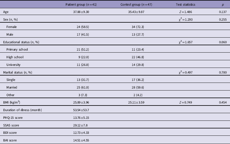
The study consisted of 88 subjects, including 41 patients and 47 healthy individuals. The demographic features of the patient and control groups are shown in Table 1. There was no statistically significant difference between the groups in terms of age, sex, BMI, marital status, and educational status (p > 0.05). The average total illness duration of the patient group was 53.54 ± 53.7 months. The scores of the psychiatric evaluation scales of the patient group are also given in Table 1.
Demographic characteristics of the two groups

BAI, Beck Anxiety Inventory; BDI, Beck Depression Inventory; BMI, body mass index; PHQ-15, Patient Health Questionnaire-15; SSAS, Somatic Symptom Amplificaiton Scale.
Values are presented as mean ± SD.
When the biochemical findings of the patient and control groups were examined, there was no statistically significant difference between the two groups in terms of liver, kidney, and thyroid function tests, lipid profiles, ferritin, folate, vitamin B12, white blood cell counts, haemoglobin, and platelet levels (p > 0.05).
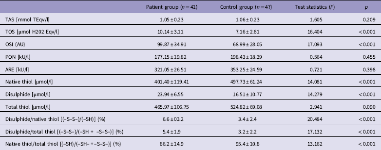
Both groups were compared in terms of oxidative stress parameters. It was found that parameters indicating oxidant molecules increased. The native thiol level of the patient group was significantly lower than that of the control group, but the disulphide level was significantly higher (p < 0.001). The TOS, OSI, disulphide/native thiol [(–S–S–)/(–SH)], and disulphide/total thiol [(–S–S–)/(–SH + –S–S–)] levels of the patient group were higher than those of the control group (p < 0.001). In addition, the native thiol/total thiol [(–SH)/(–SH– + –S–S–)] level was significantly lower for the patient group than in the control group (p < 0.001). On the other hand, no differences were found in parameters indicating antioxidant molecules. There was no statistically significant difference between the two groups for total thiol, TAS, and PON and ARE parameters (p < 0.05) (Table 2).
Comparison of the oxidative stress measurements of the patient and control groups

ARE: Arylesterase; OSI, Oxidative stress index; PON, Paraoxonase; TAS, Total antioxidant status; TOS, Total oxidant status.
Values are presented as mean ± SD.
In the examination of the relationship between the oxidative stress parameter levels of the patients and total duration of illness and symptom severity, there was no statistically significant relationship between oxidative stress parameters and the total duration of illness, somatosensory amplification scores or symptom severity (p > 0.05 for all). Given the correlation analysis, there was no statistically significant correlation between oxidative stress parameters and age, sex, and BMI (p > 0.05 for all).
Discussion
In this study, it was found that oxidative balance was disrupted in favour of oxidants in patients with somatic symptom disorder. We found that TOS, OSI, disulphide, disulphide/native thiol and disulphide/total thiol parameters were higher in patients with somatic symptom disorder than in healthy individuals. This study is the first to investigate the relationship between oxidative stress and somatic symptom disorder.
TOS, TAS, and OSI
The prominent finding of this study was that TOS and OSI levels were significantly higher in patients with somatic symptom disorder as compared with the control group. In some studies conducted with patients with somatoform disorders, it was reported that there was an increase in proinflammatory processes, and that inflammation triggered oxidative stress (Reference Rief, Hiller and Margraf26–Reference Maes, Twisk, Kubera and Ringel29). The increase in oxidant load causes a decrease in antioxidant molecules, and thus oxidative damage in many molecules such as DNA and protein (Reference Maes30).
The damaged molecules become the new target epitopes of autoimmunity. Among neurotransmitters, serotonin is also one of the targets of this autoimmunity (Reference Maes, Ringel, Kubera, Berk and Rybakowski31). It is known that the serotoninergic system inhibits the response to painful stimuli (Reference Codd, Shank, Schupsky and Raffa32). In addition to causing serotoninergic neuronal damage, oxidative stress can also decrease the production of serotonin from tryptophan by increasing the level of indoleamine 2,3-dioxygenase enzyme activity (Reference Hochstrasser, Ullrich, Sperner-Unterweger and Humpel33). Moreover, it was demonstrated that a decrease in tryptophan levels and an increase in its degradation products might cause psychological stress, fatigue, and increased pain and autonomic symptoms (Reference Davies, Hood, Argyropoulos, Morris, Bell, Witchel, Jackson, Nutt and Potokar34–Reference Huengsberg, Winer and Round36). The fact that TOS levels were found to be high for somatic symptom disorder in our study can be associated with the aforementioned proinflammatory processes in this patient group.
Although some studies expressed how the immune system and proinflammatory cytokines could cause somatisation symptoms (Reference Koyama, McHaffie, Laurienti and Coghill37,Reference Anderson, Berk and Maes38), no significant relationship has been detected between TOS levels and somatosensory amplification scores or the severity of somatic symptoms. The use of self-reporting scales to assess symptom severity could be a confounding factor concealing the relationship between them.
In this study, the TAS level of the patients with somatic symptom disorder was not different from those of the control group. Similarly, in another study, it was revealed that there was no difference in some anti-inflammatory cytokines between patients with somatisation disorder and the control group (Reference Rief, Pilger, Ihle, Bosmans, Egyed and Maes9). The TAS levels of our study group can be interpreted as a ‘rebound increase’ to compensate for the increase in oxidant molecules. However, the increased OSI ratio indicating general oxidative stress balance, demonstrates that this probable reactive increase is inadequate to balance the oxidative stress, and the oxidative balance is disrupted in favour of oxidants. The fact that no significant difference was detected in our study between the patient and control groups for PON and ARE antioxidant enzymes further shows that the status of oxidative stress in somatic symptom disorder might not be associated with the disruption in the antioxidant system, but with the increase in oxidant molecules.
Lipid peroxidation
It is known that PON and ARE enzymes inhibit lipid peroxidation by protecting high-density lipoprotein (HDL) and low-density lipoprotein cholesterol against oxidative reactions, and have antioxidant and anti-inflammatory features. It was found that the peroxidation of HDL, which is seen in some diseases characterised by oxidative damage, is related with decreased PON-1 activity (Reference Hsuchou, Kastin, Mishra and Pan28). No studies in the literature have evaluated the role of lipid peroxidation in the pathogenesis of somatic symptom disorder. However, we found that there was non-significant difference between the two groups for PON and ARE enzyme levels. This result shows that lipid peroxidation might not have a role in the aetiopathogenesis of this disorder.
Thiol-disulphide homeostasis
In this study, we also found that there was a decrease in native thiol levels and an increase in disulphide levels for the patient group. Consequently, the thiol-disulphide balance changed in favour of the reduced side. Thiols can undergo oxidation reactions with ROS, and they lose electrons from sulfhydryl (–SH) groups to reduce them. Therefore, they protect cells against oxidative damage and disulphide bonds (–S–S) are formed as a result of these reactions (Reference Erel23). Glutathione is the intracellular molecule that contains the most thiol groups and is one of the important antioxidant defense systems in the brain (Reference Zhang, Li, Geng, Duan, Guo and Cao39). It was demonstrated that there was a decrease in antioxidant molecules such as glutathione, zinc, and coenzyme Q10 in patients with depression and somatisation symptoms such as chronic fatigue syndrome (Reference Maes, Twisk, Kubera and Ringel29,Reference Bass and Murphy40).
In some studies, it was reported that the thiol-disulphide balance was disrupted in favour of disulphide, as in this study, in a variety of neurologic and psychiatric diseases such as depression, schizophrenia (Reference Topcuoglu, Bakirhan, Yilmaz, Neselioglu, Erel and Sahiner41), drug abuse (Reference Kotan, Yilmaz, Neselioglu, Erel, Okay, Kiral, Bakirhan and Goka42), attention-deficit/hyperactivity disorder (Reference Avcil, Uysal, Avcil, Alisik and Bicer43), and Alzheimer’s disease (Reference Gumusyayla, Vural, Bektas, Deniz, Neselioglu and Erel44). Despite the different groups of disorders with differing aetiologic and clinical features, there are overlapping findings regarding the disruption in thiol-disulphide balance among them, which might be due to the role of psychological and biologic stress in the aetiologies and clinical courses of the diseases.
There are certain limitations to our study. One of which is that the study was conducted in a single centre and with a relatively limited number of subjects. Another is that our research was a cross-sectional study. In addition, our findings may be inadequate to fully reflect the oxidative balance of the brain because oxidative stress parameters were only studied for serum samples. Studies with CSF or changes in receptors with regard to oxidative parameters might yield clearer results.
There are many unknown factors in terms of the aetiology, diagnosis, and treatment of somatic symptom disorder. Our study contributes to the understanding of the aetiology of somatic symptom disorder by showing that oxidative balance is disrupted in favour of oxidants in the disorder. Different therapeutic interventions that reduce oxidant load could be considered in the treatment of somatic symptom disorder. This result needs to be supported by new studies with larger sample groups. Further studies are also needed to investigate the potential effects of antioxidant treatment on oxidative stress parameters and symptoms of somatic symptom disorder.
Supplementary Material
To view supplementary material for this article, please visit https://doi.org/10.1017/neu.2018.33
Acknowledgements
This article is the result of a resident thesis and the authors would like to thank all subjects who participated in the study and the staff of the biochemistry laboratory of Ankara Ataturk Research and Training Hospital for their assistance. Authors’ contribution: All authors contributed equally to all steps of this manuscript such as the design of the study, data collection, data interpretation, literature review, content revision, and final approval.
Financial Disclosure
This study was supported by a grant given by Ankara Yildirim Beyazit University Scientific Research Project Unit with the project number 2670.
Conflicts of Interest
None.
Ethical Standards
The authors assert that all procedures that contributed to this work complied with the ethical standards of the relevant national and institutional committees on human experimentation and with the Helsinki Declaration of 1975, as revised in 2008. Approval for the study was granted by the Ankara Yildirim Beyazit University Institutional Ethics Committee, and written informed consent was obtained from all participants.