Case report
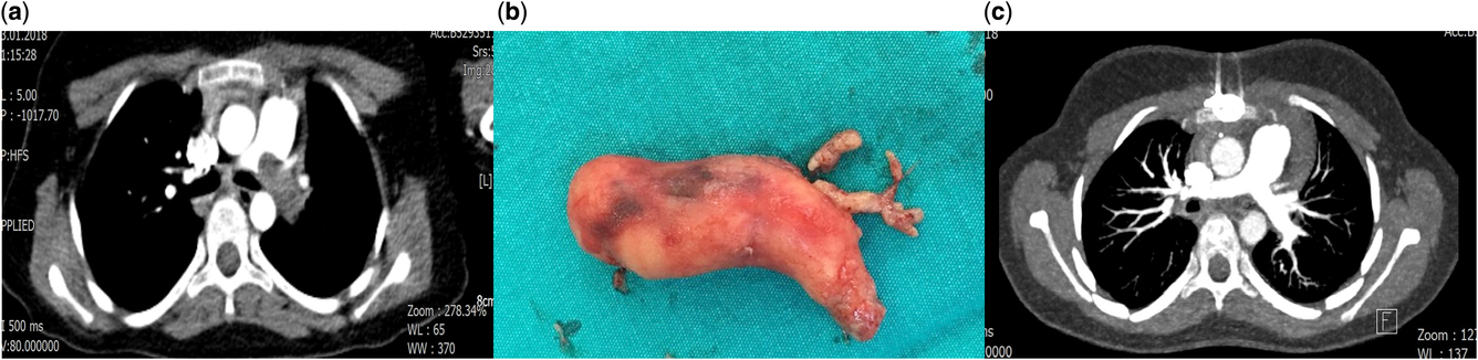
Informed consent to publish the data was taken from the parents. A 10-year-old female patient with no known risk factors for venous thromboembolism (VTE) admitted to the emergency department (ED) of another hospital with syncope. She was diagnosed as acute convulsive attack due to the normal vital signs, nonsignificant physical examination and chest X-ray. She was discharged from hospital on the same day. Two hours after discharge, she experienced the second syncope incident at home and was brought to the ED of the same hospital. The clinical data related to readmission were not obtained and she was emergently referred to our centre for further evaluation. While the patient seemed to be alert and oriented, she was sweaty, tachycardic and tachypneic (32 bpm) and had dyspnoea. Jugular venous distension was detected. She was hypotensive (75/45 mmHg) and had sinus tachycardia (135 bpm). On pulse oxymeter, oxygen saturation was 87–88%. Sudden cardiac arrest developed 15 minutes after admission. She was resucitated and responded to resuscitation after 10 minutes. Her admission arterial blood gas analysis revealed significant hypoxia and acidosis (pH 6.95, pCO2 25 mmHg, pO2 58 mmHg, sO2 86%, Lactate 11.2 mmol/L and Unconvertable OLE object of type ‘Equation.DSMT4’ removed 12.1 mmol/L). Blood biochemistry revealed mass creatinine kinase (CK)-MB 10.3 ng/ml, Troponin T (hs-T) 534.9 ng/ml, fibrinogen 318 mg/dL and d-Dimer 4.7 ug/ml. Contrast enhanced thorax computed tomography (CT) revealed massive pumonary embolism (PE) partially obstructing the main pulmonary artery (PA) lumen and extending to the left PA with total occlusion (Fig 1a).
( a ) Massive embolus within the left pulmonary artery. ( b ) Gross appearance of the embolus after embolectomy. ( c ) Postoperative control imaging showing normal pulmonary arteries.

The patient was emergently transferred to the operating theatre. Transoesophageal echocardiography (TEE) revealed dilation and dysfunction of the right ventricle (RV) and flattening the interventricular septum suggesting RV pressure overload. Following median sternotomy and cardiopulmonary bypass (CPB), the patient was cooled to 24°C. Under aortic cross-clamping and cardioplegia, pulmonary arteriotomy was made and massive thrombus was removed (Fig 1b). We preferred low-flow CPB and deep hypothermia during the removal of thrombus, as the backflow from the distal pulmonary vascular bed precluded proper visualisation of the orifices of the lobar branches. The patient revealed uneventful recovery postoperatively. Continuous infusion of standard Heparin was started as early as 6 hours after surgery. Oral anticoagulation with Warfarin and folic acid supplementation therapy were started on postoperative day 1, and she was discharged on postoperative day 20.
A complete thrombophilic study abnormal revealed the serum level of protein C 33% (70–130%), homocysteine 171.5 µg/L (4.8–7.4), lupus anticoagulant 44.8 seconds (31.4–43.4). Also, heterozygot mutation of Factor V Leiden (FVL) (G1691A) (R506Q) and heterozygot mutation of methylenetetrahydrofolate reductase (MTHFR) (A1298C) were detected. Nothing was abnormal in serum protein electrophoresis. The result of the lower extremity colour-flow venous duplex scanning was negative for deep venous thrombosis. Radiological screening for malignancy and paraneoplastic syndromes was also negative. Early postoperative CT showed normal PAs (Fig 1c). Pathologic examination revealed subacute to chronically organised thrombotic material (Fig 2). Due to the presence of the multiple risk factors for thrombosis (presence of protein C deficiency, hyperhomocysteinemia, lupus anticoagulant, heterozygot mutation of FVL and MTHFR), warfarin was given as a life-long therapy. The duration of follow up was 1 year. We repeated pulmonary CT angiography and no recurrent thrombosis was seen.
Pathologic examination of the thrombotic material.

Discussion
Pulmonary embolism is a very rare disease in children (0.14–0.9 in 100.000) and most cases are diagnosed in previously sick children and in-hospital circumstances. The majority of paediatric PE patients have at least one underlying risk factor like central venous catheter insertion, immobility, infection, malignancy and renal disease and so like. Reference Zaidi, Hutchins and Rajpurkar1 Excellent cardiopulmonary reserve of children, inability to vocalise symptoms and presence of more serious primary disorders or underlying comorbid conditions could mask the symptoms and reduces the level of suspicion by doctors in children with PE. Children and adults were also different in terms of massive PE. First, the mortality rate is higher in children, with almost 50% dying suddenly. Second, instead of deep venous thrombosis in the lower extremities, the aetiology of massive PE in children is tumour emboli in the majority. Reference Baird, Killinger and Kalkbrenner2
Our patient had multiple hereditary risk factors for PE. The impact of the hereditary causes of thrombosis on the risk of PE in children is not being as straightforward as in adult patients. Revel-Vilk et al. Reference Revel-Vilk, Chan and Bauman3 found that inherited prothrombotic coagulation proteins do not contribute significantly to the pathogenesis of VTE in children. Moreover, the deficiency of MTHFR results in hyperhomocysteinemia/homocystinuria and although an association was shown between MTHFR C677T/A1298 C combined mutations and PE in the Turkish population, the evidence on the impact of MTHFR polymorphisms on the risk of PE still remains weak. Reference Basol, Karakus and Savas4 On the other hand, in a meta-analysis by Young et al., Reference Young, Albisetti and Bonduel5 it was stated that the presence of the thrombophilia traits of antithrombin, protein C, and protein S produced the odds ratios (OR) of 8.73, 7.75 and 5.77, respectively, for paediatric VTE and revealed a significant association with the first onset of VTE, as well as recurrence. In an analysis by Kemer et al. Reference Kenet, Limperger, Shneyder and Nowak-Göttl6 it was concluded that significant associations between paediatric venous thrombosis and presence of protein C, protein S and antithrombin deficiency, factor V (F5: rs6025), factor II (F2: rs1799963) have been revealed in many studies and it was also stated that the risk is even more pronounced when combined inherited thrombophilias are involved.
In paediatric massive PE, there are several options for treatment like anticoagulation, systemic thrombolysis, embolectomy, catheter-directed therapies (thrombolytics with/without ultrasound-accelerated thrombolysis), extracorporeal life support (ECLS) or combination of these methods. In a paper by Eini et al., Reference Eini, Houri and Cohen7 41 children with massive PE were analysed and pulmonary embolectomy was performed in the 68% of the patients and 80% of them survived. In 23% of the patients, thrombolysis was used as initial treatment with an 80% failure rate. Moreover, 14% of the patients were treated with only anticoagulation and only 33% of them survived. They concluded as pulmonary embolectomy could be superior to all other treatment modalities in children with massive PE. While ECLS, as an adjunctive measure, has been used for hemodynamic stabilisation before embolectomy/thrombolysis, its impact of survival was reported to be non-significant in massive PE in adults. Reference Weinberg, Tapson and Ramzy8 In children while pharmacological thrombolysis is advised as an initial step in the management of massive PE with systemic hypotension and cardiac strain, mechanical thrombolysis and surgical embolectomy may be utilised for patients with a high risk of bleeding or with contraindications to anticoagulation, if pharmacologic treatment has failed or if the patient presents with hemodynamic collapse. Reference Baird, Killinger and Kalkbrenner2 Recently catheter-directed therapies have been emerging as an alternative therapy in children with sub-massive and massive PE and the outcome was reported to be promising. Reference Akam-Venkata, Forbes and Schreiber9
In conclusion, it seems to be essential to pay a higher level of suspicion for massive PE in children with syncope and unexplained cardiopulmonary instability. Emergent pulmonary embolectomy is a lifesaving procedure in children with massive PE and could be regarded as a first-line therapy in cases with massive PE with severe hemodynamic instability and cardiac arrest.
Acknowledgements
We thankfully acknowledge the contribution of Ass. Prof. Ipek Erbarut Seven, M.D. for pathologic examination.
Conflict of Interest
Nothing to be disclosed. We certify that this paper has not been presented in a scientific congress or meeting.