INTRODUCTION
Although details of the classification and adult anatomy and ecology of the Pinnidae have been recorded in detail (e.g. Grave, Reference Grave1911; Yonge, Reference Yonge1953; Turner & Rosewater, Reference Turner and Rosewater1958; Rosewater, Reference Rosewater1961; Buyle, Reference Buyle1992; Butler et al., Reference Butler, Vicente and De Gaulejac1993) and some of which illustrate the larval shell attached to the adult, very little has been recorded of the development and metamorphosis of the larvae. The only mention that has been traced is in a study by Shimao et al. (Reference Shimao, Chengeong, Lixuan and Qinghe1987) of the rearing of Atrina pectinata (Linné, 1767) which consists of little more than a series of figures that give scant detail of the larval morphology. Other than that, a study of the ontogeny of Jurassic bakevellids (Malchus, Reference Malchus2004) gives details of the larval shells and hinge characters of early fossil species of the Pinnidae.
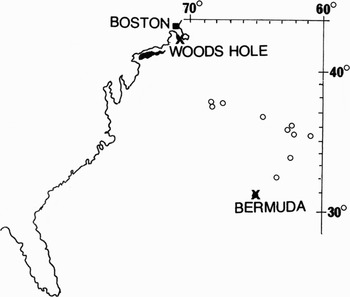
During the Woods Hole Oceanographic Institution's RV ‘Knorr’ cruise No. 38 (1974) from Bermuda to Woods Hole (Figure 1), living larvae of two species of Pinna and one of Atrina were obtained. These were maintained on-board in small aquaria, where they eventually metamorphosed and grew to small adults. By comparison with the larval shells present on young adults, three species were identified, namely Pinna rudis Linné 1758, P. carnea Gmelin 1791 and Atrina seminuda (Lamarck, 1819). Pinna rudis is present throughout the Atlantic from 40°N to 40°S, while P. carnea, although present around Madeira, is more common in the western Atlantic also from 40°N to 40°S. Atrina seminuda is restricted to the western Atlantic from 38°N to 40°S.
The positions of plankton samples containing pinnid larvae taken on RV ‘Knorr’ cruise No. 38, 1974.

The opportunity was taken to observe and record the functional morphology and also the process of metamorphosis. Although the account is largely based on P. carnea, the most common species in the samples, little difference was observed between the three species. Such differences as were observed are mentioned in the text.
MATERIALS AND METHODS
The samples were taken with a plankton net of three-quarter metre mouth diameter, which was lowered obliquely to a depth of approximately 150 m with 200 m of wire attached and towed for a duration of twenty minutes. Pinnid larvae were taken from the following stations: Station 20 (32°29.9′N 64°32.0′W) 5; Station 21 (33°50.3′N 63°24.1′W) 5; Station 22 (35°31.9′N 61°56.1′W) 3; Station 24 (35°51.6′N 62°23.3′W) 9; Station 25 (35°25.2′N 60°59.0′W) 3; Station 29 (36°03.6′N 62°06.6′W) 4; Station 32 (37°49.9′N 67°56.0′W) 18; Station 33 (37°48.2′N 67°49.1′W) 14; Station 34 (37°31.0′N 68°18.1′W) 58; and Station 35 (37°41.0′N 68°36.9′W) 23 (Figure 1). Of these, A. seminuda was only recorded at Station 29.
Some larvae were fixed in 10% formalin and later sectioned (10 µm) and stained in Ehrlich's haematoxylin, to obtain details of their internal anatomy.
RESULTS
Veligers
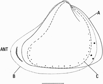
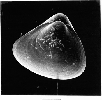
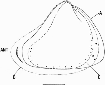
The shells of the planktonic larvae of Pinna and Atrina are triangular in outline, transparent and pale golden in colour. All are inequilateral, but Pinna more so than Atrina, with the antero-dorsal shell margin concave and the postero-dorsal margin convex. The curvature of the margins varies depending on the species (Figure 2). The shortest of the three species was P. carnea with the most fore-shortened anterior margin. It also has the most posteriorly convex shell of all three species. In addition, the larval shells of all species are slightly inequivalve with the left valve larger than the right, the difference most obvious in the umbonal region (Figure 3). The first prodissoconch is clearly visible as small circular valves at the umbo. For an account of the nomenclature and details of the developmental stages of bivalves see Ockelmann (Reference Ockelmann, Cox and Peak1965).
Comparative outlines of the left valves of the three pinnid species recorded in the samples: (A) Atrina seminuda; (B) Pinna rudis; (C) Pinna carnea. Dots indicate the position of pigment spots. Scale = 0.1 mm.
Larval shell of Atrina seminuda as seen from the left side. Note the first prodissoconch is clearly visible. Scale = 0.1 mm.

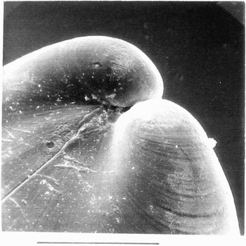
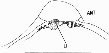
The larval hinge lies at the shell margin of the second prodissoconch immediately below the umbo and is slightly overlapped by the left valve of the first prodissoconch (Figure 4). There are multiple simple hinge teeth both anterior and posterior to the umbo. The form and number of the teeth, although similar, vary according to the species. In all the posterior teeth are shorter and more numerous than the anterior series (Figure 5). The ligament is internal and opisthodetic. It comprises inner, outer and periostracal layers. It is semicircular in cross-section and lies partly anterior to the posterior tooth series.
Detail of the larval shell of Atrina seminuda showing the overhang of the umbo of the right valve. Scale = 0.01 mm.

Detail of the left hinge of a larva of Pinna rudis. LI, Ligament.

Except dorsally, where there is some extension of the primary ligament by fused periostracum, there is no other fusion of the mantle edges; not even at the point close to the posterior mantle edge where the gill rudiment terminates. The mantle edge is typically trilobed with a well-defined periostracal groove. The inner mantle lobe contains a few muscle fibres. The middle sensory lobe is the best developed of the three with a relatively broad flattened surface that bears long cilia that are particularly obvious on the posterior margin. Pigment spots are present in the mantle of both species of Pinna (Figure 2). For the most part these lie within the sensory fold tissue although a few are internal to the mantle edge in P. rudis. In the case of P. carnea between 12 and 18 (the number increasing with age) are arranged along the length of the anterior ventral and posterior margins as a series of small black spots and which are more or less paired with those of the opposing mantle edge. In the case of P. rudis each mantle bears two or three spots that are larger than those of P. carnea and located at the posterior margin ventral to the gill junction with the mantle edge. There is also a band of black pigmented cells in the mantle close to the outer edge of the anterior adductor muscle. The larvae of Atrina seminuda had no pigmented spots in the mantle or in its margin (Figure 2).
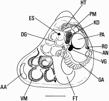
The adductor muscles of the larvae are heteromyarian; the anterior is small and bean-shaped in cross-section and the posterior is circular in cross-section and approximately twice the size of the anterior. Both muscles lie close to the shell margin, the anterior particularly so with space between it and the viscera for housing the retracted velum (Figure 6).
Larva of Pinna carnea in lateral view showing body organs and retracted velum. Scale = 0.1 mm. AA, Anterior adductor muscle; AN, anus; DG, digestive gland; ES, eye spot; FT, foot; GA, gill axis; HG, hind gut; HT, heart; KD, kidney; PA, posterior adductor muscle; PM, pedal muscle; RO, pallial organ rudiment; VG, visceral ganglion; VM, velum.

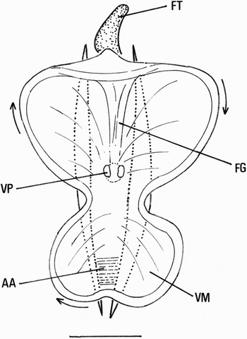
The velum, when expanded forms a flat plate that is carried parallel to the ventral margin of the shell (Figure 7). As in other teleplanktic larvae it is large, but is bilobed a less common feature of bivalve veligers. The margin of the velum has a deep food groove and is heavily ciliated, with pigment cells scattered below the ciliated epithelium. The food groove is continuous around the periphery and has sides of unequal height, the aboral (outer) edge overlapping the oral (inner). Posteriorly the edge is further extended to form a hood, below which the left and right oral margins extend down the posterior (ventral) face of the velar stalk to form a ciliated food groove to the mouth. Two ill-defined papillae are present at the junction of the velum and its stalk on either side of the food groove. The marginal cilia are extremely long and beat metachronally and clockwise round the margin when viewed from the front. Frequently the rim of the velum is turned anteriorly so that the marginal food groove faces towards the direction of movement. The marginal cilia impart a rotation that is clockwise when viewed from the frontal surface of the velum.
Shows the extent of the expanded velum adjacent to the ventral margin of the shell of Pinna carnea. Scale = 0.1 mm. AA, Anterior adductor; FG, food groove; FT, foot; VM, velum; VP, velar pit.

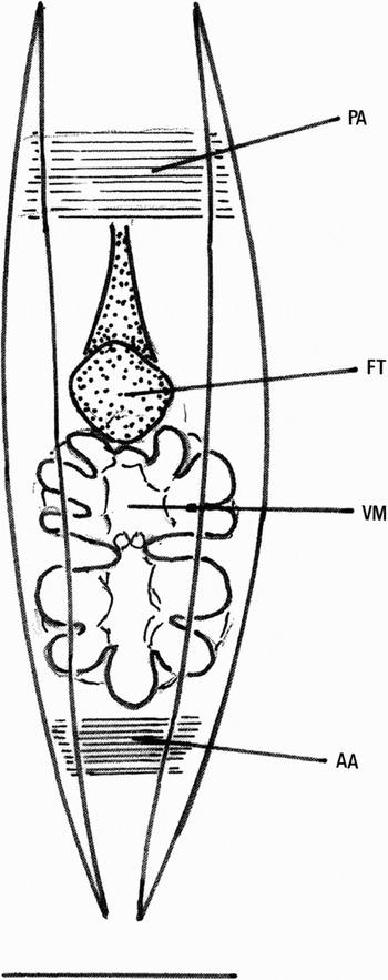
The velum has two pits one to the left and one to the right of the centre each penetrating to the ventral side of the cerebral ganglia. In addition, a stout nerve passes from each ganglion to the apical region of the velum to just below each pit. The ciliary currents on the velar surface pass from the anterior to the postero-lateral margins. When the velum is extended the viscera are moved antero-ventrally, thus vacating the umbonal region of the shell. The velar musculature consists of an array of dorso-lateral and lateral muscles that insert laterally and antero-dorsally on the shell. In transverse section they form a peripheral arc around the viscera. Anterior to the viscera the muscles form the base of the velar stalk and then fan out on either side of the mid-line within the velum itself. In the extended velum they appear as fine radiating lines. The exact number is difficult to determine in the living, moving animal but there appear to be approximately 14 on each side which are individually paired with their opposite number on either side of the midline. The folding process of the contracting velum is precise and identical in all specimens of all species observed. When retracted the velum lies posterior and partially dorsal to the anterior adductor muscle (Figure 6). On retraction, the first fold is along the line separating the two left and right lobes of the extended velum. With further contraction four circular lobes form (two on either side of the initial sagittal plane) and these turn anteriorly. With complete contraction further folding occurs, giving rise to three dorsal and two ventral subsidiary folds to the main folds. The dorsal lobes are larger than the ventral, the latter being adjacent to the mouth when fully retracted and of the ventral lobes those adjacent to the mouth are the smallest. Furthermore, because the folds curve in front of the apex, the whole retracted velum is pulled dorsally as well as posteriorly by the velar retractors so that the mouth faces inwards (Figures 8 & 9).
Shows the position of the contracted velum as seen from the ventral shell margin of Pinna carnea. Scale = 0.1 mm. AA, Anterior adductor muscle; FT, foot; PA, posterior adductor muscle; VM, velum.

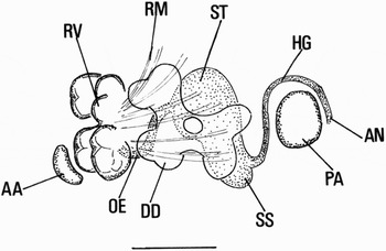
Shows the format of the alimentary canal of the early pinnid larva. Scale = 0.1 mm. AA, Anterior adductor; DD, right digestive diverticulum; HG, hind gut; OE, oesophagus; PA posterior adductor muscle; RM, velar retractor muscles; RV, retracted velar lobes; SS, combined style sac and mid-gut; ST, stomach.

The larval valves completely enclose the retracted velum and the velar cilia, when within the mantle cavity, do not beat, however, when partly retracted the cilia outwith the ventral gape continue beating. These create a current that enters the mantle cavity posteriorly and medially, and exits anteriorly ventral to the anterior adductor muscle. This takes place even though a number of gill filaments may have developed. When the retracted velum is extended the anterior lobes appear at the ventral margin slightly ahead of the posterior lobes and the cilia become active immediately they are clear of the mantle edge.
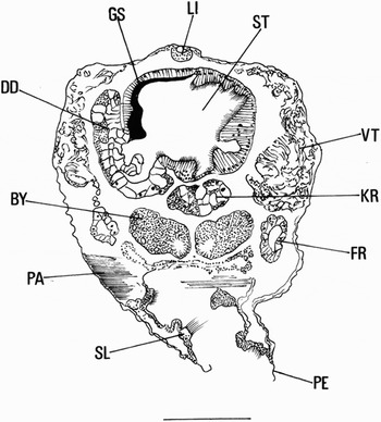
The retracted velum affects the course of the oesophagus in that it passes vertically between the velar muscles and then antero-dorsally between the anterior limits of the digestive diverticula to join the antero-ventral face of the stomach. In a specimen with the velum extended, the viscera move forward and the oesophagus takes the usual route to the stomach. The stomach is large and lies dorso-centrally within the visceral mass (Figures 9 & 10). Sectioned larvae show that there is a gastric shield that bears a large tooth below a shallow dorsal hood (Figure 10). The stomach has various ciliated folds and has two openings to the left and right digestive diverticula, the right being slightly anterior to the left. There is a short broad style sac with an even shorter, combined, mid-gut to the right of the posterior side, the latter continuing as the hind gut. The mid-gut is in the form of a ciliated groove with a typhlosole to one side, both of which originate in the stomach at a point between the entrances to the left and right ducts to the digestive diverticula. The ducts are very short, no more than 4 or 5 cell lengths. The right and left digestive diverticula originate as simple sacs that quickly become lobular and extend posteriorly and anteriorly from the duct. The posterior end is bilobed and while initially the anterior end is bilobed it later becomes quadrilobed (Figure 9). Each diverticulum pulsates in an alternate rhythm to its partner. The hind gut forms a simple loop that passes through the larval heart at a point close to the postero-dorsal mantle margin, then, continues dorsal to the posterior adductor muscle to the anus (Figure 13). The pallial organ (see page 6 and Figure 13) is not developed in the veliger, although there is a small patch of cells on the posterior side of the hind gut immediately above the anus that is the rudiment of this structure (Figure 6).
Oblique cross-section through the stomach and byssal gland of the larva of Pinna carnea. Scale = 0.1 mm. BY, Byssal gland; DD, left digestive diverticulum; FR, gill rudiment; GS, gastric shield; KR, kidney rudiment; LI, ligament; PA posterior adductor muscle; PE, periostracum; SL, sensory fold of mantle margin; ST, stomach; VT, parts of the retracted velum and velar musculature.

Kidney rudiments are present anterior to the posterior adductor muscle on either side of the byssus gland (Figure 6). They are small simple sacs, the walls of which are one cell thick and without connection with the pericardium. Similarly, the kidney aperture to the hypobranchial cavity was not observed in the larva.
The gill rudiment is present in the larva as a development of the mantle epithelium to the inside of the muscular fold at the posterior edge of the mantle cavity. Gill filaments develop precociously with as many as 17 short developing filaments being present in the veliger. These filaments correspond to the descending lamella of the inner demibranch of the adult gill. The gill axis is developed from the mantle and not the body wall, being supplied by a stout axial nerve that originates in the visceral ganglion.
The larval nervous system is well formed. The ganglia are large and the commissures are stout. The paired pedal ganglia lie close to the junction of the anterior margin of the foot with the viscera. Associated with them is a pair of large statocysts. These lie to the dorsal side of the ganglia and each opens to the mantle cavity via a fine pore. Because of the presence of the velum, the larval cerebral ganglia are widely separate from the oesophagus. The velar nerves and pits have already been referred to in the account of the morphology of the velum. The visceral ganglia are very broad and lie posterior to the byssal gland at the antero-ventral corner of the posterior adductor muscle. In late veligers there is an eyespot on each side of the body below the umbo, within the visceral mass close to the digestive diverticulum (Figure 6).
The byssus gland of P. rudis is a paired structure. Each half is pear-shaped and lies posterior within the body above the heel of the foot. In transverse section each half is oval and with a duct close to the inner margin into which discharge the intracellular ducts of the elongate conical gland cells (Figure 10). The cells situated on the dorsal side of the gland are filled with fine particles that stain heavily with haemotoxylin. Each cell has a large basal nucleus close to the outer wall of the gland. The cells to the ventral side of the gland are eosinophyllic and contain granules that are far less obvious than those of the dorsal cells. At the level of the heel of the foot the ducts join and form a single byssal duct that opens to form the ventral byssal groove of the foot. The larval foot is not particularly muscular, but is heavily ciliated on its ventral surface and on its anterior tip. The epithelial cells in the depth of the byssal groove are sparsely ciliated. There are no gland cells at the tip of the foot and, thus, the byssus thread does not terminate in an adhesive disc. The byssus gland of P. carnea, although very similar in structure to that described above, is larger with an additional concentration of gland cells lateral to the main bulk of the gland. The foot is also more robust as too are other structures such as the velar retractor muscles and the cerebral ganglia. The larval foot is very mobile and may be extended posterior to, and at the same time as, the velum.
Metamorphosis
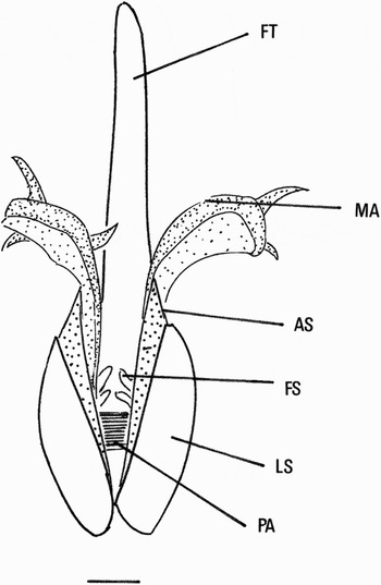
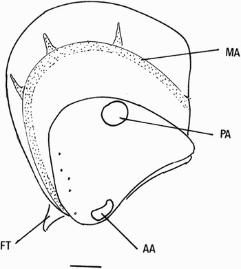
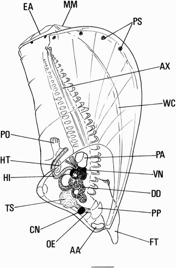
The posterior margin of the mantle thickens in the late veliger. This is particularly apparent at the point closest to the gill rudiment. Although no fusion of the opposing mantle margins occurs, this thickening delimits a dorsal exhalant area from the remainder of the mantle. At this stage the mantle can be separated from the shell margin and both extended beyond the shell margin and contracted within the shell (Figures 11 & 12). Adult shell is laid down by the extension of the mantle at the posterior and ventral margins of the larval shell. Shell deposition occurs in a stepwise fashion, being dependent on the periodic extension of the mantle edge beyond the existing shell, this then laying down a new section of shell by rapid deposition of calcium carbonate crystals within a coarse network of scleroprotein. The stepwise pattern of growth can be seen as a series of posterior ridges that are particularly well defined in the early post-larva (Figures 12 & 13). At the same time the dorsal secondary ligament extends in pace with the shell deposition. When viewed edge on, either from the ventral or dorsal side, it is obvious that, initially, there is a great disparity between the width of the larval shell and that of the newly developing adult shell. The transition is marked by a series of stepped growth increments to the posterior and ventral margins of the larval shell (Figure 12).
Initial deposit of adult shell of Pinna carnea as seen from the ventral margin and showing extended mantle. Scale = 0.1 mm. AS, Adult shell; FT, foot; FS, gill filaments; LS, larval shell; MA, mantle margin; PA, posterior adductor muscle.

Lateral view of the developing adult shell of Pinna carnea showing the retracted mantle margin. Scale = 0.1 mm. AA, Anterior adductor muscle; FT, foot; MA, mantle margin; PA, posterior adductor muscle.

Development of the adult gill and mantle structures of Pinna carnea prior to settlement. Scale = 0.1 mm. AA, Anterior adductor muscle; AX, gill axis; CN, cerebral ganglion; DD, digestive diverticulum; EA, exhalant aperture; FA, gill axis; FT, foot; HI, hind gut; HT, heart; MM, mantle margin; OE, oesophagus; PA, posterior adductor muscle; PM, pallial muscles; PO, pallial organ; PP, palps; PS, pigment spots; ST, stomach; VN, visceral ganglion; WC, waste canal.

As adult shell growth proceeds the hind gut begins to migrate posteriorly. It remains within the confines of the larval shell until the adult shell is more than three times the length of the larva (Figure 13). At this stage the velum is sloughed off as fine cellular debris. At first much of this adheres to the ventral margin of the shell, being pushed out of the mantle cavity by the foot. After the debris is cleared by the foot, the outside of the shell and the surfaces within the mantle cavity are cleaned by the mobility and ciliary activity of the tip of the foot. The gill filaments are fully functional by this stage and the cilia of the foot do not appear to assist in feeding. Instead, it forms a creeping sole that is extended from the ventral edge of the shell, the shell being held vertically above it. Byssus threads were not observed during the early stages of metamorphosis. The foot is extraordinarily mobile and it is capable of exploring the whole of the mantle space. Later, on soft sediments, the foot extends vertically into the sediment and pulls the shell upright so that the posterior margin is uppermost. If soft sediment is not available, it becomes byssally attached to the surface of the substrate and, as far as can be observed, with no effect on shell growth.
The number of functional gill filaments doubles by the time an equal length of adult shell has been added to that of the larva. At this stage 6–7 filaments remain contained within the larval shell. This doubling of the number of functional filaments may occur immediately prior to the velum being sloughed off and the new shell forming. At this stage only the first 7 or 8 pairs of filaments are functional, the remainder being bud-like rudiments. This posterior non-functional developing gill is in the form of a dorsal coil (Figure 6). Unlike the velum, the gill cilia beat while the valves are closed.
The only part of the velum that is retained following metamorphosis is the ciliated groove at the base of the velar stalk. This becomes the oral groove of the adult and the walls at the outer limits of the groove form the initial part of the rapidly developing oral palps (Figure 13). It is likely that the velar stalk muscles contribute to the musculature of the developing palps. As velar muscles atrophy there is a spectacular increase in the musculature of the developing adult and this is particularly noticeable in the case of the foot and the gill axis. In addition there is great hypertrophy of the posterior pedal retractors. At this stage there do not appear to be specific byssal muscles, but it may well be that individual fibres of the pedal retractors attach to the external wall of the gland. The anterior pedal retractors are not so obvious despite the rapid reorganization of the body following the loss of the velum. The mouth migrates close to the anterior adductor muscle, the cerebral ganglia come to lie close to the anterior part of the oesophagus and the palps develop rapidly (Figure 13). These rapid transitions are related to the change from velar to ctenidial feeding.
Apart from the continuing development of the digestive gland and the elongation of the style sac and intestine, there is a significant loss of fat globules within the cells of the digestive gland. These had a dual function; partly to assist flotation in what is a large and heavy plankter and partly as a food store to be used in metamorphosis. The loss of the fat stores signals the start of metamorphosis.
Post larva
By the time that the spat is three times the length of the shell most of the adult characters are formed (Figure 13). No anterior growth of the mantle occurs after metamorphosis. Mantle retractor muscles are quickly developed following atrophy of the velum and rapidly become functional. These muscles correspond to the external radiations of the shell. By the time the post-larva is four times the length of the larval shell the mantle rejection tract and ridge are visible (Figure 13). The gills develop rapidly and new filaments are added at the posterior limit of the axis, the four posterior filaments being non-functional. The mantle margins dorsal to the attachment of the gill are extended as a triangular flap, forming a short exhalant aperture (Figure 13). There is no mantle fusion to separate the exhalant from the inhalant areas. However, the hypobranchial cavity is separated from the mantle cavity by ciliary junctions uniting the ctenidia in the mid-line, the ctenidia and mantle ridge laterally and the membranes posterior to the ctenidia, as described and figured in detail by Yonge (Reference Yonge1953). The outer demibranch is not formed until some 35 filaments of the inner demibranch have been formed. The outer demibranch develops in the same manner as the inner demibranch. Plicae were not formed during the time of these observations, though filament rudiments are present as a row of thickened pads dorsal to the gill axis (Figure 13). After metamorphosis, the gill axis extends posteriorly from a point dorsal to the mouth and the gill functions in the manner described for mature specimens (Yonge, Reference Yonge1953). Amoebocytes could be seen circulating in the vessels of the gill, particularly within the axial vessel. Their movement is in the form of an oscillation with the forward movement carrying them half as much again as the backward movement, thus producing a net forward flow. Although individual gill filaments can contract, the main movement of the gill involves the axial retractor muscles. The latter, together with the pallial muscles, can retract the mantle and gills by a third of their length from the anterior margin of the shell. At the posterior end of each gill axis there are two projections a little larger than the most posterior filament rudiment, the function of which is presumed to be sensory (Figure 13).
The palps become moderately large with the tips extending to the third or fourth filament. They constantly move ventral to the gill. The gut changes little in form as metamorphosis takes place. The stomach appears to be non-contractile, whereas the right and left lobes of the digestive diverticula contract at approximately half the rate of the heart beat (approximately 1 beat per 3 seconds). Peristalsis of the hind gut forms solid ovoid faecal pellets that are voided dorsal to the gill. The hind gut is capable of distending to three times its resting diameter. Voided faeces may be assisted on their passage to the exhalent aperture by the pallial organ (Figure 12). The pallial organ is a development of the rectal wall (not the mantle: Grave, Reference Grave1911; Yonge, Reference Yonge1953). It develops after metamorphosis, is contractile and possibly may be involved in removing shell debris, prior to repair in these easily damaged fragile shells (Yonge, Reference Yonge1953). The digestive gland becomes more ramified yet remains contractile. The left and right halves alternate in their contractions. Two consecutive contractions occur with every heart beat. The rate of beat is approximately one beat every 0.35 seconds. The heart stops beating whenever the mantle and gill contract, presumably because otherwise the heart would be functioning against back pressure. Curiously, there was no clear indication where the blood rests when the mantle contracts. Yonge (Reference Yonge1953) reported that in large specimens, excess blood collects in the pallial organ, however, in recently metamorphosed specimens this was not observed.
Following metamorphosis the paired kidneys become interconnected. In addition, a reno-pericardial duct and an excretory pore develop. The excretory pore opens at the dorsal margin of the posterior adductor muscle.
The foot, when retracted, is usually held anterior to the palps between the viscera and the anterior adductor muscle.
DISCUSSION
It is evident that the most remarkable aspect of the development and metamorphosis of the pinnid larva is the restriction of the growth of the adult shell to the postero-dorsal and ventral margins of the larval shell. Growth of the adult shell is rapid and erosion of the larval shell occurs soon after settlement and byssal attachment, as a result few adult shells are found with the remains of the larval shell. Nevertheless, the anterior adductor survives by posterior growth increments onto the slender antero-ventral margin of the adult shell.
The other remarkable aspect of the larva is its ability to house the whole of the retracted velum within its shell. As mentioned, this requires adjustment of the anterior part of the body and gut in order to accommodate the velum in the antero-dorsal region of the mantle cavity. Furthermore, the ability to restrict ciliary activity to only those parts of the velum that extend beyond the shell boundary is noteworthy, particularly as this does not apply to the gill cilia which continue to beat when the larval and juvenile shells are closed. However, it should be noted that the fragile and frequently repaired adult shells tend not to be capable of closing their valves completely.
ACKNOWLEDGEMENTS
I wish to record many thanks to Miss Tracy Price who so greatly assisted in preparing the manuscript for publication. Many thanks are also due to Dr Ami Scheltema who kindly supplied taxonomic information on the Atlantic deep water species of the Pinnidae.