Introduction
Sudden cardiac death is the leading cause of death in youth participating in sports, with an estimated incidence of 1 in 50,000 to 100,000 athletes annually. Reference Harmon, Asif and Maleszewski1 Most cases of sudden cardiac death are due to undiagnosed structural or electrical cardiac abnormalities such as hypertrophic cardiomyopathy or ion channelopathies, which may be detected on electrocardiograms. Reference Harmon, Asif and Maleszewski1 Pre-participation evaluations are a common tool to identify at-risk youth prior to sport participation.
The benefits of electrocardiogram screening in paediatric athletes are still unclear. In 2014, the American Heart Association did not support the use of mandatory electrocardiogram screening for paediatric sports participation evaluations. Reference Maron, Friedman and Kligfield2 Instead, the American Heart Association supports the use of electrocardiogram as a follow-up to an abnormal 14-point screening questionnaire. Reference Maron, Friedman and Kligfield2 This is based on concerns about false positives, cost-effectiveness, and the low overall incidence of sudden cardiac death. Reference Schmehil, Malhotra and Patel3
However, previous published studies compared the 14-point screening questionnaire to electrocardiogram screening and found that the 14-point questionnaire’s sensitivity, specificity, and positive predictive value were substantially lower. Reference Williams, Pelto and Toresdahl4 Longitudinal data regarding electrocardiogram screening utility in healthy paediatric athletes are lacking; therefore, it is unknown whether electrocardiogram screening should be universal or targeted for specific subgroups. While some electrocardiogram abnormalities may be benign and physiologic adaptations to athletic training, others may indicate evolving pathologies with future clinical implications. Understanding physiologic versus pathologic findings can help determine if universal, repeated electrocardiogram screening is warranted or whether more targeted strategies suffice.
Our study differs in that it provides a multi-year follow-up on electrocardiogram screening in a real-world cohort of competitive youth soccer athletes. We evaluate not only incidence but also recurrence of electrocardiogram abnormalities across years, alongside associated demographic factors. We hypothesise that electrocardiogram abnormalities in paediatric athletes are infrequent and non-recurrent, and that minor abnormal findings in one year are not predictive of future abnormalities.
Materials and methods
Data source and study period
We conducted a single-centre retrospective cohort study using data collected from the electronic medical records of athletes in MLS Sporting Kansas City’s youth soccer programme. All electrocardiograms were obtained and interpreted by paediatric cardiologists at Children’s Mercy Hospital between January 2021 and December 2024.
Study population
Participants were eligible if they were aged 11–18 years and underwent at least one pre-participation electrocardiogram screening during the study period. Patients were excluded if their electrocardiogram data were incomplete or uninterpretable.
Primary outcome
The primary outcome was the incidence of at least one abnormal electrocardiogram finding, as defined by standard paediatric electrocardiogram interpretation criteria.
Secondary outcomes
Secondary outcomes included: the recurrence of electrocardiogram abnormalities across multiple years, type and frequency of electrocardiogram abnormalities, and demographic (e.g. age, race, family history, and medication usage) association with electrocardiogram findings. Age was measured at the time of initial electrocardiogram screening. Race/ethnicity was categorised as African American, Hispanic, Multi-racial, Caucasian, or Asian. Known family history of sudden cardiac death, hypertrophic cardiomyopathy, or channelopathy was determined by “yes” or “no” questionnaire response. Prescription medications were reviewed.
Statistical analysis
Analysis was conducted by examining all electrocardiograms (n = 368) to assess the consistency of abnormal findings across time. Descriptive statistics were used to summarise the frequency and types of electrocardiogram abnormalities. For comparisons between patients with and without electrocardiogram abnormalities, Fisher’s exact test was used for categorical variables, and t-tests were used for continuous variables. Changes in electrocardiogram abnormalities were further examined among the subset of participants (n = 92) with more than one year of electrocardiogram data. Statistical significance was defined as a two-sided p-value < 0.05.
IRB statement
This study was reviewed and approved with exempt status by the Children’s Mercy Hospital Institutional Review Board (IRB #2098408).
Results
Cohort description
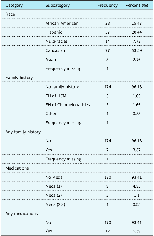
A total of 368 electrocardiograms were reviewed from 182 unique male youth soccer athletes aged 11–18 years who were screened between January 2021 and December 2024. The average age of the patients at their initial screening was 13.61 years (SD: 1.96). Demographic characteristics of the study population are summarised in Table 1, including race/ethnicity, family history of cardiac conditions, and medication use. A total of 181 patients had a complete race/ethnicity record, with the largest group being Caucasian (53.59%), followed by Hispanic (20.44%), African American (15.47%), Multi-racial (7.73%), and Asian (2.76%).
Baseline characteristics of study cohort (n = 182)

Key: Medications are coded as 0 = None, 1 = albuterol, 2 = stimulant, and 3 = Selective serotonin reuptake inhibitor.
Incidence and types of ECG abnormalities
Sixty-eight of the 368 electrocardiograms (18.5%) had at least one abnormal finding (95% CI: 14.5–22.4%). The most frequently observed abnormalities were left ventricular hypertrophy (n = 32, 8.70%), right axis deviation (n = 23, 6.25%), and left axis deviation (n = 5, 1.36%). Other abnormalities included premature ventricular contractions (n = 4, 1.09%), right ventricular hypertrophy (n = 2, 0.54%), 1st-degree heart block (n = 1, 0.27%), supraventricular ectopy (n = 1, 0.27%), right bundle branch block (n = 1, 0.27%), T wave inversion in more than one lead (n = 1, 0.27%), and deep Q waves (n = 1, 0.27%). No patients were found to have a delta wave/preexcitation pattern or left bundle branch block, prolonged QTc or a Brugada pattern. Among the 68 electrocardiograms with abnormalities, left ventricular hypertrophy was the most common finding (n = 32, 47.06%), followed by right axis deviation (n = 23, 33.82%).
Longitudinal findings
Among the 92 patients with more than one year of data available, 16 (17%) had no abnormality in the first year but developed an abnormality at a subsequent screening. Eight (9%) had an abnormality in the first year that did not recur in subsequent years. Four (4%) had abnormalities reported every year. Thirteen (14%) had “back and forth” responses, with abnormal status changing from year to year.
Association with demographic and clinical variables
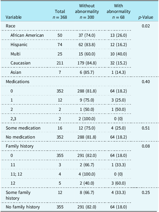
The average age of patients with an abnormal electrocardiogram (14.4 years, SD 1.9) was not statistically different from that of patients with a normal electrocardiogram (14.1 years, SD 2.0) (p = 0.22). There was a statistically significant association between race and the presence of an electrocardiogram abnormality (p = 0.02) (Table 2). A higher proportion of African American (26.0) and Multi-racial (40.0) athletes had abnormalities compared to Caucasian (15.2) and Hispanic (16.2) athletes. No statistically significant associations were found between electrocardiogram abnormalities and either medication use (p = 0.40) or family history of early cardiac death or genetic conditions (p = 0.08) (Table 2).
Association of demographic and clinical variables with ECG abnormalities (Visit level)

The total number of ECGs with abnormalities at the visit level was 68. Percentages are calculated based on the total number of visits within each subcategory. The p-values are for the overall comparison within each main variable category and the summary categories.
Clinical implications
The majority of the electrocardiogram findings were benign or normal physiologic variants. Among athletes with abnormal electrocardiogram findings, additional cardiac evaluation was performed as clinically determined by the paediatric cardiologist after review of all clinical information (history, examination, family history). Three athletes underwent ambulatory Holter monitoring, all of which were normal. Seven athletes underwent echocardiography, of which six were normal, and one demonstrated left ventricular hypertrophy without other abnormalities. One athlete underwent an exercise stress test with normal results. No clinically significant cardiac disease was identified, and all athletes were cleared for continued sports participation.
Discussion
This retrospective study provides data on the incidence and longitudinal nature of electrocardiogram abnormalities in a U.S. based cohort of male youth soccer athletes. Our primary finding is that the incidence of electrocardiogram abnormalities was similar to that noted in other studies (18.5%) in this healthy cohort, and most importantly, these abnormalities did not correlate with any significant underlying cardiac pathology upon further clinical evaluation. Reference Caramoci, Smaranda, Drăgoiu and Bădărău5 This supports the American Heart Association’s stance against universal electrocardiogram screening for paediatric athletes. The key strength of our study is longitudinal data. We observed that electrocardiogram findings can change over time, with new abnormalities appearing in subsequent screenings for some athletes (17%), while initial abnormalities resolved in others (9%). Only 4% of athletes had consistent abnormalities across all years of screening, suggesting that a one-time electrocardiogram may have limited predictive value for long-term risk assessment in this population. The nonrecurrent nature of the findings suggests these initial abnormalities are benign physiological adaptations to training and/or growth rather than evolving pathologies.
Our findings that a higher proportion of African American and Multi-racial athletes had electrocardiogram abnormalities are consistent with existing literature. Reference Magalski, Maron and Main6,Reference Ozo and Sharma7 The high rate of left ventricular hypertrophy in our abnormal electrocardiogram cohort (47.06%) and the lack of clinically significant findings on follow-up evaluations further support this notion of a physiological hypertrophy. It is also important to consider the sport-specific nature of our cohort. Soccer is a predominantly endurance-based sport, and sustained aerobic training is known to produce physiologic adaptations, including increased QRS voltage, sinus bradycardia, left ventricular hypertrophy, and repolarisation changes on electrocardiogram. Reference Małek, Dotka, Brzezińska, Pyndryk, Materkowski and Wiecha8 Many of the abnormalities observed in our cohort, particularly left ventricular hypertrophy and axis deviations, are well-recognised manifestations of the athletic heart rather than markers of underlying disease. These findings likely reflect physiologic athletic remodelling rather than disease, highlighting the importance of nuanced electrocardiogram interpretation.
This study provides multi-year, real-world screening data in a U.S. pediatric soccer population, which is lacking in current studies. However, limitations must be noted. This was a single-centre, male-only cohort, which may limit generalisability to female athletes or other sports. The retrospective design also limited the uniformity of follow-up evaluation and diagnostic testing.
Conclusions
In conclusion, our data support that universal electrocardiogram screening of healthy paediatric athletes, even in the competitive arena, is of limited value due to the low incidence of abnormalities and the low predictive value of these findings for significant underlying cardiac disease. Our findings suggest that many of the identified electrocardiogram abnormalities are transient or benign and likely represent physiological changes. Future research is needed to help identify independent predictive risk factors for clinically significant electrocardiogram abnormalities and to determine if targeted, rather than universal, electrocardiogram screening protocols would be more effective and efficient.
Acknowledgements
None.
Financial support
None.
Competing interests
None.