

Emotional development is an integral part of the development and maintenance of social relationships, and the ability to recognize facial expressions is a basic aspect of emotional understanding and social cognition. The development of emotion plays an important role in the development and maintenance of social relationships. In that sense, facial expressions represent important clues to the meanings within social conversations and provide a basis on which to build greater emotional comprehension (Denham, Reference Denham1998). Individuals with neuromuscular diseases, such as myotonic dystrophy (Kobayakawa, Tsuruya, Takeda, Suzuki, & Kawamura, Reference Kobayakawa, Tsuruya, Takeda, Suzuki and Kawamura2010) or Duchenne’s muscular dystrophy (Hinton, Fee, De Vivo, & Goldstein, Reference Hinton, Fee, De Vivo and Goldstein2007), show impaired recognition of facial emotions, and thus, may have more trouble accurately interpreting the emotions of others.
Myasthenia gravis (MG) is an autoimmune disorder of neuromuscular transmission that is characterized by increased fatigability of voluntary muscles (Kuks & Oosterhuis, Reference Kuks, Oosterhuis and Kaminski2003). MG frequently involves the muscles that control eye and eyelid movements and facial expressions and is the most common disorder of the neuromuscular junction, with a prevalence of approximately 15 per 100,000 individuals. In young adults, MG is more common in females, with a female-to-male ratio of approximately 2:1; however, this ratio is reversed in older adults (Philips, Reference Philips2003). An increased incidence of psychiatric disorders, depressive and anxiety disorders, epilepsy, multiple sclerosis, and electroencephalographic abnormalities has also been noted in patients with MG (Bartel & Lotz, Reference Bartel and Lotz1995; Keesey, Reference Keesey1999; Ybarra et al., Reference Ybarra, Kummer, Comini, Oliveira, Gómez and Texeira2011).
MG autoantibodies alter the postsynaptic nicotinic acetylcholine receptors (nAChRs) of muscles (Meriggioli & Sanders, Reference Meriggioli and Sanders2009). Although events leading to a loss of nAChRs at the neuromuscular junction in MG are well-established, little is known regarding the effects of this loss on the central nervous system, as previously suggested by Kojevnikov (Reference Kojevnikov1986). A study two years later by Tucker, Roeltgen, Wann, and Wertheimer (Reference Tucker, Roeltgen, Wann and Wertheimer1988) supported the hypothesis that myasthenia is associated with central cholinergic effects manifested by cognitive dysfunction, such as memory impairment. Additionally, Graef, Schönknecht, Sabri, and Hegerl (2011) stated that cholinergic receptor manipulations have an effect on cognition, emotion, and brain electrical activity. Nicotinic receptor manipulations can regulate not only memory and attentional processes in humans, but also mood states such as depression and anxiety. Emotional functioning in MG, however, remains unclear.
Authors such as Sitek, Bilinska, Wieczorek, and Nyka (Reference Sitek, Bilinska, Wieczorek and Nyka2009) interpreted the cognitive dysfunction identified in MG patients as an effect of muscle fatigability rather than central nervous system involvement. Indeed, significant excessive daytime sleepiness resulting from sleep disturbances can also impair memory and the performance of myasthenia patients on neuropsychological tests (Keesey, Reference Keesey1999). This author proposed four alternative hypotheses to possibly explain the neuropsychological deficits in MG patients: nocturnal respiratory problems, nonspecific immunological processes, increased mental fatigue, and central cholinergic deficit manifestation.
In accordance with the cholinergic hypothesis, the present study examined the emotional functioning of myasthenic patients with respect to two components of theory of mind: the ability to interpret different emotions expressed on one’s face and empathy. There is not research in myasthenia gravis about these important components. These are important domains of social cognition, which contribute to effective social competence and emotional understanding. In contrast, impaired ability in emotional recognition has been studied extensively in other related domains, including Alzheimer’s disease (McLellan, Johnson, Dalrymple-Alford, & Poeter, Reference McLellan, Johnson, Dalrymple-Alford and Poeter2008), Huntington’s disease (Grey, Young, Barker, Curtis, & Gibson, Reference Grey, Young, Barker, Curtis and Gibson1997), and the use of scopolamine, an anticholinergic agent (Kamboj & Curran, Reference Kamboj and Curran2006a). The effects on cognitive functioning are likely related to an acute reduction in central cholinergic neurotransmission (Kamboj & Curran, Reference Kamboj and Curran2006b).
We hypothesised that myasthenia is related to impaired facial emotional recognition ability. This dysfunction could be related to deficits in cholinergic neurotransmissions and their manifestation in damaged facial muscles. Facial muscle expression may influence the ability to recognize emotions accurately. Because facial mimicry has been shown to play an important role in the perception of emotions in others (Adolphs, Damasio, Tranel, Cooper, & Damasio, Reference Adolphs, Damasio, Tranel, Cooper and Damasio2000), the aim of this study was to determine whether people with myasthenia are impaired in this task and whether their performance is related to gender, age, disease duration, and empathy level.
Methods
Participants
A sample of 35 individuals with MG and 36 healthy controls participated in the study. All MG patients (10 males and 25 females) were recruited from the Myasthenia Gravis Association of Spain and came from different regions of that country. The diagnosis of MG was confirmed by means of electromyography, the presence of anti-nicotinic antibodies in the serum, and a positive Tensilon Test. Patients averaged 50 years of age (SD = 12.28; range 18–69), 14 years of education (SD = 3.21; range 10–19), and 8 years (SD = 6.50) since MG onset. Using Osserman’s classification system (Osserman, Reference Osserman1958), 7 patients were classified as Class I, 16 patients as Class IIa, 9 patients as Class IIb, and 3 as Class III. A total of 17 patients (48%) were thymectomised. The majority of participants had an autoimmune myasthenia (32/35). The inclusion criteria included the following: 1) a confirmed MG diagnosis; 2) age > 18 years; 3) agreement to participate in the study via a signed informed consent form; and 4) ability to manipulate a computer. Patients were excluded if they had a psychiatric disorder (DSM IV-TR), neurological disorder other than MG, head trauma, or severe visual limitations.
The healthy control group consisted of 36 subjects (11 males and 25 females) matched to the MG group with respect to age, gender, and education level. The control group averaged 53 years of age (SD = 11.37; range 30–78) and 15 years (SD = 2.5; range 10–19) of education. Some controls were recruited from adult education courses at the University of Deusto (n = 22), while others were relatives of patients with MG or other diseases (n = 14). The inclusion criteria included the following: 1) age > 18 years; 2) agreement to participate in the study via a signed informed consent form; and 3) ability to manipulate a computer. As with the MG group, participants were excluded if they had a psychiatric disorder, neurological disorder, head trauma, or severe visual limitations.
Each participant completed the task in Spanish. All of them gave informed consent. Participants from both groups volunteered to complete the study without compensation, and the study was conducted in compliance with ethical research standards.
Instruments
Two instruments were used in this study: The Feel Test (Facially Expressed Emotion Labelling; Kessler, Bayerl, Deighton, & Traue, Reference Kessler, Bayerl, Deighton and Traue2002) and the Interpersonal Reactivity Index (IRI; Davis, 1980).
The Feel Test is a computer-based test that measures one’s ability to recognize basic, facially expressed emotions. The test consists of 42 photographs, developed by Matsumoto and Ekman (Reference Matsumoto and Ekman1988), each of which shows a person’s face expressing one of six basic emotions (anger, fear, sadness, happiness, surprise, or disgust). Each picture is presented on a computer screen for 300 ms. Subjects must then classify the respective emotion by clicking on the appropriate label. The 42 pictures together encompass seven separate examples of all six emotions. Subjects have to decide quickly and accurately which of the six emotions they have just seen in a forced-choice format. The Feel Test yields a score for the total numbers of correct answers and errors, in addition to the reaction time for each emotion. The Feel Test is the most reliable of its kind (α = 0.77), and over the years, data from over 600 healthy subjects have been collected (Hoffman, 2010).
The IRI test is a 28-item measure of general empathic tendencies that assesses both cognitive and emotional components of empathy. Items are evaluated on a 5-point Likert scale ranging from 0 (does not describe me well) to 4 (describes me very well). This instrument contains four subscales: perspective taking (tendency to spontaneously adopt the psychological point of view of others; fantasy (tendency to transpose oneself imaginatively into the feelings and actions of fictitious characters in books, movies, and plays); empathic concern (other-oriented feelings of sympathy and concern for unfortunate others); and personal distress (self-oriented feelings of personal anxiety in tense interpersonal settings). Each subscale contains seven items, and item scores are summed within each subscale to obtain the subscale score (score range for each scale = 0–28). Higher scores are indicative of greater perspective taking, fantasy, empathic concern, and personal distress. The Spanish version of the IRI has shown psychometric properties similar to those of the original version. It was validated with one sample of parents (n = 629) and two samples of college students (n = 1997 and n = 515). Alpha coefficients ranged from 0.75–0.78 for the original version (Davis, 1980) and from 0.63–0.80 for the Spanish version (Pérez-Albéniz, Paúl, Montes, & Torres, Reference Pérez-Albéniz, Paúl, Montes and Torres2003).
Procedure
The clinical and control groups were first invited to participate in the study, and an explanation of the aims was then provided. The experiment was conducted in a silent room at one of two different sites: University of Deusto and the Reference Centre for Rare Diseases in Spain. After the experimenter provided verbal instructions, the participants completed a sociodemographic questionnaire, the IRI, and the Feel Test. During the Feel Test, each participant sat approximately 50 cm from a computer screen and was given limited time to complete the task. In total, 42 pictures were shown, and seven examples of all six emotions were used. Each participant was asked to label the emotion presented in each picture. Specifically, subjects had to decide quickly and accurately which of the six basic emotions they had just seen in a forced-choice format. No feedback was given regarding correctness of the responses. The participants were asked to label the emotion presented in each picture.
Statistical analysis
Prior to data analysis, all variables were checked for normality using the Kolmogorov-Smirnov test (p > 0.05). All of the data were normally distributed; therefore, parametric statistics were utilized. A Student’s t-test or analysis of variance (ANOVA) was used to compare group mean values, and Pearson’s coefficients were calculated to further examine relationships among variables. The Mann-Whitney U-test, a non-parametric test, was performed for small samples. Effect sizes were calculated using Cohen’s d (Cohen, Reference Cohen1962). We calculated all significance thresholds using a two-tailed test, and the significance level was set at 0.05.
Results
Facial emotion recognition: differences between groups
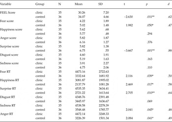
In the Feel Test, the participants in the clinical group obtained significantly lower total scores on average than controls, t(58) = −2.63, p = .01, d = .62. The clinical group also obtained lower scores for pictures depicting fear, t(69) = −1.98, p = .05, d = .47 and surprise, t(44) = −3.66, p = .001, d = .88, (see Table 1).
Correct scores from the Feel Test and corresponding reaction times. Differences between the clinical and control groups

Note:
*p < .05, ** p < .01, d Cohen’s size effect. (RT = reaction time).
With regard to the reaction time test, the myasthenic group had slower reaction times for each emotion except disgust p < .05; d = .48–.64.
For the MG group, correlation analyzes were conducted to examine the relationships of scores from the Feel Test and time since MG diagnosis with Osserman classification scores. No statistically significant correlations were detected.
Facial emotion recognition: Differences between genders
A Mann-Whitney U-test was applied to analyze the effect of gender on the Feel Test scores. Scores from females (n = 50) were significantly higher than those from males (n = 21) for the Feel Test total score, as well as for the anger and sadness pictures (p < .05). No differences between females and males were found in reaction times. In males specifically, no differences were found between the clinical (n = 11) and control (n = 10) groups. However, females in the control group (n = 25) scored significantly higher than females in the clinical group (n = 25) for surprise, U = 163, z = 1.556, p = .001, and sadness, U = 213.5, z = .849, p = .05.
Facial emotion recognition: Differences related to age
A Pearson correlation was computed to examine the relationship between age and the Feel Test total scores. In the myasthenic group, significant negative correlations were found between age and fear, r = −.466, p = .005, happiness, r = −.430, p = .01, anger, r = −.374, p = .027, surprise, r = −.517, p = .001, and sadness, r = −.562, p = .01. In the control group, age correlated significantly only with anger, r = −.415, p = .012, and sadness, r = −.537, p = .001.
Facial emotion recognition and empathy
On the IRI, a Student’s t-test revealed that patients with myasthenia received significantly lower empathy scores than controls, t(57) = −2.296, p = .025, d = .56. Additionally, in the myasthenic group, the empathy factor correlated significantly with the total Feel score, r = .342, p = .001, anger, r = .344, p = .043, and disgust, r = .488, p = .003. In contrast, in the control group, there was only a significant correlation between empathy and disgust, r = .492, p = .004.
Discussion
The aim of this study was to investigate the ability to recognize facial emotions in a group of myasthenic patients compared with those of a group of healthy controls. Findings revealed a facial recognition deficit in the myasthenic group, particularly for fear and surprise. Likewise, compared with controls, patients with myasthenia had significantly longer response times to accomplish the emotion recognition task.
Similar results have been found for people with other neuromuscular disorders and rare diseases. For example, patients with myotonic dystrophy have demonstrated a lower sensitivity to facial expressions of disgust, fear, and anger compared with healthy controls (Winblad, Hellström, Lindberg, & Hansen, Reference Winblad, Hellström, Lindberg and Hansen2006; Kobayakawa et al., Reference Kobayakawa, Tsuruya, Takeda, Suzuki and Kawamura2010). Other studies on patients with diseases such as Prader-Willi syndrome have also revealed deficits in the recognition of emotions in facial expressions (Whittington & Holland, Reference Whittington and Holland2011). These deficits in emotional recognition ability may be associated with how brain functioning affects emotional processing, as suggested by studies of individuals with other neuromuscular diseases, such as myotonic dystrophy (Kobayakawa et al., Reference Kobayakawa, Tsuruya, Takeda, Suzuki and Kawamura2010), or a chromosomal disorder involving monosomy X, particularly Turner syndrome (Mazzola et al., Reference Mazzola, Seigal, MacAskill, Corden, Lawrence and Skuse2006).
MG autoantibodies attack the postsynaptic nicotinic acetylcholine receptor of the muscle affecting cholinergic transmission (Meriggioli & Sanders, Reference Meriggioli and Sanders2009). Changes in cognition can result from cholinergic receptor manipulation or from cholinergically induced changes in vigilance or affective state. In particular, α4β2 and α7 nicotinic acetylcholine receptors affect several brain regions, such as the amygdala, hippocampus, prefrontal cortex, thalamus, striatum, and ventral segmental area. Clinical data have emphasized the role of this receptor in attention, learning, and memory (Levin, MecClernon, & Rezvani, Reference Levin, MecClernon and Rezvani2006; Miwa, Freedman, & Lester, Reference Miwa, Freedman and Lester2011; Thomsen, Hansen, Timmerman, & Mikkelsen, Reference Thomsen, Hansen, Timmerman and Mikkelsen2010). For instance, scopolamine is one drug that can cause an acute reduction in central cholinergic transmission. Kamboj and Curran (Reference Kamboj and Curran2006a) found that scopolamine-induced cholinergic hypofunction selectively impairs the recognition of human facial expressions, particularly anger and disgust. According to these authors, the effect of scopolamine on emotional recognition could reflect an inhibition of cholinergic neurotransmission in the basal ganglia, with scopolamine´s effect mediated through the amygdala.
Meynert’s nucleus provides the major cholinergic input to the cerebral cortex. For example, a consistent finding in the brains of Alzheimer’s disease patients is the loss of these cells with a corresponding reduction of cortical acetylcholine (Mesulam, Reference Mesulam2004). There is also loss of cholinergic neurons in regions of the brain known to be involved in learning and memory (Clader & Wang, Reference Clader and Wang2005). Although the related literature is limited and contains many methodological deficits, McLellan et al.’s (Reference McLellan, Johnson, Dalrymple-Alford and Poeter2008) review concluded that patients with Alzheimer’s demonstrate poorer recognition of facial expressions compared with a control group. Huntington’s disease is similarly associated with cholinergic hypofunction, and emotion recognition impairments have been broadly studied in these patients (Aviezer et al., Reference Aviezer, Bentin, Hassin, Meshino, Kennedy, Grewal and Moskovitch2009; Grey et al., Reference Grey, Young, Barker, Curtis and Gibson1997; Kipps, Duggins, McCusker, & Calder, Reference Kipps, Duggins, McCusker and Calder2007; Sprengelmeyer et al., Reference Sprengelmeyer, Young, Sprengelmeyer, Calder, Rowland, Perrett and Lange1997).
In the present study, the clinical group showed significantly lower scores than the controls in fear and surprise. This dysfunction could be related to the affection in facial muscles of myasthenic patients. The embodied-cognition theory assumes that the use of emotion concepts and the perception of particular emotional expressions both involve simulation of the corresponding emotion. When viewing an emotionally ambiguous face, participants should simulate the emotional expression associated with the emotional concept that accompanies the face. Individuals spontaneously respond facially to others’ emotional expressions with a mimicry response (Adolphs et al., Reference Adolphs, Damasio, Tranel, Cooper and Damasio2000; Halberstadt, Winkielman, Niedenthal, & Dalle, Reference Halberstadt, Winkielman, Niedenthal and Dalle2009).
According to the locationist model, the amygdala is the most important structure in the brain’s fear circuit (Ledoux, 2007; Lindquist, Kober, Bliss-Moreau, & Feldman, Reference Lindquist, Wager, Kober, Bliss-Moreau and Feldman2012). Few studies have focused on recognizing the facial expression of surprise. This expression shares similarities with the fear expression; specifically, the inner brow is raised, the outer brow raises the eyes, and the mouth is wide open with the upper eyelid raised. For this reason, fear and surprise can be confounded (Ekman, Reference Ekman1993). It is quite likely that the amygdala is involved in the processing of surprise, such as in the case of fear (Posamienter & Abdi, Reference Posamienter and Abdi2003). Moreover, the amygdala and limbic brain can be related to nicotinic acetylcholine receptor activity as well (Levin et al., Reference Levin, MecClernon and Rezvani2006; Miwa et al., Reference Miwa, Freedman and Lester2011; Thomsen et al., Reference Thomsen, Hansen, Timmerman and Mikkelsen2010).
A selective deficit in the recognition of fearful facial expressions has been reported in patients with bilateral amygdala damage (Adolphs et al., Reference Adolphs, Gosselin, Buchannan, Tranel, Schyns and Damasio2005). While no study to date has linked myasthenia to deficits in the amygdala, the amygdala could nevertheless be structurally or functionally compromised in this disease. A neuroimaging study may be considered in a future project, and further studies should consider these variables as they relate to impairment in emotional recognition ability.
Concerning one’s age and the ability to recognize facial expressions, there is a significantly more negative association between these variables in the myasthenic group than in the controls. Specifically, the older a subject is, the less accurate this person will be in the recognition of fear, happiness, anger, surprise, and sadness. According to a review article by Mao, Mo, Qin, Lai, and olde Hartman (2010), patients who have suffered from the disease longer are older and have more severe symptoms. Thus, symptom severity along with increased age could be related to the performance of myasthenic patients.
This study documented a slower processing of emotional facial expressions in myasthenic patients compared with the control group. While these reaction times are indicators of cognitive processing speed, these results must be considered in the context of myasthenia pathology and motor performance (Sitek et al., Reference Sitek, Bilinska, Wieczorek and Nyka2009). Similar results have been found in multiple sclerosis patients, although this disease clearly and severely affects the central nervous system (Jehna et al., Reference Jehna, Neuper, Petrovic, Wallner-Blazek, Schmidt, Fuchs and Enzinger2010).
Another variable examined in this study was empathy. The results revealed that patients with myasthenia achieved significantly lower empathy scores than the controls. Although there were statistically significant differences between groups, both scores were similar to the mean and standard deviation of the scores derived from the Spanish adaptation of the IRI (Pérez-Albéniz et al., Reference Pérez-Albéniz, Paúl, Montes and Torres2003).
This study has some limitations, the first of which is related to its relatively small sample size. However, MG is an uncommon disease, and this study included one of the largest cohorts of MG patients in the literature and is the first known study to examine the recognition of facial emotions. Further studies should consider the effect of medication, such as anticholinesterase (pyridostigmine) and immunosuppressive therapies, on facial emotion recognition. The possible treatments are different in each patient and must be analyzed in detail. Additional cognitive and affective variables should also be investigated.
In conclusion, this study suggests that a reduced ability to recognize emotions, particularly fear and surprise, exists in myasthenic individuals, adding a new cognitive dimension to the disease. Perhaps these findings can help guide future studies of the cholinergic system’s influence on facial emotion recognition in MG. Psychosocial intervention in myasthenic patients that includes social cognition aspects, such as facial recognition, could be considered.