Kawasaki disease, one of the most common childhood vasculitides, is emerging as the leading cause of acquired heart disease in children globally. Intravenous immunoglobulin, the gold standard treatment for Kawasaki disease, reduces the risk of coronary artery abnormalities to <5%. Coronary artery abnormalities have been reported to regress in size after 4–8 weeks of an acute episode but it may take several years for coronary artery diameters to return to normal. Reference McCrindle, Rowley and Newburger1 However, myointimal proliferation may continue to progress leading to decreased coronary artery flow reserve and vascular reactivity. Children with persistent coronary artery abnormalities are at a higher risk of ischaemic heart disease. Besides, premature atherosclerosis may lead to the development of ischaemic heart disease in children with Kawasaki disease even in the absence of overt coronary artery abnormalities. Reference Fukazawa2 Although the exact mechanism of evolution of accelerated atherosclerosis in Kawasaki disease is still not elucidated, endothelial dysfunction and ongoing chronic inflammation are important contributing factors.
Various non-invasive tools have been utilised to determine the vascular health in patients with Kawasaki disease including pulse-wave velocity, arterial stiffness, flow-mediated dilatation, endothelial peripheral artery tonometry (endo-PAT), and common carotid artery intima-media thickness. While flow-mediated dilatation, endo-PAT, and pulse-wave velocity are functional tests of endothelial integrity, carotid artery intima-media thickness is a surrogate anatomical marker of ongoing atherosclerosis. Although several studies have assessed endothelial dysfunction in Kawasaki disease, marked variability in results from these studies precludes derivation of definitive conclusions. Only a few narrative/systematic reviews have addressed this contentious issue before. Reference Chen, Curtis, Dahdah, Kowalski, Cheung and Burgner3,Reference Dietz, Tacke, Hutten and Kuijpers4 The only systematic review on the said topic, which included 30 studies, was published in 2015. Reference Dietz, Tacke, Hutten and Kuijpers4 Since then, numerous studies have been performed that have assessed long-term endothelial function in patients with Kawasaki disease. Besides, meta-analysis for certain outcome measures (e.g. PWV, endo-PAT) had not been possible due to the sparsity of relevant studies published till that time. We, hence, performed an updated systematic review (incorporating data from 49 studies) and meta-analyses (including PWV, endo-PAT) to assess long-term vascular function in patients with Kawasaki disease.
Methods
Protocol of this systematic review and meta-analyses is registered at PROSPERO: CRD 42021277854.
Type of studies
Observational studies that have assessed vascular microanatomy/physiology by pulse-wave velocity, arterial stiffness, flow-mediated dilatation, carotid artery intima-media thickness, endo-PAT, nitroglycerine-mediated dilatation, and positron emission tomography were included. Studies that evaluated these parameters during acute, convalescent or within 1 year of Kawasaki disease diagnosis were excluded.
Participants
Children with a diagnosis of Kawasaki disease (according to the American Heart Association or Japanese criteria of Kawasaki disease) were selected as cases. Participants (without underlying heart disease) matched for age and sex were included as controls.
Primary outcome
Difference in pulse-wave velocity between controls and patients with Kawasaki disease (1 year after the onset of the said illness).
Secondary outcomes
Difference in flow-mediated dilatation, arterial stiffness, endo-PAT, nitroglycerine-mediated dilatation, carotid artery intima-media thickness, and myocardial flow reserve between controls and patients with Kawasaki disease (1 year after the onset of the said illness).
Search strategy
We systematically searched PubMed, Embase, and Web of Science databases for relevant literature published from 1965 to May 2021 (Supplementary Figure 1). Following Medical Subject Headings keywords (combined with appropriate Boolean operators) were used for literature search: ‘Mucocutaneous lymph node syndrome’, ‘Kawasaki disease’, ‘Kawasaki syndrome’, ‘vascular stiffness’, ‘elasticity’, ‘carotid intima media thickness’, ‘pulse wave velocity’, ‘pulse wave analysis’, ‘peripheral artery tonometry’, ‘manometry’, ‘nitroglycerine mediated vasodilatation’, ‘flow mediated vasodilatation’, ‘compliance’, ‘endothelium’, ‘tomography emission computed’, ‘nuclear magnetic resonance’, ‘vascular’, and ‘positron emission tomography’. No language restriction was applied. Two authors (PP, AZB) identified the relevant studies to be included in the systematic review.
Data extraction
Two authors (PP, AZB) retrieved the following data from each study: name of the author(s), country, year of publication, design/methodology of the study, number of participants, methods of recruitment, risk of bias (age, sex, diagnosis, and sample size), time to enrolment, presence/absence of coronary artery abnormalities, and outcomes.
Assessment of study quality (risk of bias)
Two review authors (PP, RRD) assessed the study quality (Supplementary Table 1) by using the modified Newcastle Ottawa Scale (NOS) for observational studies. Reference Wells, Shea and O’Connell5 Disagreements between the two review authors were resolved through discussion with the third author (AZB).
Data synthesis
Data were analysed using Review Manager (RevMan, v.5.3, The Nordic Cochrane Centre, Cochrane Collaboration, Copenhagen, Denmark) Software. 6 For meta-regression, Comprehensive Meta-analysis Software (version 3) was used. Data from included studies were pooled and expressed as mean and standard deviation (SD) or mean difference (MD) with 95% confidence interval. A p-value of <0.05 was considered statistically significant. Heterogeneity between studies was assessed by Cochrane’s Q (χ2 p < 0.10) statistic and quantified by using I2. An I2 value of ≥50% indicated ‘substantial heterogeneity’, and ≥75% indicated ‘considerable heterogeneity’. Reference Higgins and Green7 Causes of heterogeneity were explored by sensitivity analysis and meta-regression. We used the random-effects model to pool the results.
Publication Bias
A funnel plot was created for all outcomes to detect publication bias. Besides visual inspection of the funnel plots, Egger regression, and Begg & Mazumdar tests were utilised to objectively assess publication bias (Supplementary Figure 2).
Grade of evidence
We used GRADE profiler software (v.3.2) to assess the certainty of evidence. Reference Schünemann, Brożek, Guyatt and Oxman8 This software uses five parameters for rating the certainty of evidence: risk of bias, inconsistency of results or unexplained heterogeneity, indirectness of evidence, imprecision of results, and publication bias.
Results
Of 2280 citations, 49 case–control studies (Supplementary Figure 1) were included. Characteristics of included studies have been detailed in Supplementary Tables 2 to 9. Outcomes of these studies, the majority of which have been performed in Asian countries, are elaborated below:
Pulse wave velocity
Thirteen studies have assessed pulse-wave velocity at different vascular sites in patients with Kawasaki disease (Supplementary Table 2). Reference Chien, Huang and Weng9–Reference Cheung, T-c, Tam, Ho and Chau21 These studies have included 937 cases and 833 controls. Two studies have measured aortic pulse-wave velocity, five have assessed ankle-brachial pulse-wave velocity, four studies have measured brachio-radial pulse-wave velocity, and two have assessed carotid-femoral pulse-wave velocity. Of these, nine studies noted a significantly higher pulse-wave velocity in patients with Kawasaki disease as compared to controls. We separately pooled the results of studies measuring ankle-brachial and brachio-radial pulse-wave velocity. As two studies Reference Chien, Huang and Weng9,Reference Lee, Ahn, You and Hong19 have provided measurements of both left and right brachial-ankle pulse-wave velocity (Supplementary Table 2), we calculated the pooled mean and standard deviation prior to analysis. Similarly, one study Reference Niboshi, Hamaoka, Sakata and Yamaguchi11 provided separate measurements for males and females which were pooled before analysis. Notably, three of the four studies assessing brachio-radial pulse-wave velocity have been performed by the same author (Cheung et al. Reference Y-f, Wong and Ho13,Reference Cheung, Ho, Tam and Yung17,Reference Cheung, T-c, Tam, Ho and Chau21 ]).
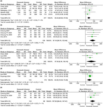
A significant difference in ankle-brachial pulse-wave velocity between patients with Kawasaki disease and controls was noted [MD 39.34 (95% CI 20.86 to 57.83) cm/sec; p = <0.0001] (Fig 1A). There was no heterogeneity for this outcome variable (I2 = 0%), and no publication bias was observed (Supplementary Figure 2A). Similarly, brachio-radial pulse-wave velocity was also significantly higher in patients with Kawasaki disease [MD 87.10 (95% CI 50.76 to 123.44) cm/sec; p = <0.00001, I2 = 0%] (Fig 1B). Additionally, ankle-brachial pulse-wave velocity was significantly higher (with considerable heterogeneity) in patients with Kawasaki disease and coronary artery abnormalities as compared to controls [MD 67.26 (95% CI 8.36 to 126.16) cm/sec; p = 0.03, I2 = 75%] (Fig 1C). After sensitivity analysis (excluding the study by Chien et al. Reference Chien, Huang and Weng9 ), heterogeneity was obviated while the results remained statistically significant [MD 44.38 (95% CI 15.48 to 73.29) cm/sec; p = 0.003, I2 = 0%] (Fig 1D). The brachio-radial pulse-wave velocity was also significantly higher in patients with Kawasaki disease and coronary artery abnormalities [MD 107.92 (95% CI 68.43 to 147.41) cm/sec; p = <0.0001, I2 = 1%] (Fig 1E).
Forest plots comparing PWV in patients with KD to controls; ( a ) Ankle-brachial PWV; ( b ) brachio-radial PWV; ( c ) ankle-brachial PWV in patients with KD and CAAs compared to controls; ( d ) forest plots comparing ankle-brachial PWV in patients with KD and CAAs to controls after sensitivity analysis; ( e ) brachio-radial PWV in patients with KD and CAAs compared to controls.

Flow-mediated dilatation
Nineteen studies have evaluated flow-mediated dilatation in patients with Kawasaki disease (Supplementary Table 3). Reference Niboshi, Hamaoka, Sakata and Yamaguchi11,Reference Borzutzky, Gutiérrez and Talesnik22–Reference Ghelani, Singh and Manojkumar39 Time to enrolment of subjects in studies varied from 3.7 to 24.1 years after diagnosis of Kawasaki disease. These studies have enrolled 520 cases and 439 controls. In eight studies, flow-mediated dilatation was measured in right brachial artery; while in two studies, it was measured in left brachial artery. In the remaining studies, it was not mentioned which artery was included (Supplementary Table 3). Of the 19 studies, 1 was excluded from the meta-analysis as flow-mediated dilatation had been analysed in the convalescent phase. Reference Ghelani, Singh and Manojkumar39 Similarly, two studies were not included in data synthesis as data has been represented as median. Reference McCrindle, McIntyre, Kim, Lin and Adeli33,Reference Duan, Du, Wang and Jia36 In 14 studies, flow-mediated dilatation was significantly different in patients with Kawasaki disease as compared to controls; while in the rest, there was no significant difference (Supplementary Table 3).
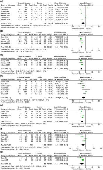
As significant heterogeneity amongst studies was noted, we decided not to pool the results. We carried out subgroup analysis including studies that have measured flow-mediated dilatation in the right brachial artery and studies that have mentioned brachial artery flow-mediated dilatation (without mentioning the side). A significant difference in right brachial artery flow-mediated dilatation was noted between patients with Kawasaki disease and controls [MD −3.83% (95% CI −6.72 to −0.94); p = 0.009] (Fig 2A). However, considerable heterogeneity was noted (I2 = 93%). The funnel plot did not reveal any publication bias (Supplementary Figure 2B). After sensitivity analysis (excluding Borzutzky et al. Reference Borzutzky, Gutiérrez and Talesnik22 , Kadono et al. Reference Kadono, Sugiyama and Hoshiai30 , Laurito et al. Reference Laurito, Stazi and Delogu31 ), heterogeneity was reduced to 24% while the results remained statistically significant [MD −6.42% (95% CI −7.46 to −5.38); p = <0.00001] (Fig 2B). Patients with Kawasaki disease and coronary artery abnormalities also showed a significant difference as compared to controls [MD −6.27% (95% CI −7.45 to −5.08); p = <0.00001, I2 = 38%] (Fig 2C).
Forest plots comparing FMD in patients with KD to controls; ( a ) right brachial artery FMD; ( b ) forest plot showing right brachial artery FMD after performing sensitivity analysis; ( c ) right brachial artery FMD in patients with KD and CAAs compared to controls; ( d ) brachial artery FMD; ( e ) forest plot showing brachial artery FMD after performing sensitivity analysis; ( f ) brachial artery FMD in patients with KD and CAAs compared to controls; ( g ) brachial artery FMD in patients with KD and CAAs compared to controls after performing sensitivity analysis.

Subgroup analysis on brachial artery flow-mediated dilatation also showed a significant difference with considerable heterogeneity and absence of publication bias [MD −3.28% (95% CI −5.48 to −1.08); p = 0.003, I2 = 83% (Figure 2D, Supplementary Figure 2C). Heterogeneity reduced to 40% after sensitivity analysis while the results remained statistically significant (excluding Silva et al. Reference Silva, Maeno, Hashmi, Smallhorn, Silverman and McCrindle38 , Sabri et al. Reference Sabri, Tavana, Ahmadi and Mostafavy37 , Parihar et al. Reference Parihar, Singh, Vignesh, Gupta and Rohit35 ) [MD −5.08% (95% CI −6.66 to −3.50; p = <0.00001] (Fig 2E). Also, brachial artery flow-mediated dilatation in children with Kawasaki disease and coronary artery abnormality was significantly different than in the controls [MD −2.92% (95% CI −5.80 to −0.04); p = 0.05); I2 = 85%] (Fig 2F). Heterogeneity reduced to 59% following sensitivity analysis and the results continued to be statistically significant [MD −5.54 (95% CI −9.10 to −1.98); p = 0.002] (Fig 2G).
Since heterogeneity remained substantial despite sensitivity analysis, we carried out metaregression to identify the underlying cause of heterogeneity. Age, sex, presence of coronary artery abnormalities, and duration to enrolment in the study were selected as covariables. However, none of the covariates was associated with heterogeneity (p = 0.31, 0.22, 0.69, and 0.29, respectively).
Arterial stiffness
Vascular stiffness in patients with Kawasaki disease has been evaluated by 14 studies Reference Y-f, Wong and Ho13,Reference AlHuzaimi, Al Mashham, Potts, De Souza and Sandor14,Reference Cheung, Ho, Tam and Yung17,Reference Duan, Du, Wang and Jia25,Reference Ikemoto, Ogino, Teraguchi and Kobayashi27,Reference Liu, Huang, Liang and Ma32,Reference Parihar, Singh, Vignesh, Gupta and Rohit35,Reference Duan, Du, Wang and Jia36,Reference Gupta, Singh, Gupta, Suri and Rohit40–Reference Cheung, O. and Woo45 (Supplementary Table 4). Except for 2, Reference AlHuzaimi, Al Mashham, Potts, De Souza and Sandor14,Reference Gupta-Malhotra, Gruber and Abraham41 all studies are from Asian countries. These studies have included 595 patients and 400 controls. Interval between diagnosis of Kawasaki disease and enrolment in the study varied from 3.1 to 20 years. Five studies documented no significant difference in arterial stiffness between the groups, whereas nine studies showed increased arterial stiffness. Of the 14 studies, 10 measured carotid artery stiffness, 3 measured aortic stiffness, and 1 study measured brachial artery stiffness. Due to the methodological difference among studies, we only pooled the stiffness index measured on carotid arteries. Amongst these, we excluded (Cheung 2008, Reference Cheung, O. and Woo45 Duan 2014 Reference Duan, Du, Wang and Jia36 ) from the meta-analysis as data were represented in median.
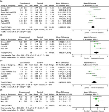
Carotid artery stiffness index was significantly different between patients with Kawasaki disease and controls [MD 0.35 (95% CI 0.11 to 0.59); p = 0.005]. However, substantial heterogeneity was observed for this outcome (I2 = 81%) (Fig 3A). The funnel plot also revealed publication bias (Supplementary Figure 2D). After sensitivity analysis (excluding Gupta-Malhotra et al., Reference Gupta-Malhotra, Gruber and Abraham41 Oguri et al, Reference Oguri, Nakamura and Tamanuki43 Ikemoto et al, Reference Ikemoto, Ogino, Teraguchi and Kobayashi27 and Parihar et al Reference Parihar, Singh, Vignesh, Gupta and Rohit35 ), heterogeneity reduced to 48% and significant difference in carotid artery stiffness between Kawasaki disease and controls was still evident [MD 0.69 (95% CI 0.41 to 0.96); p = <0.00001] (Fig 3B). Additionally, patients with Kawasaki disease and coronary artery abnormalities were found to have a significant difference in arterial stiffness in comparison to controls [MD 0.47 (95% CI 0.11 to 0.83); p = 0.009] with considerable heterogeneity (I2 = 84%) (Fig 3C). Following sensitivity analysis heterogeneity was obviated while the results remained significant [MD 0.93 (95% CI 0.65 to 1.20); p = <0.00001] (Fig 3D).
Forest plots comparing carotid artery stiffness in patients with KD to controls; ( a ) patients with KD compared to controls; ( b ) forest plot comparing patients with controls after performing sensitivity analysis; ( c ) carotid artery stiffness in patients with CAAs compared to controls; ( d ) forest plot comparing patients with CAAs to controls after performing sensitivity analysis.

Peripheral artery tonometry
Endo-PAT was assessed in four studies Reference Pinto, Laranjo, Paramés, Freitas and Mota-Carmo46–Reference Tobayama, Takahashi and Fukunaga49 with variable outcomes (Supplementary Table 5). These studies included 279 patients with Kawasaki disease and 150 controls. Two studies documented decreased reactive hyperaemia in patients with Kawasaki disease as compared to controls Reference Pinto, Laranjo, Paramés, Freitas and Mota-Carmo46,Reference Pinto, Gomes, Loureiro, Laranjo, Timóteo and Carmo47 while the remaining two studies showed no significant difference. Reference Selamet Tierney, Gal and Gauvreau48,Reference Tobayama, Takahashi and Fukunaga49 The latter studies also included patients with coronary artery abnormalities while the former two studies did not.
Meta-analysis showed no significant difference of RH-PAT index between patients with Kawasaki disease and controls with considerable heterogeneity [MD −0.16 (95% CI −0.52 to 0.20); p = 0.38, I2 = 90%] (Supplementary Figure 3A). On pooling the two studies from Portugal by Pinto et al, Reference Pinto, Laranjo, Paramés, Freitas and Mota-Carmo46,Reference Pinto, Gomes, Loureiro, Laranjo, Timóteo and Carmo47 the RH-PAT index was significantly decreased in patients with Kawasaki disease [MD −0.47 (95% CI −0.68 to −0.25); p = <0.0001, I2 = 33%] (Supplementary Figure 3B).
Nitroglycerine mediated dilatation
Eleven studies have assessed nitroglycerine-mediated dilatation in patients with Kawasaki disease (Supplementary Table 6). Reference Deng, Li, Xiang, Chang and Li23–Reference Duan, Du, Wang and Jia25,Reference Ishikawa and Iwashima28,Reference Ishikawa and Seki29,Reference McCrindle, McIntyre, Kim, Lin and Adeli33–Reference Duan, Du, Wang and Jia36,Reference Silva, Maeno, Hashmi, Smallhorn, Silverman and McCrindle38,Reference Ghelani, Singh and Manojkumar39 Except for three studies, all have been performed in Asian countries. These studies have included 302 patients with Kawasaki disease and 242 controls. Except for the study by Duan et al, Reference Duan, Du, Wang and Jia25 all studies showed no significant difference in nitroglycerine-mediated dilatation between patients with Kawasaki disease and controls. We excluded four studies from the meta-analysis; of these, data were presented as median in two studies, Reference McCrindle, McIntyre, Kim, Lin and Adeli33,Reference Duan, Du, Wang and Jia36 one study had been done during the convalescent stage of Kawasaki disease, Reference Ghelani, Singh and Manojkumar39 and one study nitroglycerine-mediated dilatation was measured in patients only. Reference Parihar, Singh, Vignesh, Gupta and Rohit35
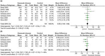
No difference in nitroglycerine-mediated dilatation between Kawasaki disease and controls was noted [MD 0.05% (95% CI −1.49 to 1.59); p = 0.95], and no heterogeneity was noticed (I2 = 0%) (Fig 4A). Besides, no publication bias was observed for this outcome (Supplementary Figure 2E). Similarly, no significant difference in nitroglycerine-mediated dilatation between patients with Kawasaki disease plus coronary artery abnormalities and controls was noted [MD −0.72% (95% CI −3.20 to 1.76); p = 0.95, I2 = 49%] (Fig 4B).
Forest plots comparing NMD between patients with KD and controls; ( a ) patients compared to controls; ( b ) patients with CAAs compared to controls.

Common carotid artery intima-media thickness
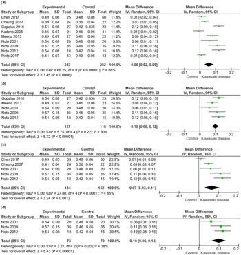
Twenty-five studies (Supplementary Table 7) have assessed carotid artery intima-media thickness in patients with Kawasaki disease. Reference Y-f, Wong and Ho13,Reference Shah, Christov and Mukasa16,Reference Lee, Ahn, You and Hong19,Reference Chen, Zannino, Curtis, Cheung and Burgner20,Reference Duan, Du, Wang and Jia25,Reference Ikemoto, Ogino, Teraguchi and Kobayashi27–Reference Laurito, Stazi and Delogu31,Reference Noto, Okada and Karasawa34,Reference Parihar, Singh, Vignesh, Gupta and Rohit35,Reference Gupta-Malhotra, Gruber and Abraham41–Reference Oguri, Nakamura and Tamanuki43,Reference Cheung, O. and Woo45,Reference Pinto, Gomes, Loureiro, Laranjo, Timóteo and Carmo47,Reference Selamet Tierney, Gal and Gauvreau48,Reference Dietz, Tacke and de Groot50–Reference Dietz, Tacke and Gort56 The majority of studies on carotid artery intima-media thickness have been carried out in Asian countries, and a few are from the Caucasian population. We excluded Wu et al Reference Wu, Kuo, Tain, Lin, Kuo and Chien55 from meta-analysis as carotid artery intima-media thickness was assessed during the acute stage. Additionally, two studies were excluded as data have been presented as median (Cheung et al 2008, Reference Cheung, O. and Woo45 Shah et al 2015 Reference Shah, Christov and Mukasa16 ). Furthermore, two studies by Dietz et al Reference Dietz, Tacke and de Groot50,Reference Dietz, Tacke and Gort56 were excluded as internal carotid artery and carotid bulb IMT were also included (besides carotid artery intima-media thickness) in the final average measurement. Finally, 20 studies were selected for data synthesis comprising 951 patients with Kawasaki disease and 622 controls. In 12 studies, no significant difference in carotid artery intima-media thickness was observed between patients of Kawasaki disease and controls. However, eight studies showed a significant difference. We did not pool the results of these 20 studies due to variability in the methodology (Supplementary Table 8) of carotid artery intima-media thickness measurement (especially, final measure and laterality). We, hence, carried out a sub-group analysis.
In the nine studies measuring mean of the maximums farwall carotid artery intima-media thickness, patients with Kawasaki disease had a significantly higher carotid artery intima-media thickness [MD 0.06 (95% CI 0.02 to 0.09) mm; p = 0.0006] (Fig 5A). However, heterogeneity was considerable (I2 = 88%) and publication bias was also noted (Supplementary Figure 2F). The heterogeneity was trivial after performing sensitivity analysis while results remained significant [MD 0.10 (95% CI 0.08 to 0.12) mm; p = <0.00001, I2 = 30%] (Fig 5B). Patients with Kawasaki disease and coronary artery abnormalities were also noted to have significantly higher carotid artery intima-media thickness than controls but with considerable heterogeneity [MD 0.07 (95% CI 0.03 to 0.11) mm; p = 0.0001; I2 = 86%] (Fig 5C). On sensitivity analysis, heterogeneity reduced to 38% while the results remained significant [MD 0.10 (95% CI 0.06 to 0.13) mm; p = <0.00001) (Fig 5D).
Mean of maximum CIMT in patients with KD as compared to controls; ( a ) forest plot comparing patients with controls; ( b ) comparison of maximum CIMT in patients with KD and controls after sensitivity analysis; ( c ) forest plot comparing patients with KD and CAAs to controls; ( d ) comparison of maximum CIMT in patients with KD plus CAAs and controls after sensitivity analysis.

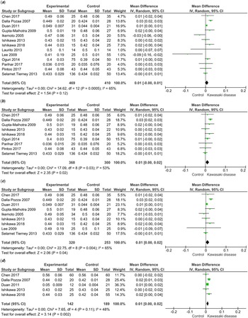
In 13 studies, the final measure was explicitly stated to be mean, mean of means, or it was unavailable (however, likely to be mean) (Supplementary Table 8). Meta-analysis showed similar carotid artery intima-media thickness in patients with Kawasaki disease and controls [MD 0.01 (95% CI −0.00 to 0.01) mm; p = 0.12] (Fig 6A). However, heterogeneity was substantial (I2 = 65%). No publication bias was noted on the funnel plot (Supplementary Figure 2G). After sensitivity analysis heterogeneity was reduced to 53% while the mean carotid artery intima-media thickness difference assumed statistical significance [MD 0.01 (95% CI 0.00 to 0.02) mm; p = 0.02] (Fig 6B). Carotid artery intima-media thickness in patients with Kawasaki disease and coronary artery abnormalities was slightly higher compared to controls [MD 0.01 (95% CI 0.00 to 0.02) mm; p = 0.04, I2 = 65%] (Fig 6C). Following sensitivity analysis, heterogeneity was reduced to 48% and the carotid artery intima-media thickness difference remained statistically significant [MD 0.01 (95% CI 0.00 to 0.02) mm; p = 0.002, I2 = 48%] (Fig 6D). On meta-regression analysis, sex and presence of coronary artery abnormalities associated with heterogeneity (p values of 0.04 and 0.02, respectively), while the other two covariables did not (age, p = 0.97; time to enrolment, p = 0.46).
Mean CIMT in patients with KD as compared to controls; ( a ) forest plot comparing patients with controls; ( b ) comparison of mean CIMT in patients with KD and controls after sensitivity analysis; ( c ) forest plot comparing patients with KD and CAAs to controls; ( d ) comparison of maximum CIMT in patients with KD plus CAAs and controls after sensitivity analysis.

Positron emission tomography
Positron emission tomography has been used to assess endothelial function in seven studies Reference Furuyama, Odagawa and Katoh57–Reference Ohmochi, Onouchi, Oda and Hamaoka63 (Supplementary Table 9). Amongst these, three were case–control studies, three cross-sectional studies, and one was a case report. Compared to controls, the myocardial flow reserve was found to be significantly lower in patients with Kawasaki disease. However, we did not pool the findings because of significant methodological variations amongst studies.
Quality of studies
Of the 49 studies included, 31 were of good quality, 11 were of fair quality, and 7 were of poor quality (Supplementary Table 1). This suggests that >80% of studies were above average quality. In the studies that scored poor, the major reason was the participant selection domain (selection of cases and controls). Majority of good and fair quality studies had an issue with the comparability domain.
Grading of evidence
The evidence generated was of ‘very low quality’ for all the outcomes except pulse-wave velocity (which was of ‘moderate quality’). A detailed analysis of the summary of evidence is provided in Supplementary Table 10.
Discussion
Summary of the main results
In this meta-analysis, we analysed the surrogate microanatomical and physiological markers of vascular function in patients with Kawasaki disease over the long term. The pulse-wave velocity was found to be significantly higher in patients with Kawasaki disease (with or without coronary artery abnormalities) than in controls. Similarly, arterial stiffness was higher in patients with Kawasaki disease. Patients with Kawasaki disease were also noted to have a slightly higher carotid artery intima-media thickness. Patients with Kawasaki disease (with or without coronary artery abnormalities) had a lower flow-mediated dilatation compared to controls; however, substantial heterogeneity was noted even after sensitivity analysis. On metaregression analysis, none of the selected covariates were associated with heterogeneity. Although decreased myocardial flow reserve in positron emission tomography has been documented in most studies, meta-analysis was not possible for this outcome measure due to methodological differences. No significant difference in nitroglycerine-mediated dilatation and endo-PAT was noted between patients and controls.
Overall completeness and applicability of evidence
Strengths of our meta-analysis include an extensive analysis of studies that have assessed long-term vascular function (including pulse-wave velocity and endo-PAT) in patients with Kawasaki disease. Prior to our study, pulse-wave velocity and endo-PAT have not been subjected to a meta-analysis. We separately analysed studies employing different sites or methodology for a particular outcome measure; for example, in the case of pulse-wave velocity, we performed meta-analysis for both ankle-brachial and brachio-radial pulse-wave velocity; for flow-mediated dilatation, right brachial artery flow-mediated dilatation and brachial artery (unspecified laterality) flow-mediated dilatation were analysed separately; and in case of carotid artery intima-media thickness, both mean of means carotid artery intima-media thickness and mean of maximums carotid artery intima-media thickness were studied. Our meta-analysis also has some limitations. Considerable heterogeneity for outcome measures such as carotid artery intima-media thickness and flow-mediated dilatation was noted. Even after performing meta-regression, subgroup and sensitivity analysis, the cause(s) of heterogeneity remained largely unexplained. We could not perform a meta-analysis on PET and endothelial function due to significant variability in the study methodology. We also did not include biomarkers of endothelial dysfunction (e.g. circulating endothelial cells/progenitors, receptor expression, etc.) in this systematic review.
As most of the studies assessing long-term vascular function in Kawasaki disease are from Asia, it may not be prudent to extrapolate these outcomes to other ethnicities. Additionally, most studies have a limited sample size with a case-control study design and absence of longitudinal data. Overall, the quality of evidence was ‘very low’ for most outcome measures.
Implications and caveats
Pulse-wave velocity is a non-invasive modality to detect arterial stiffness and can be measured at multiple sites. To date, no meta-analysis has been reported on this surrogate marker of vascular dysfunction in Kawasaki disease. Measurement of carotid-femoral pulse-wave velocity is the gold standard and predicts adverse cardiovascular events. Reference Laurent, Cockcroft and Van Bortel64 However, only two studies have measured carotid-femoral pulse wave velocity in patients with Kawasaki disease. Reference Shah, Christov and Mukasa16,Reference Chen, Zannino, Curtis, Cheung and Burgner20 Considering the methodological differences amongst studies determining pulse-wave velocity, we only pooled results of studies measuring ankle-brachial and/or brachio-radial pulse-wave velocity. The pulse-wave velocity was found to be significantly higher in patients with Kawasaki disease than in controls. Conversely, the two studies measuring carotid-femoral pulse-wave velocity in patients with Kawasaki disease noted no significant difference in pulse-wave velocity. Besides, it is important to note that the predictive value of pulse-wave velocity for cardiovascular outcomes is lower when measured in peripheral arteries as compared to central arteries. Reference Yu and McEniery65 Nonetheless, increased pulse-wave velocity in patients with Kawasaki disease signifies increased arterial stiffness and higher shear pressure. Higher shear stress has the potential to cause endothelial injury which may initiate the atherosclerotic process earlier in patients with Kawasaki disease compared to healthy individuals. Moreover, our meta-analysis also showed an increased carotid artery stiffness index in patients with Kawasaki disease which was in agreement with previously published meta-analyses. Reference Dietz, Tacke, Hutten and Kuijpers4
Flow-mediated dilatation is an indicator of endothelial function (vasodilation due to nitric oxide generated by the endothelium post transient ischaemic challenge) measured ultrasonically. Similar to the present meta-analyses, Dietz et al documented lower flow-mediated dilatation in patients with Kawasaki disease compared to controls, although with considerable heterogeneity. Reference Dietz, Tacke, Hutten and Kuijpers4 On meta-regression, no relationship with the selected covariates and heterogeneity was found (by Dietz et al), which was in concurrence with our study. Reference Dietz, Tacke, Hutten and Kuijpers4 Zeng et al and Zhang et al have also noted similar results with heterogeneity. The reason(s) for the heterogeneity remain unascertained. Reference Zeng, Chen, Zhang and Ji66,Reference Zhang, Xu, Xie, Huang, Shen and Xiao67 Nonetheless, these results suggest the presence of endothelial dysfunction in the peripheral vasculature in patients with Kawasaki disease even in the absence of coronary artery abnormalities. In our meta-analysis, we also noted patients with Kawasaki disease (with or without coronary artery abnormalities) to have a similar nitroglycerine-mediated dilatation compared to controls. The presence of normal nitroglycerine-mediated dilatation (measuring vasodilation due to extraneously administered nitric oxide) in patients with Kawasaki disease suggests normal vascular smooth muscle function and, taken together with the results of abnormal flow-mediated dilatation, also reflects the presence of endothelial dysfunction in Kawasaki disease.
Carotid artery intima-media thickness is a microanatomical marker of endothelial homeostasis. An increase in carotid artery intima-media thickness is believed to be associated with coronary artery disease in adults. Reference van der Meer, Bots, Hofman, del Sol, van der Kuip and Witteman68 However, the value of carotid artery intima-media thickness in predicting future adverse cardiovascular events is controversial. Reference Lorenz, Polak and Kavousi69,Reference Goldberger, Valle, Dandekar, Chan, Ko and Nallamothu70 A large meta-analysis has demonstrated the lack of an increased risk of adverse cardiovascular events in adults with increased carotid artery intima-media thickness. Reference Lorenz, Polak and Kavousi69 Conversely, an earlier meta-analysis reported a higher likelihood of developing a non-fatal myocardial infarction in patients with increased carotid artery intima-media thickness. Reference Goldberger, Valle, Dandekar, Chan, Ko and Nallamothu70 Similar to the results of our study, Dietz et al Reference Dietz, Tacke, Hutten and Kuijpers4 noted slightly increased mean carotid artery intima-media thickness (MD of 0.01 cm) in patients with Kawasaki disease (with or without coronary artery abnormalities). The meta-analysis of maximum carotid artery intima-media thickness (five studies) highlighted the conflicting results obtained in various studies. In our updated meta-analysis of nine studies measuring maximum carotid artery intima-media thickness, a significantly higher value was noted in patients with Kawasaki disease (MD of 0.06 mm). Nonetheless, the small difference in carotid artery intima-media thickness values in patients with Kawasaki disease and healthy controls needs to be interpreted with caution given the axial resolution of high-frequency linear probes (used to measure carotid artery intima-media thickness) is 0.01–0.03 cm. Reference O'Leary and Bots71 Besides, the lower limit of 95% CI (for MD) in both the studies for mean carotid artery intima-media thickness was very close to 0 (in fact, rounded up to 2 decimal points to 0.00). Furthermore, in all meta-analyses (ours, [Reference Dietz, Tacke, Hutten and Kuijpers4], and [Reference Zeng, Chen, Zhang and Ji66]), significant heterogeneity amongst studies was noted. Unlike previous studies (wherein none of the predefined covariables associated with heterogeneity), sex and coronary artery abnormalities associated with heterogeneity on meta-regression analysis in our study. The pathological correlate of increased carotid artery intima-media thickness (intimal proliferation or premature atherosclerosis) in patients with Kawasaki disease is also unclear. Absence of lipid-laden material on histopathological examination of the intima of patients with Kawasaki disease suggests role of the former mechanism in the pathogenesis of carotid artery intima-media thickness. Reference Chen, Curtis, Dahdah, Kowalski, Cheung and Burgner3 Taken together, interpretation of increased carotid artery intima-media thickness in the long-term in patients with Kawasaki disease is problematic.
Although early atherosclerotic coronary heart disease has been reported in patients with Kawasaki disease and coronary artery abnormalities, whether Kawasaki disease in childhood is associated with a statistically significant elevation in risk of CVAs or premature CVAs in adulthood is also not clear. This is due to the paucity of longitudinal studies on this subject. To the best of our knowledge, no studies have calculated the standardised incidence ratio of CVAs in patients who have had Kawasaki disease (in comparison to controls). Besides, the confounding influence of traditional cardiovascular risk factors also needs to be taken into consideration while assessing the risk of future CVAs in patients with Kawasaki disease. Lastly, the importance of altered flow-mediated dilatation, arterial stiffness, pulse-wave velocity, and carotid artery intima-media thickness in predicting CVAs in adults who have had Kawasaki disease also remains unquantified. The conclusion of increased risk of CVAs based on altered surrogate markers of endothelial function in patients with Kawasaki disease would, thus, largely remain conjectural.
Future research implications
Considering the heterogeneity in the published literature on long-term endothelial dysfunction in patients with Kawasaki disease, future research on this aspect must ensure a uniform (and standardised) methodology. These include patient inclusion criteria (encompassing consideration of other cardiovascular risk factors as well), time interval after diagnosis of Kawasaki disease, and techniques for assessing endothelial dysfunction and outcome variables. There is a need to carry out prospective long-term follow-up studies on surrogate markers of endothelial dysfunction in patients with Kawasaki disease and their correlation with (the risk of) cardiovascular events. Besides, the confounding effect of traditional cardiovascular risk factors like obesity, hypertension, and smoking also would need to be minimised or obviated. Lastly and, perhaps, most importantly standardised incidence ratio of CVAs in patients who have had Kawasaki disease (in comparison to controls) needs to be determined to definitely quantify the increased risk of CVAs in patients with Kawasaki disease. As the discovery of Kawasaki disease has completed 50 years (and, hence, the oldest patients with Kawasaki disease would be in their 50s or 60s), such studies would hopefully be published in near future.
Conclusions
A ‘very low quality’ evidence suggests endothelial dysfunction in patients with Kawasaki disease even in the absence of overt coronary artery abnormalities. It may be prudent to advise long-term follow-up for all patients with Kawasaki disease. Additionally, avoiding additional cardiovascular risk factors (e.g., obesity, hypertension, smoking) may need to be emphasised. In the absence of longitudinal data, the implications of altered endothelial function are still unclear and remain a subject of further research.
Supplementary material
To view supplementary material for this article, please visit https://doi.org/10.1017/S1047951122002906
Author contribution statement
PKP: Writing of initial draft of the manuscript, editing and revision of the manuscript, review of literature, data acquisition and analysis, and final approval.
AZB: Writing of initial draft of manuscript, editing and revision of the manuscript, review of literature, data analysis, and final approval.
RRD: Writing of initial draft of manuscript, editing and revision of the manuscript, review of literature, data acquisition and analysis, and final approval.
SM: Editing and revision of the manuscript, review of literature, data acquisition and analysis, and final approval.
AKJ: Inception of the idea, evaluation and critical revision of the manuscript, supervision of data acquisition and analysis, and final approval.
SS: Evaluation and critical revision of the manuscript, overall supervision of manuscript preparation, and final approval.
Acknowledgements
None.
Financial support
This research received no specific grant from any funding agency, commercial, or not-for-profit sectors.
Conflict of interest
None.
Ethical standards
As this manuscript pertains to literature review and analysis, specific ethics approval is not mandated.
Registration
PROSPERO (CRD 42021277854).