The target article suggests that the symptoms of autism spectrum disorders (ASD) and psychotic-spectrum disorders (PSD) are mediated by altered biases toward paternally or maternally expressed genes, respectively. This hypothesis fits well with the existing literature, and provides a potential genotype that acts together with brain development and environmental factors to create the complex phenotypes that define these disorders.
Though understanding the genetic underpinning of these disorders is of critical importance, diagnosis usually occurs around age 2–3 in ASD and during adolescence or later in PSD. Thus, the phenotypic presentation is a result of the complex interaction among genetic risk factors, brain development, and environment, and ultimately the manifestation of brain plasticity. It is worth considering whether the mechanisms of plasticity might be normal, simply acting upon a genetically determined abnormal substrate, or may themselves be pathological, thus contributing to progressively dysfunctional states.
There are several pieces of evidence to support abnormal plasticity in ASD. First, the developmental trajectory of brain size abnormalities suggests a dynamic underlying process. Second, studies have found decreased cortico-cortical and long-range connections and an increase in intracortical and superficial connections. Such patterns of abnormal connectivity might be the result of increased ability to form new connections and/or a decreased ability to prune superfluous ones. Abnormal plasticity is also consistent with the phenotype of PSD, which has also been reported to have abnormal connectivity patterns. Though there are inconsistencies in the findings, one study finds increased corpus callosum size with decreased local connections (Siekmeier & Hoffman Reference Siekmeier and Hoffman2002). Additionally, the lower levels of BDNF in schizophrenia (Moises et al. Reference Moises, Zoega and Gottesman2002; Palomino et al. Reference Palomino, Vallejo-Illarramendi, González-Pinto, Aldama, González-Gómez, Mosquera, González-García and Matute2006; Weickert et al. Reference Weickert, Hyde, Lipska, Herman, Weinberger and Kleinman2003) may also be related to abnormal plasticity. A recent study suggests that there is a relationship between having a specific polymorphism in the BDNF gene and experience-dependent motor plasticity (Kleim et al. Reference Kleim, Chan, Pringle, Procaccio, Jimenez and Cramer2006).
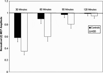
Abnormal plasticity provides a plausible, parsimonious, and testable mechanism, consistent with both the main proposal of the target article, as well as the core phenotypes of ASD and PSD. Studies are currently underway to evaluate the role of plasticity in the etiology of ASD using transcranial magnetic stimulation (TMS). TMS paradigms can capture aspects of homosynaptic and heterosynaptic plasticity (see Hallett Reference Hallet2007 for a review). A paradigm known as theta burst stimulation (TBS) is well-suited for investigating homosynaptic plasticity and allows a rapid conditioning of the motor cortex that produces controllable, consistent, and long-lasting effects on motor physiology and behavior. In typical individuals, TBS for 20–90 seconds results in suppression of the motor evoked potential (MEP) for up to 30 minutes. Preliminary data suggest this effect is greater in both degree and duration in individuals with ASD. (See Fig. 1.)
Bars represent the standardized (Z score) MEP amplitude at 30, 60, 90, and 120 minutes following continuous theta burst stimulation (cTBS) for the control and ASD sample. cTBS induces an LTD-like phenomenon that can be quantified as a reduction in the amplitude of the MEP. Error bars represent the standard error of the mean. Note the greater degree and longer duration of suppression in the ASD group.

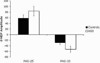
Paired associative stimulation (PAS) is well-suited for investigations of heterosynaptic plasticity and ability for the motor cortex to form long term potentiation (LTP) and long term depression (LTD) in response to sensory input. PAS involves stimulation of the median nerve combined with TMS over the motor cortex at variable interstimulus intervals (ISI). At 25 msec ISI the MEP shows facilitation, while an ISI of 10 msec inhibits the MEP. Both effects are amplified in individuals with ASD. (See Fig. 2.)
Bars represent the average percent change in MEP amplitude following paired associative stimulation (PAS) (with an ISI of 25 msec and 10 msec) for the control and ASD sample. PAS25 results in facilitation, while PAS10 results in suppression of the MEP. Note the greater degree of both facilitation and suppression in the ASD group. Error bars represent the standard error of the mean.

Abnormal patterns of plasticity may be leading to the abnormal connectivity in ASD, which in turn may be mediating the deficits in social and communicative skills (as these skills require synchronization of neuronal populations across relatively disparate regions of the cortex) as well as the restricted, repetitive, and stereotyped patterns of behaviors, interests, and activities. Abnormal plasticity may also lead to increased long-range connections in PSD, perhaps contributing to the complex social delusions, enhanced imagination and creativity, and “loose” associations between words and aspects of the environment in this population.
Though the genetic predisposition toward maternally or paternally expressed genes might predispose a child for developing these disorders, the recent increase in incidence can better be accounted for by a change in environment rather than an increase in the penetrance of the genetic risk factor. Children growing up in recent years are being exposed to an ever more diverse and stimulating environment packed with stimuli to process from very early on. In a child who is predisposed to increased plasticity, this overly stimulating environment could lead to deleterious consequences. In such a setting, ASD genetic risk factors may lead to a facilitated, insufficiently controlled reshaping of neural connections. The child may develop local hyperconnectivity, a surge in brain size, and an inefficient processing system for incoming sensory stimuli. If, however, this child is placed in a very structured environment and develops a specific interest, this enhanced plasticity may actually lead to savant abilities.
One can also consider the opposite scenario with a child who is genetically at risk for developing PSD with an increased bias toward effects of genes with maternal expression leading to uncontrollable increased global connectivity. Later in life when more associative long-range connections are maturing, abnormal plasticity in this population would lead to the characteristic positive symptoms of PSD.
TMS can be used not only to measure the degree of plasticity in a given system, but also as a valuable tool for clinicians developing therapeutic interventions for these disorders. Specifically, repetitive TMS (rTMS) has been shown to produce long-term (several-month) changes in cortical plasticity and has been effective in other brain diseases, for example, depression.
In conclusion, Crespi & Badcock (C&B) present an interesting hypothesis for genetic factors that may lead to ASD and PSD. We suggest that abnormal plasticity may be the common underlying mechanism that ultimately leads to many of the core behavioral deficits which define these disorders. The manifestation of the specific pattern of abnormal plasticity may lie in the timing of the hyperplastic response. Specifically, if hyperplasticity is promoted early in life, this may preferentially affect local circuits, resulting in ASD. However, PSD may result from abnormal plasticity during adolescence and young adulthood, leading to promotion of long-range hyperplasticity. Such conceptualization provides an appealing account for the increase in incidence of ASD and PSD in our highly stimulating society. Finally, modulation of plasticity may provide an effective means to treat these disorders.