Introduction
Oocyte development in mammals starts early in fetal development and is arrested in the diplotene of the prophase stage of first meiosis cleavage within the ovarian follicle. When the follicle is recruited, the oocyte enters the growth phase in which it increases in volume, and undergoes replication and redistribution of cytoplasmic organelles (Picton et al., Reference Picton, Briggs and Gosden1998). Oocyte meiosis progression and developmental competence during folliculogenesis is influenced by the local microenvironment formed by companion somatic cells, called cumulus oophorus (Sanchez and Smitz, Reference Sanchez and Smitz2012). The communication between oocyte and its cumulus cells is critical for the development and functions of both cell types (Eppig, Reference Eppig2001; Matzuk et al., Reference Matzuk, Burns, Viveiros and Eppig2002; Gilchrist et al., Reference Gilchrist, Ritter and Armstrong2004). For instance, the removal of cumulus cells before in vitro maturation inhibits oocyte developmental competence (Vozzi et al., Reference Vozzi, Formenton, Chanson, Senn, Sahli, Shaw, Nicod, Germond and Haefliger2001). Similar results were obtained when the interaction of both cells was disrupted using gap junction inhibitors (Atef et al., Reference Atef, Francois, Christian and Marc-Andre2005).
In the later stages of follicular development, ovulation is the result of a long and orchestrated process to release the competent oocyte which is fertilizable, followed by normal embryo development and eventually the birth of a healthy offspring (Labrecque and Sirard, Reference Labrecque and Sirard2014). The transcriptome dynamics in oocytes (Fair et al., Reference Fair, Carter, Park, Evans and Lonergan2007; Regassa et al., Reference Regassa, Rings, Hoelker, Cinar, Tholen, Looft, Schellander and Tesfaye2011) and cumulus cells (Assidi et al., Reference Assidi, Dieleman and Sirard2010; Regassa et al., Reference Regassa, Rings, Hoelker, Cinar, Tholen, Looft, Schellander and Tesfaye2011; Nivet et al., Reference Nivet, Vigneault, Blondin and Sirard2013) before and after the maturation process revealed a spatiotemporal regulation of gene expression within bovine follicular cells. The differentially expressed genes in oocytes and cumulus cells cultured without their surrounding cumulus cells and oocyte cytoplasm, respectively, indicated the molecular cross-talk between the oocytes and surrounding cumulus cells (Regassa et al., Reference Regassa, Rings, Hoelker, Cinar, Tholen, Looft, Schellander and Tesfaye2011). Similar to the mRNAs, our previous studies revealed the microRNAs transcript abundance in oocytes (Tesfaye et al., Reference Tesfaye, Worku, Rings, Phatsara, Tholen, Schellander and Hoelker2009; Abd El Naby et al., Reference Abd El Naby, Hagos, Hossain, Salilew-Wondim, Gad, Rings, Cinar, Tholen, Looft, Schellander, Hoelker and Tesfaye2013) and cumulus cells (Abd El Naby et al., Reference Abd El Naby, Hagos, Hossain, Salilew-Wondim, Gad, Rings, Cinar, Tholen, Looft, Schellander, Hoelker and Tesfaye2013) during oocyte maturation and their potential dependency on each other for proper expression of the miRNAs.
During in vitro maturation (IVM) of the oocyte, bovine cumulus cells are able to produce and to secrete steroid hormones (Mingoti et al., Reference Mingoti, Garcia and Rosa-e-Silva2002). The inhibition of endogenous steroid production during maturation drastically decreased the percentage of mature oocytes (MII stage) and suppressed cumulus expansion in bovine cumulus–oocyte complexes (COCs) (Wang et al., Reference Wang, Isobe, Kumamoto, Yamashiro, Yamashita and Terada2006; Pan et al., Reference Pan, Toms, Shen and Li2015). As reported previously, progesterone is one of the steroid hormones that are produced and secreted by cumulus cells to support oocyte meiosis resumption (van Tol et al., Reference van Tol, van Eijk, Mummery, van den Hurk and Bevers1996; Choi et al., Reference Choi, Carnevale, Seidel and Squire2001; Ježová et al., Reference Ježová, Scsuková, Nagyová, Vranová, Procházka and Kolena2001; Yamashita et al., Reference Yamashita, Shimada, Okazaki, Maeda and Terada2003; Shimada et al., Reference Shimada, Yamashita, Ito, Okazaki, Kawahata and Nishibori2004b; Montano et al., Reference Montano, Olivera and Ruiz-Cortes2009; Aparicio et al., Reference Aparicio, Garcia-Herreros, O’Shea, Hensey, Lonergan and Fair2011; Nagyová et al., Reference Nagyová, Camaioni, Scsuková, Mlynarcikova, Procházka, Nemcova and Salustri2011; Nagyová et al., Reference Nagyová, Scsuková, Nemcova, Mlynarcikova, Yi, Sutovsky and Sutovsky2012) under the stimulation of FSH and LH (van Tol et al., Reference van Tol, van Eijk, Mummery, van den Hurk and Bevers1996; Choi et al., Reference Choi, Carnevale, Seidel and Squire2001; Shimada and Terada, Reference Shimada and Terada2002; Shimada et al., Reference Shimada, Yamashita, Ito, Okazaki, Kawahata and Nishibori2004b). The important role of progesterone in bovine follicular development has been indicated by the higher progesterone levels in the follicular fluid of mature oocytes compared with that in immature oocytes (Grimes and Ireland, Reference Grimes and Ireland1986). In addition, progesterone is believed to promote oocyte maturation in pig (Yamashita et al., Reference Yamashita, Shimada, Okazaki, Maeda and Terada2003), mouse (Jamnongjit et al., Reference Jamnongjit, Gill and Hammes2005) and bovine in a dose-dependent manner (Siqueira et al., Reference Siqueira, Barreta, Gasperin, Bohrer, Santos, Buratini, Oliveira and Goncalves2012) during IVM. Conversely, the inhibition of progesterone synthesis resulted in a negative effect on cumulus cells expansion, oocyte maturation rate, ovulation rate and subsequent embryonic development in mouse (Sirotkin, Reference Sirotkin1992; Panigone et al., Reference Panigone, Hsieh, Fu, Persani and Conti2008; Aparicio et al., Reference Aparicio, Garcia-Herreros, O’Shea, Hensey, Lonergan and Fair2011; Siqueira et al., Reference Siqueira, Barreta, Gasperin, Bohrer, Santos, Buratini, Oliveira and Goncalves2012), porcine (Shimada and Terada, Reference Shimada and Terada2002; Shao et al., Reference Shao, Markstrom, Friberg, Johansson and Billig2003; Shimada et al., Reference Shimada, Yamashita, Ito, Okazaki, Kawahata and Nishibori2004b; Kawashima et al., Reference Kawashima, Okazaki, Noma, Nishibori, Yamashita and Shimada2008) and bovine (Roh et al., Reference Roh, Batten, Friedman and Kim1988; Shao et al., Reference Shao, Markstrom, Friberg, Johansson and Billig2003; Shimada et al., Reference Shimada, Nishibori, Yamashita, Ito, Mori and Richards2004a, 2004c; Wang et al., Reference Wang, Isobe, Kumamoto, Yamashiro, Yamashita and Terada2006; Aparicio et al., Reference Aparicio, Garcia-Herreros, O’Shea, Hensey, Lonergan and Fair2011; O’Shea et al., Reference O’Shea, Hensey and Fair2013).
Previously, we have shown that miR-20a is differentially expressed in bovine granulosa cells derived from the different sized follicles at the late phase of bovine oestrous cycle (Gebremedhn et al., Reference Gebremedhn, Salilew-Wondim, Ahmad, Sahadevan, Hossain, Hoelker, Rings, Neuhoff, Tholen, Looft, Schellander and Tesfaye2015). Subsequently we showed that miR-20a overexpression and knockdown of its target genes (PTEN and BMPR2) in cultured bovine granulosa cells promoted cell proliferation and suppressed cell differentiation (Andreas et al., Reference Andreas, Hoelker, Neuhoff, Tholen, Schellander, Tesfaye and Salilew-Wondim2016). In addition, the progesterone levels in spent medium of granulosa cell culture were elevated in that experiment. This finding was supported by the cross-talk between PTEN/PI3K/AKT and BMP-SMAD signalling pathways in progesterone synthesis (Chang et al., Reference Chang, Cheng, Klausen and Leung2013; Hosoya et al., Reference Hosoya, Otsuka, Nakamura, Terasaka, Inagaki, Tsukamoto-Yamauchi, Hara, Toma, Komatsubara and Makino2015; Luo et al., Reference Luo, Zhao, Jin, Tao, Zhu, Wang, Hemmings and Yang2015). However, the potential involvement of miR-20a during oocyte maturation has not been reported to date. Therefore, here we aimed to investigate the potential involvement of miR-20a in bovine oocytes maturation in vitro. Subsequently we found that miR-20a expression during IVM process was increased and decreased in cumulus cells and oocytes, respectively. We also observed that miR-20a expression in cumulus cells and oocytes was regulated by the presence or absence of their companion cells. Moreover, our experiments provide evidence that the oocyte maturation progression during IVM could be triggered by the modulation of miR-20a expression in its surrounding somatic cells.
Materials and methods
Cumulus–oocyte complexes collection and in vitro oocyte maturation
Bovine ovaries, as a source of cumulus–oocyte complexes (COCs), were obtained from a local slaughterhouse and transported to the laboratory within 2 h of slaughter in a thermo-flask that contained a 0.9% saline solution. The COCs were aspirated from healthy small follicles (2–8 mm follicle diameter). Good quality and morphologically uniform COCs (oocytes with a homogenous, evenly granulated ooplasm, and surrounded by at least three layers of cumulus cells) were selected in this study. The selected COCs were washed with TCM-199 medium before being placed in culture to obtain matured oocytes or were directly frozen as immature COCs (germinal vesicle; GV). The COCs were cultured in groups of 50 in 400 µl of maturation medium (modified Parker medium (MPM) supplemented with 12% oestrus cow serum and 10 µg/ml Follitropin®) under mineral oil in 5-well dishes. Maturation was performed for 22 h at 39°C in a humidified atmosphere with 5% (v/v) CO2 in air. Spent medium of in vitro maturation medium was collected for progesterone assay. The cumulus cells and oocytes from immature and matured group of COCs were separated by gentle pipetting in TCM-199 medium supplemented with hyaluronidase (1 mg/ml; Millipore Sigma). The complete removal of cumulus cells was assessed by observing denuded oocytes under a stereomicroscope. After transferring the denuded oocytes into a new tube containing 10 µl 1× phosphate-buffered saline (PBS), the cumulus cells were isolated by gentle centrifugation. The cumulus cell pellet was resuspended using 50 µl lysis buffer (0.8% Igepal, 40 U RNasin and 5 mM DTT). The cumulus cells, oocytes and spent medium were snap frozen in liquid nitrogen and stored at −80°C until further analysis. Matured oocytes (metaphase II stage; MII) were indicated by the presence of the first polar body as seen under an inverted microscope. The total numbers of recovered and matured oocytes after in vitro maturation (IVM) were recorded. The maturation rate was calculated from the number of matured oocytes compared with the total number of recovered oocytes.
Cumulus cells and denuded oocytes culture
To investigate the effect of oocytes on cumulus cells microRNA expression and vice versa, cumulus cells and oocytes were cultured in the presence or absence of their companion cells. For this, cumulus cells and oocytes from 50 collected COCs were separated in TCM-199 medium supplemented with hyaluronidase (1 mg/ml). The cumulus cells and denuded oocytes were cultured for 22 h in the maturation medium at 39°C in 5% (v/v) CO2 incubator, as described in the previous section. Cumulus cells and denuded oocytes were collected and stored at −80°C until further analysis. The cumulus cells and oocytes obtained from cultured COCs were used as controls.
MicroRNA and siRNA transfection
To investigate the function of miR-20a in oocyte maturation, the collected COCs were cultured in a group of 50 in 5-well dishes containing 400 µl maturation medium, as described above. An equal concentration (50 nM) of miRCURY LNA™ miR-20a mimic, miR-20a inhibitor or corresponding negative controls (mimic NC and inhibitor NC) was transfected into the appropriate well using Lipofectamine® 2000 reagent. Transfected COCs were cultured for 22 h at 39°C in a humidified atmosphere with 5% (v/v) CO2 in air. To validate the function of miR-20a, siRNA against BMPR2 gene were used to knockdown the expression of the BMPR2 gene (Andreas et al., Reference Andreas, Hoelker, Neuhoff, Tholen, Schellander, Tesfaye and Salilew-Wondim2016).
Total RNA isolation and cDNA synthesis
Total RNA from cumulus cells was isolated using an miRNeasy® mini kit following the manufacturer’s protocol, while oocyte total RNA extraction was performed using the PicoPure® RNA isolation kit. The quality and quantity of extracted RNA were determined using a NanoDrop 8000 spectrophotometer (Thermo Scientific). For gene expression analysis, equal amounts of total RNA (100 ng from cumulus cell and 50 ng from oocyte total RNA) were reverse transcribed using the RevertAid first stand cDNA synthesis kit (Life Technologies GmbH) according to the manufacturer’s protocol. For microRNA expression analysis, the cDNA was synthesized from 50 ng and 25 ng of total RNA from cumulus cells and oocytes, respectively, using the Universal cDNA synthesis kit (Exiqon) following the manufacturer’s instructions.
MicroRNA and mRNA quantitative PCR analysis
Quantitative PCR (qPCR) analysis of several candidate genes and miR-20a expression were performed using the iTaq™ Universal SYBR® Green Supermix and ExiLENT SYBR® Green master mix, respectively, in the Applied Biosystems® StepOnePlus™ system. The primers for gene expression analysis (Table 1) were tested using qualitative PCR followed by sequencing analysis using the GenomeLab™ GeXP Genetic Analysis System, while microRNA primers were purchased from Exiqon. In addition, the specificity of amplification in qPCR processes was indicated by a single melting curve generated at the end of the qPCR protocol. The relative expression levels of candidate genes and miR-20a were analyzed using comparative Ct (2-ΔΔCt) methods (Livak and Schmittgen, Reference Livak and Schmittgen2001). The expression levels of β-ACTIN and 5S rRNA were used to normalize the candidate genes and miR-20 expression, respectively.
List of primers used for candidate genes expression analysis in bovine cumulus cells and oocytes

Progesterone measurement
Progesterone levels in oocyte maturation medium were measured using a progesterone enzyme-linked immunosorbent assay kit (ELISA) kit (ENZO Life Sciences). Prior to measuring the progesterone level, the spent maturation medium was diluted 1:1000 in 1× PBS. Progesterone levels were measured using the progesterone assay kit according to the manufacturer's instructions, and the 405 nm optical density (OD) was detected using a Synergy™ H1 Multi-Mode Reader.
Data analysis
All quantitative data are presented as mean ± standard error of the mean (SEM). Data were obtained from three replicates and each replicate was derived from 50 COCs. Statistical significance of the data was analyzed using either t-test or one-way analysis of variance (ANOVA) methods (Prism® software version 5.02; GraphPad). The P-values are indicated in the corresponding figure legend.
Results
Temporal expression of miR-20a during in vitro maturation
To investigate the temporal expression of miR-20a in cumulus cells and oocytes during IVM, first we collected COCs from small healthy follicles at the GV stage. Parts of these COCs were used as the immature (GV) group, while the others were matured (MII). The cumulus cells and oocytes from both immature and mature groups were investigated separately. The qPCR analysis showed that miR-20a expression was significantly higher (P < 0.05) in cumulus cells of matured COCs compared with those cumulus cells from GV stage COCs. Conversely, the expression of miR-20a was lower in MII oocytes compared with the GV stage oocytes (Fig. 1).
Temporal alteration of miR-20a expression in cumulus cells and oocytes during maturation from immature (GV) and matured (MII) oocyte stages. The expression of 5S rRNA was used as internal control. Data are shown as mean ± SEM (two-tailed t-test; n = 3; *P < 0.05; ***P < 0.001).

The role of oocytes and cumulus cells factors in expression of miR-20a in cumulus cells and oocytes
To investigate whether the expression of miR-20a in cumulus cells and oocytes was affected by the presence or absence of their companion cells, we cultured cumulus cells (without oocytes) and denuded oocytes. Both cumulus cells and oocytes derived from cultured COCs were used as controls. Results showed that the expression of miR-20a in cumulus cells derived from cultured COCs was relatively higher (P = 0.0543) compared with those from cultured cumulus cells only. Conversely, denuded oocytes cultured without the surrounding cumulus cells showed higher expression of miR-20a compared with those oocytes derived from cultured COCs (Fig. 2).
Relative expression level of miR-20a in cumulus cells and oocytes cultured with or without their companion cells. The expression of 5S rRNA was used as an internal control. Data are shown as mean ± SEM (two-tailed t-test; n = 3; *P < 0.05).

The effect of miR-20a modulation in cumulus cells and oocytes
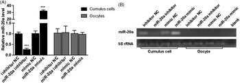
To investigate the role of miR-20a in oocyte maturation, we first studied the feasibility of miR-20a overexpression or inhibition during IVM using 50 nM of miR-20a mimic or inhibitor. As a negative control, cultured COCs were transfected using the same amount of mimic or inhibitor negative controls (mimic NC or inhibitor NC). The qPCR analysis revealed a significant increase (P < 0.001) of miR-20a expression in cumulus cells from COCs transfected with miR-20a mimic compared with mimic NC. Conversely, the transfection of miR-20a inhibitor resulted in decreased (P < 0.001) miR-20a expression in cumulus cells compared with the inhibitor NC group (Fig. 3A). However, neither miR-20a mimic nor inhibitor transfection had an effect on the miR-20a expression in the oocytes. We observed a very negligible level of miR-20a expression in oocytes as indicated by larger Ct values (beyond 35 cycles), and it was further evidenced by electrophoresis of PCR products on 1.5% agarose gel (Fig. 3B).
The effect of transfection is restricted in the cumulus cells. The expression of miR-20a in cumulus cells and oocytes derived from COCs transfected with miR-20a mimic, inhibitor and corresponding controls (A). Agarose gel (1.5%) electrophoresis of miR-20a amplification products in cumulus cells and oocytes derived from COCs transfected with miR-20a mimic, inhibitor and corresponding controls (B). The miR-20a expression level was compared with corresponding negative controls (mimic NC or inhibitor NC) and the expression of 5S rRNA was used as an internal control for qPCR analysis. Data are shown as mean ± SEM (two-tailed t-test; n = 3; ***P < 0.001).

miR-20a overexpression during IVM increased oocyte maturation rate
We next studied the effect of miR-20a expression on maturation rate of oocytes. Based on the presence of the first polar body in MII oocytes under an inverted microscope the maturation rates of oocytes in different treatment groups was determined. The maturation rates in medium supplemented with inhibitor NC, miR-20a inhibitor, mimic NC or miR-20a mimic were (mean ± SEM) 72.1 + 0.83, 72.4 ± 1.77, 71.3 ± 0.32, and 74.8 ± 1.72, respectively. We observed that miR-20a overexpression during IVM resulted in an increased oocyte maturation rate (P < 0.05). However, the transfection of miR-20a inhibitor had no effect on maturation rate at all (Fig. 4).
miR-20a overexpression in cumulus cells increased oocyte maturation rate. The maturation rate was compared with corresponding negative controls (mimic NC or inhibitor NC). Data are shown as mean ± SEM (two-tailed t-test; n = 3; *P < 0.05).

miR-20a modulation altered expression of oocyte maturation-related genes
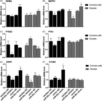
To study whether the effect of miR-20a on the maturation rate was accompanied by the changes in the expression of oocyte maturation marker genes, we analyzed the expression level of genes related to the oocyte competence (INHBA, MAPK1 and PTGS2), cumulus cells expansion (PTX3 and EGFR) and cell cycle regulator (CYCB2) in cumulus cells and oocytes (Fig. 5). We found that decreased miR-20a induced decreased expression of INHBA, MAPK1, PTGS2 and EGFR genes in cumulus cells. Conversely, increased miR-20a induced the expression of EGFR, INHBA and CYCB2 genes in cumulus cells. In addition, miR-20a inhibitor transfection during IVM resulted in decreased expression of MAPK1 (P < 0.05) and EGFR (P < 0.05) genes, but increased the expression of PTGS2 gene in oocytes. miR-20a overexpression using miR-20a mimic transfection exhibited an increased expression of INHBA (P < 0.01), MAPK1 (P < 0.05) and PTX3 (P < 0.05), but decreased the expression of PTGS2 (P < 0.001) genes in oocytes.
Expression of INHBA (A), MAPK1 (B), PTGS2 (C), PTX3 (D), EGFR (E) and CYCB2 (F) in cumulus cells and oocytes derived from COCs transfected with miR-20a mimic, inhibitor or corresponding negative controls (NC). The expression level of β-ACTIN was used as an internal control. Data are compared with corresponding negative controls (mimic NC or inhibitor NC) and shown as mean ± SEM (two-tailed t-test; n = 3; *P < 0.05; **P < 0.01; ***P < 0.001).

miR-20a enhanced oocyte maturation through cumulus cell progesterone biosynthesis by targeting PTEN and BMPR2 genes
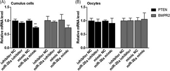
Parallel with the oocyte maturation, the spent medium of IVM was collected and analyzed for the level of progesterone released during the maturation process by the cumulus cells. Even though not statistically significant, overexpression and inhibition of miR-20a relatively increased (P = 0.0936) and decreased (P = 0.0993) the progesterone levels in spent medium, respectively (Fig. 6A). This result was further accompanied by the increase in expression of progesterone synthesis-related genes, namely CYP11A1 (P < 0.05; Fig. 6B) and STAR (P < 0.01; Fig. 6C). In addition, elevated progesterone levels in spent medium due to overexpression of miR-20a was also accompanied by a reduction in mRNA expression of the PTEN (P < 0.01) gene in cumulus cells. Despite not being statistically significant, BMPR2 gene expression in cumulus cells derived from cultured COCs transfected with miR-20a mimic was relatively lower compared with the control cohort (Fig. 7A). There was no difference in terms of PTEN and BMPR2 gene expression in oocytes between miR-20a mimic or inhibitor compared with their negative control counterparts (Fig. 7B).
miR-20a overexpression during IVM elevated cumulus cell progesterone synthesis. The effect of miR-20a mimic and inhibitor during IVM on progesterone level in spent medium (A). The expression of CYP11A1 (B) and STAR (C) mRNA in cumulus cell transfected with miR-20a mimic, inhibitor or corresponding negative controls (NC). Data are shown as mean ± SEM (two-tailed t-test; n = 3; *P < 0.05; **P < 0.01). The expression level of β-ACTIN was used as an internal control in mRNA analysis.

miR-20a overexpression during in vitro oocyte maturation reduced cumulus cell PTEN and BMPR2 genes expression. The expression of PTEN (A) and BMPR2 (B) genes in cumulus cells derived from COCs transfected with miR-20a mimic, inhibitor or corresponding controls. mRNA expression of PTEN and BMPR2 genes in oocytes derived from COCs transfected with miR-20a mimic, inhibitor or corresponding controls (C). mRNA and protein expression levels were compared with negative controls (mimic NC or inhibitor NC). β-ACTIN was used to normalized mRNA expression. The expression levels of mRNAs are shown as mean ± SEM (two-tailed t-test; n = 3; **P < 0.01).

BMP15 supplementation during IVM reduced oocyte maturation rate and progesterone synthesis
Here, we investigated the effect of BMP15 treatment on the maturation and progesterone synthesis. We supplemented maturation medium with different doses of BMP15 (0, 10 ng/ml, 50 ng/ml and 100 ng/ml). The maturation rates of cultured oocytes supplemented with 0, 10, 50 and 100 ng of BMP15 were (mean ± SEM) 81.6 ± 0.41, 71.7 ± 0.93, 68.8 ± 0.43, and 65.7 ± 1.24, respectively. We found that BMP15 supplementation at concentration 10 ng/ml increased (P < 0.05) BMPR2 mRNA expression (Fig. 8A). We also found that BMP15 supplementation (10–100 ng/ml) during IVM reduced (P < 0.01) the oocyte maturation rate (Fig. 8B). However, there was no difference in progesterone levels in spent medium upon BMP15 supplementation (Fig. 8C). We further analyzed the expression levels of oocyte maturation (Fig. S1) as well as progesterone synthesis marker genes (Fig. S2) in cumulus cells derived from COCs treated with BMP15.
BMP15 supplementation reduced maturation rate and progesterone synthesis. Expression of BMPR2 mRNA level in cumulus cells (A). Effect of BMP15 during IVM on oocyte maturation rate (B). Progesterone level in spent medium after IVM supplemented with BMP15 (C). The expression level of β-ACTIN was used as an internal control for mRNA analysis. Data are compared with BMP15 0 ng/ml and shown as mean ± SEM (one-way ANOVA; Tukey’s post-hoc test; n = 3; small letters indicate P < 0.05; capital letter P < 0.01).

miR-20a rescued the BMP15-treated oocytes by promoting maturation rate and progesterone synthesis
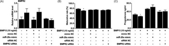
To investigate whether miR-20a overexpression could rescue the effect of BMP15 treatment on COCs physiology, we transfected miR-20a mimic to COCs cultured in the presence of BMP15. Even though not statistically significant, we found that BMP15 (10 ng/ml) relatively increased the expression of BMPR2 gene in cumulus cells. However, neither miR-20a mimic nor BMPR2 siRNA transfection could reduce the expression of BMPR2 gene in cumulus cells from BMP15-supplemented COCs (Fig. 9A). In addition, we found that 10 ng/ml BMP15 supplementation during IVM reduced (P < 0.05) the maturation rate (Fig. 9B) as well as progesterone levels in spent medium (Fig. 9C). However, we showed that miR-20a or BMPR2 siRNA transfection rescued the reduced maturation rate and progesterone levels in spent medium in COC cultures supplemented with BMP15. These results were also supported by the expression of oocyte maturation (Fig. S3) and progesterone synthesis marker genes (Fig. S4).
The transfection of miR-20a mimic and BMPR2 siRNA could not rescue the effect of BMP15 during IVM. Effect of miR-20a and BMPR2 siRNA transfection on BMPR2 expression at mRNA (A). Maturation rate of bovine oocyte transfected with miR-20a mimic and BMPR2 siRNA during IVM (B). Progesterone levels in spent medium transfected with miR-20a mimic and BMPR2 siRNA (C). The expression level of β-ACTIN was used as an internal control for mRNA expression analysis. Data are compared with their corresponding controls and shown as mean ± SEM (two-tailed t-test; n = 3; *P < 0.05).

Discussion
The oocyte maturation process is complex and requires an integration of endocrine, paracrine, juxtacrine and autocrine signalling pathways (Takahashi et al., Reference Takahashi, Morrow, Wang and Dey2006). This process involves an interaction between the oocyte and surrounding cumulus cells. In our previous studies, we have shown the differential expression of mRNAs (Regassa et al., Reference Regassa, Rings, Hoelker, Cinar, Tholen, Looft, Schellander and Tesfaye2011) and microRNAs (Abd El Naby et al., Reference Abd El Naby, Hagos, Hossain, Salilew-Wondim, Gad, Rings, Cinar, Tholen, Looft, Schellander, Hoelker and Tesfaye2013) in cumulus cells before and after the in vitro maturation process, which could confirm the idea that signals released from somatic cells stimulated meiotic progression and oocyte maturation (Chen et al., Reference Chen, Torcia, Xie, Lin, Cakmak, Franciosi, Horner, Onodera, Song, Cedars, Ramalho-Santos and Conti2013). In this study, we observed a differential expression pattern of miR-20a in cumulus and oocytes after IVM in which expression was increased in cumulus cells and decreased in oocytes following 22 h IVM. The depletion of miR-20a in oocytes after IVM, like most maternal transcripts, has also been reported in human oocytes (Xu et al., Reference Xu, Wang, Ding, Li, Gu and Zhou2011).
Bidirectional communication between the gamete and the surrounding somatic cells is essential for proper maturation of oocytes, fertilization and further embryonic development (Buccione et al., Reference Buccione, Schroeder and Eppig1990a; Eppig, Reference Eppig2001; Matzuk et al., Reference Matzuk, Burns, Viveiros and Eppig2002; Gilchrist et al., Reference Gilchrist, Ritter and Armstrong2004). For instance, oocyte-secreted factors (OSFs) such as GDF9 and BMP15 are believed to regulate key cumulus cell functions (Buccione et al., Reference Buccione, Vanderhyden, Caron and Eppig1990b; Vanderhyden et al., Reference Vanderhyden, Caron, Buccione and Eppig1990; Eppig et al., Reference Eppig, Wigglesworth, Pendola and Hirao1997; Joyce et al., Reference Joyce, Clark, Pendola and Eppig2000; Li et al., Reference Li, Norman, Armstrong and Gilchrist2000; Gilchrist et al., Reference Gilchrist, Ritter and Armstrong2001; Otsuka and Shimasaki, Reference Otsuka and Shimasaki2002; Tanghe et al., Reference Tanghe, Van Soom, Nauwynck, Coryn and de Kruif2002; Gilchrist et al., Reference Gilchrist, Morrissey, Ritter and Armstrong2003, Reference Gilchrist, Ritter and Armstrong2004; Eppig et al., Reference Eppig, Pendola, Wigglesworth and Pendola2005; Hussein et al., Reference Hussein, Froiland, Amato, Thompson and Gilchrist2005; Sugiura et al., Reference Sugiura, Pendola and Eppig2005; Gilchrist et al., Reference Gilchrist, Ritter, Myllymaa, Kaivo-Oja, Dragovic, Hickey, Ritvos and Mottershead2006). We have previously shown that the presence or absence of oocyte cytoplasm or the cumulus cells during IVM resulted in altered expression of several mRNAs (Regassa et al., Reference Regassa, Rings, Hoelker, Cinar, Tholen, Looft, Schellander and Tesfaye2011) and of microRNAs (Abd El Naby et al., Reference Abd El Naby, Hagos, Hossain, Salilew-Wondim, Gad, Rings, Cinar, Tholen, Looft, Schellander, Hoelker and Tesfaye2013) expression in cumulus cells or in oocytes. In the present study, miR-20a expression in cumulus cells cultured without oocyte cytoplasm was relatively lower compared with cumulus cells cultured in the presence of oocyte cytoplasm. Conversely, miR-20a expression in denuded oocytes was higher compared with those oocytes derived from intact COCs. These findings indicated cross-talk between oocyte and cumulus cells in the regulation of miR-20a expression in both types of cells.
With regards to microRNA, several studies have demonstrated the role of microRNAs in oocyte developmental competence (Yao et al., Reference Yao, Liang, Liang, Yin, Lu, Lian, Wang and Sun2014; Pan et al., Reference Pan, Toms, Shen and Li2015). Following transfection of the miR-20a mimic or inhibitor it was possible to confirm that changes in expression of endogenous miR-20a was evident only in cumulus cells, but not in the oocyte cytoplasm. This could be due to the barrier effect of the zona pellucida surrounding the oocyte cytoplasm. Similar observations were also reported in studies using lentivirus transduction (Pan et al., Reference Pan, Toms, Shen and Li2015). In the present study, overexpression of miR-20a during IVM resulted in increased oocyte maturation rate, while no difference was found in maturation rate when miR-20a expression was inhibited. The analysis of oocyte developmental competence-related genes revealed that miR-20a overexpression increased the expression of INHBA, EGFR and CYCB2 genes in cumulus cells. The expression of oocyte competence marker genes in oocytes has never been reported before. However, regardless of the statistical significance level, we found a similar pattern in the expression of oocyte competence marker genes in cumulus cells and oocytes. This result was expected, as some mRNA transcripts could be transferred from cumulus cells to the oocytes through gap junctions during the oocyte development process. Here, we suggest that miR-20a regulates oocyte maturation and the expression of oocyte developmental competence-related genes.
Progesterone synthesis during oocyte maturation process is essential for oocyte meiosis resumption and subsequent oocyte maturation processes (van Tol et al., Reference van Tol, van Eijk, Mummery, van den Hurk and Bevers1996; Choi et al., Reference Choi, Carnevale, Seidel and Squire2001; Ježová et al., Reference Ježová, Scsuková, Nagyová, Vranová, Procházka and Kolena2001; Yamashita et al., Reference Yamashita, Shimada, Okazaki, Maeda and Terada2003; Shimada et al., Reference Shimada, Yamashita, Ito, Okazaki, Kawahata and Nishibori2004b; Montano et al., Reference Montano, Olivera and Ruiz-Cortes2009; Aparicio et al., Reference Aparicio, Garcia-Herreros, O’Shea, Hensey, Lonergan and Fair2011; Nagyová et al., Reference Nagyová, Camaioni, Scsuková, Mlynarcikova, Procházka, Nemcova and Salustri2011, Reference Nagyová, Scsuková, Nemcova, Mlynarcikova, Yi, Sutovsky and Sutovsky2012). Interestingly, progesterone levels were found to be higher in follicular fluid obtained from follicles containing matured oocytes compared with those with immature ones (Grimes and Ireland, Reference Grimes and Ireland1986). Administration of progesterone during in vitro maturation promoted oocyte maturation and induced nuclear maturation in pig (Yamashita et al., Reference Yamashita, Shimada, Okazaki, Maeda and Terada2003), mouse (Jamnongjit et al., Reference Jamnongjit, Gill and Hammes2005) and bovine (Siqueira et al., Reference Siqueira, Barreta, Gasperin, Bohrer, Santos, Buratini, Oliveira and Goncalves2012) in a dose-dependent manner. Conversely, the inhibition of progesterone synthesis was shown to have a negative effect on oocyte meiosis resumption, cumulus cell expansion, final oocyte maturation, ovulation and number of ovulated oocytes and subsequent embryonic development in mouse (Sirotkin, Reference Sirotkin1992; Aparicio et al., Reference Aparicio, Garcia-Herreros, O’Shea, Hensey, Lonergan and Fair2011; Siqueira et al., Reference Siqueira, Barreta, Gasperin, Bohrer, Santos, Buratini, Oliveira and Goncalves2012), porcine (Shimada and Terada, Reference Shimada and Terada2002; Shao et al., Reference Shao, Markstrom, Friberg, Johansson and Billig2003; Shimada et al., Reference Shimada, Yamashita, Ito, Okazaki, Kawahata and Nishibori2004b) and bovine (Roh et al., Reference Roh, Batten, Friedman and Kim1988; Shao et al., Reference Shao, Markstrom, Friberg, Johansson and Billig2003; Shimada et al., Reference Shimada, Nishibori, Yamashita, Ito, Mori and Richards2004a, Reference Shimada, Yamashita, Ito, Okazaki, Kawahata and Nishibori2004c; Wang et al., Reference Wang, Isobe, Kumamoto, Yamashiro, Yamashita and Terada2006; Aparicio et al., Reference Aparicio, Garcia-Herreros, O’Shea, Hensey, Lonergan and Fair2011; O’Shea et al., Reference O’Shea, Hensey and Fair2013). Here we showed that progesterone level tended to be regulated by modulation of miR-20a during the IVM process. This result was accompanied by the expression of progesterone synthesis marker genes, namely CYP11A1 and STAR (Nuttinck et al., Reference Nuttinck, Guienne, Clément, Reinaud, Charpigny and Grimard2008). Similarly, the selective knockdown of PTEN and BMPR2 genes in cultured granulosa cells confirmed the role of miR-20a in progesterone biosynthesis (Andreas et al., Reference Andreas, Hoelker, Neuhoff, Tholen, Schellander, Tesfaye and Salilew-Wondim2016). Moreover, we observed that higher progesterone levels in miR-20a expression-induced cumulus cells was consistent with the increase in oocyte maturation rate in miR-20a mimic-transfected COCs, which suggested that miR-20a overexpression in cumulus cells promoted oocyte maturation by increasing cumulus cell progesterone synthesis.
Several studies have shown the role of specific microRNAs in regulating granulosa cell proliferation, apoptosis and estradiol synthesis (Dai et al., Reference Dai, Sun, Fang, Zhang, Wu, Jiang, Ding, Yan and Hu2013; Jiang et al., Reference Jiang, Huang, Li, Chen, Chen, Zhao and Yang2015; Wang et al., Reference Wang, Li, Zhang, Xing, Gao and Wu2016). However, only few studies have reported the function of microRNA in regulating oocyte maturation (Pan et al., Reference Pan, Toms, Shen and Li2015) and cumulus cells expansion (Yao et al., Reference Yao, Liang, Liang, Yin, Lu, Lian, Wang and Sun2014). Recently, we have shown that miR-20a directly targets PTEN and BMPR2, to regulate their expression both at the mRNA and protein levels in cultured granulosa cells (Andreas et al., Reference Andreas, Hoelker, Neuhoff, Tholen, Schellander, Tesfaye and Salilew-Wondim2016). Similarly, here we showed that overexpression of miR-20a in bovine cumulus cells also suppressed the expression of PTEN and BMPR2 genes, suggesting the post-transcriptional regulation of these two genes by miR-20a in cumulus cells such as in cultured granulosa cells.
To validate the function of miR-20a on cumulus cell-regulated oocyte maturation and progesterone synthesis, we cultured COCs in maturation medium supplemented with BMP15 and miR-20a mimic or BMPR2 siRNA. BMP15 is one of the OSFs that determines the function of granulosa cells (Li et al., Reference Li, Norman, Armstrong and Gilchrist2000). A dose-dependent effect of BMP15 supplementation on oocyte maturation rate during IVM has been observed. Concomitantly, supplementation with 10 ng/ml of BMP15 during IVM resulted in increased BMPR2 expression and a decrease in the maturation rate and progesterone levels in spent medium. Similar phenomena have been reported using human granulosa cells in which BMP15 suppresses progesterone production by downregulating the activity of the STAR gene (Chang et al., Reference Chang, Cheng, Klausen and Leung2013). Furthermore, we demonstrated that COCs transfected with miR-20a or BMPR2 siRNA in BMP15-supplemented maturation medium had the same oocyte maturation rate and progesterone levels in spent medium compared with those transfected with miR-mimic NC or siRNA NC. Therefore, we suggested that miR-20 overexpression could rescue the negative effects of BMP15 on oocyte maturation and progesterone synthesis.
In conclusion, our work has demonstrated that modulation of miR-20a expression in cumulus cells regulates oocyte maturation by stimulating cumulus cell progesterone synthesis by suppressing the expression of BMPR2 gene. In addition, the expression of several cumulus expansion-, oocyte maturation- and cell cycle-related genes in both cumulus cells and oocytes suggested the role of miR-20a during oocyte maturation progression. The findings of the present study offer new insight into the mechanisms of action of microRNAs in oocytes surrounding cumulus cells and their regulatory role in oocyte developmental competence.
Supplementary material
To view supplementary material for this article, please visit https://doi.org/10.1017/S0967199420000933
Conflict of interest
The authors declare no conflicts of interest
Financial support
This research was financially supported by the German Research Foundation (DFG) with grant number TE-589/5–1.
Ethical standards
Not applicable