Introduction
Galectins belong to an evolutionary ancient family of glycan-binding proteins which bind β-galactosides, such as lactose and N-acetyllactosamine, either in free form or as components of glycoproteins or glycolipids (Massa et al., Reference Massa, Cooper, Leffler and Barondes1993). Galectins have been isolated from fungi, invertebrates, fish, amphibians, birds and mammals. Galectins are involved in important intracellular processes; they regulate signalling pathways and cell interactions (Vasta, Reference Vasta2012). They also mediate host defence by acting as pattern recognition receptors (Vasta, Reference Vasta2009), killing microbes (Stowell et al., Reference Stowell, Arthur, Dias-Baruffi, Rodrigues, Gourdine, Heimburg-Molinaro, Ju, Molinaro, Rivera-Marrero, Xia, Smith and Cummings2010) and by regulating immune responses. Various aspects of galectins have recently been reviewed by a number of groups (Shi et al., Reference Shi, Xue, Su and Lu2018; Modenutti et al., Reference Modenutti, Capurro, Di Lella and Marti2019) but the interaction of nematode galectins and host galectins has not been explored.
Structure of host galectins
There are 17 distinct galectins identified in mammals and they are labelled gal-1 to gal-17. Generally, galectins with the same name in mammals are homologous; gal-1 in humans is homologous to gal-1 in mice. However, gal-11 and gal-15, both in sheep, are probably variants of the same protein. There are two different proteins called galectin-14; one from sheep and one in humans. The sheep molecule was the first protein to be called galectin-14 (Dunphy et al., Reference Dunphy, Barcham, Bischof, Young, Nash and Meeusen2002). In comparison with the human galectins, the ovine gal-14 sequence is most similar to galectin-9 with 57% amino acid identity. Functionally, ovine gal-14 is most similar to human galectin-10 because both gal-10 and gal-14 function similarly in eosinophils (Young et al., Reference Young, Barcham, Kemp, Dunphy, Nash and Meeusen2009). However, amino acid similarity with gal-10 is only 25% (Dunphy et al., Reference Dunphy, Barcham, Bischof, Young, Nash and Meeusen2002). The second protein to be called gal-14 was originally called placental protein 13-like because of its similarity to placental protein 13 (Yang et al., Reference Yang, Ying, Yuan, Chen, Meng, Wang, Xie and Mao2001a). It is part of a cluster of 5 galectin genes on human chromosome 19 (Than et al., Reference Than, Romero, Goodman, Weckle, Xing, Donga, Xua, Tarquini, Szilagyi, Gale, Hou, Tarca, Kim, Kim, Haidarian, Uddin, Bohn, Benirschke, Santolaya-Forgas, Grossman, Erez, Hassan, Zavodszky, Pap and Wildman2009). Gal-5 is found in rat erythrocytes but has not been reported in humans (Gitt et al., Reference Gitt, Wiser, Leffler, Herrman, Xia, Massa, Cooper, Lusis and Barondes1995). Galectin-6 is only found in mice and is a recent duplication of gal-4 (Gitt et al., Reference Gitt, Colnot, Poirier, Nani, Baronde and Leffler1998; Cooper, Reference Cooper2002). Galectins in other classes of vertebrates with the same name are not necessarily homologous; gal-1 in humans need not correspond to gal-1 in a fish species.
There is considerable diversity among galectins in their structure and function. Most galectins show less than 40% amino acid identity to each other (Fig. S1). Some galectins occur in the cytoplasm while others are secreted by the cell and act extracellularly. However, no galectin contains a secretion signal peptide (Cooper and Barondes, Reference Cooper and Barondes1999). Galectin synthesis occurs on free polyribosomes prior to export through a noncanonical Golgi-independent pathway (Lindstedt et al., Reference Lindstedt, Apodaca, Barondes, Mostov and Leffler1993). Some galectins such gal-1 and gal-3 are expressed in cells of the myeloid and lymphoid lineages while other galectins like gal-7 and gal-12 are only expressed in specific tissues.
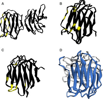
Galectins vary in size from 14 to 39 KDa. Structurally, galectins are classified into three types: prototype, chimaeric or tandem repeat. Figure 1 shows an example of each type of galectin. Gal-1, gal-2, gal-7 (Saussez and Kiss, Reference Saussez and Kiss2006), gal-10 (Ackerman et al., Reference Ackerman, Liu, Kwatia, Savage, Leonidas, Swaminathan and Acharya2002), gal-11, gal-13 (Than et al., Reference Than, Romero, Goodman, Weckle, Xing, Donga, Xua, Tarquini, Szilagyi, Gale, Hou, Tarca, Kim, Kim, Haidarian, Uddin, Bohn, Benirschke, Santolaya-Forgas, Grossman, Erez, Hassan, Zavodszky, Pap and Wildman2009) ovine gal-14, human gal-14, gal-16 and gal-17 are prototype galectins that each have one carbohydrate recognition domain (CRD). They are usually synthesised as monomers and non-covalently homodimerize.
Molecular structure of human and parasitic galectins showing the similarity in structure. (A) The prototypic human galectin 1(PDB 2 km2) shown as a dimer. (B) The chimaeric human galectin 3 (PDB 3ZSL). (C) The tandem repeat human galectin 4. (D) The predicted structure of the tandem repeat galectin 1 from T. circumcincta (O01410) using Phyre2 (Kelley et al., Reference Kelley, Mezulis, Yates, Wass and Sternberg2015). The human galectin structures were determined but the parasite galectin is a predicted structure. To emphasize this difference, the parasite galectin has been coloured differently, the N terminal CRD is shown in blue while the C terminal CRD is shown in grey. In all three figures of human galectin, the amino acids responsible for binding beta-galactosides have been highlighted but the amino acids responsible for binding glycans have not been determined for the parasite galectin. The structures were visualised in EzMol (Reynolds et al., Reference Reynolds, Islam and Sternberg2018).

The only chimaeric galectin is gal-3 which is produced as a 35KDa monomer with 250 amino acids and one CRD (Fig. 1). The monomers can polymerize to form oligomers after binding of the CRD (Modenutti et al., Reference Modenutti, Capurro, Di Lella and Marti2019).
Galectin-4, −6, −8, −9 and −12 are tandem repeat galectins with two distinct CRDs joined by a linker peptide. The CRDs in the same molecule differ in their binding affinities (Gitt et al., Reference Gitt, Colnot, Poirier, Nani, Baronde and Leffler1998; Cooper, Reference Cooper2002; Huflejt and Leffler, Reference Huflejt and Leffler2004). Galectin-8 has a high affinity for 3-O-sulfated or 3-O-sialylated glycoconjugates and a Lewis X-containing glycan and this affinity is largely due to the N-terminal CRD (Ideo et al., Reference Ideo, Seko, Ishizuka and Yamashita2003, Reference Ideo, Matsuzaka, Nonaka, Seko and Yamashita2011). The linker of gal-9 is of various sizes in humans. The long linker has 58 amino acids, the medium linker has 26 amino acids and the short linker is only 14 amino acids (Hirashima et al., Reference Hirashima, Kashio, Nishi, Yamauchi, Imaizumi, Kageshita, Saita and Nakamura2002). Alternative splicing creates at least 5 splice variants (Heusschen et al., Reference Heusschen, Schulkens, van Beijnum, Griffioen and Thijssen2014). Gal-12 has an unusual C-terminal domain and many usually conserved residues are absent (Yang et al., Reference Yang, Hsu, Yu, Ni and Liu2001b). Uniquely among galectins, gal-12 preferentially recognises 3-fucosylated structures (Maller et al., Reference Maller, Cagnoni, Bannoud, Sigaut, Pérez Sáez, Pietrasanta, Yang, Liu, Croci and Di Lella2020).
Structurally, the CRDs of galectins are arranged in a tightly folded conserved beta-sandwich structure formed by a six-stranded sheet (S1–S6) and a five stranded sheet (F1–F5). Figure 1 shows the structure of gal-1 (Kishor et al., Reference Kishor, Ross and Blanchard2018), gal-3 (Saraboji et al., Reference Saraboji, Hakansson, Genheden, Diehl, Qvist, Weininger, Nilsson, Leffler, Ryde, Akke and Logan2012) and gal-4 (Rustiguel et al., Reference Rustiguel, Soares, Meisburger, Davis, Malzbender, Ando, Dias-Baruffi and Nonato2016). The carbohydrate-binding amino acids are in strands S4–S6 (Rini and Lobsanov, Reference Rini and Lobsanov1999; Loris, Reference Loris2002) and they have been highlighted (Fig. 1).
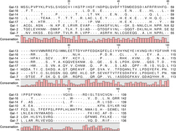
Figure 2 shows the alignment of the prototypic human galectins gal-1, 2, −7, −10, −13 and −14. There is considerable diversity in the amino acid sequences. The amino acids implicated in binding to carbohydrate in galectin-1 (Kishor et al., Reference Kishor, Ross and Blanchard2018) have been identified as His44, Asn46, Arg48, His52, Asn61, Trp68, Glu71 and Arg73. Of these eight sites, only Trp68 is conserved among all the prototypic galectins. Clearly, the amino acids involved in binding carbohydrate are not identical in each galectin and these differences presumably influence the specificity of carbohydrate-binding (Modenutti et al., Reference Modenutti, Capurro, Di Lella and Marti2019).
Multiple sequence alignment of prototypic human galectins that show differences in the amino acid sequences among galectins. The accession numbers were gal-1 (P09382), gal-2 (P05182), gal-7 (P47929), gal-10 (Q05315), gal-13 (Q9UHV8), gal-14 (Q8TCE9), gal-16 (A8MUM7) and gal-17 (Q6ZW74).

Function of host galectins
Galectins bind glycans on glycoproteins and glycolipids and mediate a wide variety of cellular processes including cellular interactions, intracellular signalling and host defence against infection (Nielsen et al., Reference Nielsen, Stegmayr, Grant, Yang, Nilsson, Boos, Carlsson, Wood R, Unverzagt, Leffler and Wandall2018). Of particular relevance to parasitologists, several galectins play key roles in immune regulation (Sato et al., Reference Sato, St-Pierre, Bhaumik and Nieminen2009). Gal-1 mediates T lymphocyte apoptosis (Perillo et al., Reference Perillo, Pace, Seilhamer and Baum1995) as does gal-2 (Sturm et al., Reference Sturm, Lensch, Andre, Kaltner, Wiedenmann, Rosewicz, Dignass and Gabius2004). Gal-2 has also been shown to inhibit the development of the parasitic nematode Ascaris suum as well as the free-living nematode Caenorhabditis elegans by binding to the galactoseβ1-4fucose epitope (Takeuchi et al., Reference Takeuchi, Tamura, Ishiwata, Hamasaki, Hamano, Arata and Hatanaka2019).
Gal-3 binds IgE and promotes mast cell degranulation (Dumic et al., Reference Dumic, Dabelic and Flogel2006; Henderson and Sethi, Reference Henderson and Sethi2009) while gal-9 has a high affinity for IgE, which is the most heavily glycosylated immunoglobulin.
Gal-11 has only been reported in three species of Bovidae (cattle, sheep and goats). It is an inducible galectin present in the cytoplasm and nucleus of epithelial cells of the ovine gastrointestinal tract during infection with H. contortus; uninfected animals lack gal-11 (Dunphy et al., Reference Dunphy, Balic, Barcham, Horvath, Nash and Meeusen2000). Protein production is restricted to the epithelial cells lining the gastrointestinal system, as no changes in gal-11 expression level are noticed after infection with the bovine lungworm Dictyocaulus viviparus (Hoorens et al., Reference Hoorens, Rinaldi, Mihi, Dreesen, Grit, Meeusen, Li and Geldhof2011). Increased expression of gal-11 is observed 2–7 days after challenge infection with H. contortus (Robinson et al., Reference Robinson, Pleasance, Piedrafita and Meeusen2011). Increased levels of gal-11 were observed in the late phase of infection or after repeated infection of animals (da Souza et al., Reference da Souza, Lambert, Nishi, Benavides, Berne, Madruga and de Almeida2015). Gal-11 binds to molecules from the L4 and adult stages of H. contortus, but not to the L3 stage and is associated with inhibited larval development of H. contortus (Preston et al., Reference Preston, Beddoe, Walkden-Brown, Meeusen and Piedrafita2015). Sheep possess two gal-11 variants (Sakthivel et al., Reference Sakthivel, Preston, Gasser, Costa, Hernandez, Shahine, Shakif-Azam, Lock, Rossjohn, Perugini, Gonzalez, Meeusen, Piedrafita and Beddoe2020). Only one variant is able to form polymers and only this variant reduced the development of L3–L4 (Sakthivel et al., Reference Sakthivel, Preston, Gasser, Costa, Hernandez, Shahine, Shakif-Azam, Lock, Rossjohn, Perugini, Gonzalez, Meeusen, Piedrafita and Beddoe2020). It also damaged L4 during in vitro exposure (Sakthivel et al., Reference Sakthivel, Preston, Gasser, Costa, Hernandez, Shahine, Shakif-Azam, Lock, Rossjohn, Perugini, Gonzalez, Meeusen, Piedrafita and Beddoe2020). The targets of gal-11 and ovine gal-14 have been investigated by affinity chromatography combined with mass spectrometry and over 30 molecules from adult H. contortus have been identified for each galectin (Sakthivel et al., Reference Sakthivel, Swan, Preston, Shakif-Azam, Faou, Jiao, Downs, Rajapaksha, Gasser and Piedrafita2018).
Ovine galectin-14 is produced by eosinophils in ovine gastrointestinal mucus (Young et al., Reference Young, Barcham, Kemp, Dunphy, Nash and Meeusen2009). It is homologous and functionally similar to human galectin-10 (Ackerman et al., Reference Ackerman, Liu, Kwatia, Savage, Leonidas, Swaminathan and Acharya2002). Ovine gal-14 levels are increased 7 days after H. contortus infection and are temporally associated with parasite expulsion (Robinson et al., Reference Robinson, Pleasance, Piedrafita and Meeusen2011). In addition, the level of abomasal ovine gal-14 is correlated with the parasitic burden in sheep infected with H. contortus (da Souza et al., Reference da Souza, Lambert, Nishi, Benavides, Berne, Madruga and de Almeida2015); animals with low parasite burdens express lower levels of gal-14. Ligands for gal-11 and gal-14 have been identified in L4 and adult stage extracts (Sakthivel et al., Reference Sakthivel, Littler, Shahine, Troy, Johnson, Rossjohn, Piedrafita and Beddoe2015). Increased levels of gal-15 are observed in the abomasal mucosa of the resistant Canarian Hair Breed compared to the susceptible Canarian Sheep breed (Guo et al., Reference Guo, Gonzalez, Hernandez, McNeilly, Corripio-Miyar, Frew, Morrison, Yu and Li2016). Proteomic analysis of the gastric mucosal wash showed production of Gal-14 and Gal-15 in sheep challenged with T. circumcinta but not in uninfected animals (Athanasiadou and Huntley, Reference Athanasiadou and Huntley2008).
Mucus is a poorly understood aspect of host immunity. Nematodes encounter mucus very early in infection and lie within the superficial mucus during the parasitic phase. The properties of mucus are determined by the high molecular weight heavily glycosylated mucins which make up about 10% of the weight of the mucus (Miller, Reference Miller1987). Other constituents include proteins, glycolipids and DNA (Miller, Reference Miller1987). Interactions among large molecules and with water determine mucus viscosity. Mucus viscosity increases following nematode infection and this may also be influenced by the addition of immunoglobulins and other molecules (Miller, Reference Miller1987) and of increased sulphation (Hasnain et al., Reference Hasnain, Dawson, Lourie, Hutson, Tong, Grencis, McGuckin and Thornton2017). Disulphide bridges can affect the gel-like nature of mucus (Miller, Reference Miller1987). Interestingly, mice that lack the mucin Muc5a (Hasnain et al., Reference Hasnain, Evans, Roy, Gallagher, Kindrachuk, Barron, Dickey, Wilson, Wynn and Grencis2011) or the sulphate transporter Sat1 (Hasnain et al., Reference Hasnain, Dawson, Lourie, Hutson, Tong, Grencis, McGuckin and Thornton2017) are unable to expel Trichuris muris. The production of gal-11 corresponds with increased mucus stickiness (Robinson et al., Reference Robinson, Pleasance, Piedrafita and Meeusen2011); possibly gal-11 is cross-linking the glycans on mucins as shown for gal-1 (Wasano and Hirakawa, Reference Wasano and Hirakawa1997) and this would impede nematode motility.
Nematode galectins
The model non-parasitic nematode C. elegans has 12 galectins encoded by genes lec-1 to lec-12 although other galectins may exist (Nemoto-Sasaki et al., Reference Nemoto-Sasaki, Hayama, Ohya, Arata, Kaneko, Saitou, Hirabayashi and Kasai2008). A genome search found 38 sequences with one or more galectin-like domains in C. elegans but only 13 sequences were found in the transcriptome (Bauters et al., Reference Bauters, Naalden and Gheysen2017). Fewer galectins have so far been identified among the parasitic nematodes. The nematode galectins are not orthologous to the mammalian galectins (Houzelstein et al., Reference Houzelstein, Goncalves, Fadden, Sidhu, Cooper, Drickamer, Leffler and Poirier2004). However, they do appear similar in structure (Fig. 1).
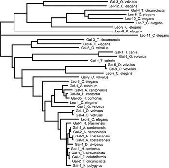
Figure 3 shows a tree of the nematode galectins. The tree was created in Geneious Prime as a neighbour-joining tree from the protein sequences using Jukes-Cantor genetic distances. There are no close homologues to some of the C. elegans galectins such as lec-6, −9 and −11. Nomenclature is not comparable within the parasitic nematodes; e.g. gal-1 from Teladorsagia circumcincta (Greenhalgh et al., Reference Greenhalgh, Loukas and Newton1999) is not orthologous to gal-1 from Toxocara canis (Fig. 3).
Phylogenetic tree of nematode galectins created by using Jukes-Cantor distances and the neighbour-joining method. The tree demonstrates that the nematode galectins fall into multiple groups with varying degrees of similarity to the C. elegans galectins. The accession numbers were A. caninum gal-1 (AOA368GH13), A. cantonensis gal-1 (AOA158PB17) gal-2 (G1EUS2), gal-3 (G1EUS1), A. costaricensis gal-1 (A0A0R3PHH7), gal-2 (A0A3P7H3L8), D. viviparus gal-1 (AOAOD8Y2F3), H. contortus gal-1 (O76646), gal-3a (Q9NJV1), gal-3b (O44126), H. polygyrus gal-1 (AOA3P8DGW1), N. brasiliensis gal-1 (A0A0N4Y6Y9), O. volvulus gal-1 (Q25597), gal-2 (A0A044VHG4), gal-3 (A0A044UJD3), gal-4 (A0A044QNW3), gal-5 (A0A044STF4), gal-6 (A0A044VFH7), gal-7 (A0A044R2B2), gal-8 (O96928), gal-9 (A0A044VGC2) T. canis gal-1 (A0A0B2VDY3), T. circumcincta gal-1 (O01410), gal-2 (O01412), gal-3 (A0A2G9UTC0), gal-4 (A0A2G9V3L5), T. colubriformis gal-1 (Q7KPD1), T. spiralis (A0A0V1B757), C. elegans Lec-1 (P36573), Lec-2 (Q20684), Lec-3 (I2HAG1), Lec-4 (Q18625), Lec-5 (G5EFI4), Lec-6 (Q9N384), Lec-7 (Q09605), Lec-8 (Q09610), Lec-9 (G5EC10), Lec-10 (G5EBV4), Lec-11 (Q94215) Lec-12 (Q5ZR27).

Interactions between host and nematode galectins
After exsheathing in the sheep rumen, incoming H. contortus larvae produce numerous galectin-like proteins (Hewitson et al., Reference Hewitson, Grainger and Maizels2009). Similarly, galectins and members of the venom allergen family are major components of excretory-secretory products of incoming T. circumcincta larvae in sheep (Craig et al., Reference Craig, Wastling and Knox2006). Several mammalian galectins are also upregulated in parasitic infections: gal-1, gal-3, gal-9, gal-11, ovine gal-14 and gal-15. The simultaneous appearance of both host and parasite galectins following gastrointestinal nematode infection provides an opportunity for these molecules to interact. Nematode galectins may have evolved not only to regulate cellular processes but also to mimic host galectin (Tang et al., Reference Tang, Gao, Rosa, Abubucker, Hallsworth-Pepin, Martin, Tyagi, Heizer, Zhang and Bhonagiri-Palsikar2014).
The nematode Strongyloides ratti has seven identified galectins named after the C. elegans galectins (Ditgen et al., Reference Ditgen, Anandarajah, Reinhardt, Younis, Witt, Hansmann, Lorenz, Garcia-Hernandez, Paclik, Soblik, Jolodar, Seeberger, Liebau and Brattig2018). Sr gal-1 is predominantly expressed in free-living adult females while Sr gal-3 is predominantly expressed in parasitic females. Both galectins may bind to host mucosal cells and trigger the release of the Th2 cytokines thymic stromal lymphopoietin and IL-22 (Ditgen et al., Reference Ditgen, Anandarajah, Reinhardt, Younis, Witt, Hansmann, Lorenz, Garcia-Hernandez, Paclik, Soblik, Jolodar, Seeberger, Liebau and Brattig2018). Galectin from Trichinella spiralis has also been implicated in the invasion of host mucosal cells (Xu et al., Reference Xu, Yang, Yang, Jiang, Liu, Zhang, Cui and Wang2018).
Vaccination with a combination of recombinant rHco-gal-m/f proteins (from male and female) can confer partial protection to homologous infection in goats (Yanming et al., Reference Yanming, Ruofeng, Muleke, Guangwei, Lixin and Xiangrui2007). However, vaccination with Hco-gal- 2 does not provide strong protection in sheep (Newlands et al., Reference Newlands, Skuce, Knox, Smith and Smith1999). The eosinophil-specific chemokinetic activity of H. contortus infective L3 larvae is mediated by nematode galectins in a carbohydrate-dependent manner and may mimic the activity of host galectin 9 (Vasta, Reference Vasta2009). In mice, vaccination against Angiostrongylus cantonensis galectin inhibits the immune response to subsequent infection with A. cantonensis (Yan et al., Reference Yan, Shi, Zu, Shen, Chen, Zhao, Li, Yan and Huang2018). Angiostrongylus cantonensis gal-1 causes apoptosis of macrophages by binding to Annexin A2 and activating the apoptotic signalling pathway (Shi et al., Reference Shi, Xiao, Xie, Shi, Zhang, Leavenworth, Yan and Huang2020).
The changes in mucus following infection are at least partly host-mediated and widely assumed to benefit the host (Knight et al., Reference Knight, Griffith, Pemberton, Pate, Guarneri, Anderson, Talbot, Smith, Waddington and Fell2011; Hasnain et al., Reference Hasnain, Dawson, Lourie, Hutson, Tong, Grencis, McGuckin and Thornton2017). However, H. contortus produces a mucin-like molecule and vaccination with parasite-derived extracts containing this molecule significantly enhances protection (Piedrafita et al., Reference Piedrafita, de Veer, Sherrard, Kraska, Elhay and Meeusen2012). This mucin exists on the external surface of H. contortus L3 and interacts with host mucus. This molecule is hypothesized to increase the viscosity of mucus and vaccination against this molecule is expected to reduce mucus viscosity.
Teladorsagia circumcincta galectins strongly bind to or are bound by sheep antibody (McCririe et al., Reference McCririe, Bairden, Britton, Buitkamp, McKeand and Stear1997). Similarly, galectin from Onchocerca volvulus has been shown to bind IgE (Klion and Donelson, Reference Klion and Donelson1994). The binding of heavily glycosylated immunoglobulin IgE by both host and parasite galectin will influence mast cell degranulation. Mast cell degranulation is mediated by cross-linking IgE bound to the high-affinity IgE receptor-FcεRI present on the mast cell surface (Turner and Kinet, Reference Turner and Kinet1999). Upon degranulation, mast cells release vasoactive mediators and constrict blood vessels, contract gut smooth muscle, increase mucus production and upregulate proinflammatory factors such as histamine, tryptase, chymase, prostaglandins and leukotrienes, matrix metalloproteinase 9, platelet-activating factor and cytokines mainly IL-4, IL-10 and TNF-a, that expel the adult nematodes (Pennock and Grencis, Reference Pennock and Grencis2006). Resistant sheep with higher concentrations of degranulated mast cells (globule leucocytes) had lower numbers of T. circumcincta following reinfection (Stear et al., Reference Stear, Bishop, Doligalska, Duncan, Holmes, Irvine, McCririe, McKellar, Sinski and Murray1995). Mast cells from Gal-3 deficient mice release less Interleukin 4 and less histamine during mast cell degranulation (Chen et al., Reference Chen, Sharma, Yu, Zuberi, Weng, Kawakami, Kawakami, Hsu and Liu2006).
Most research has assumed that modulating the induction of immune responses is the key mechanism. However, in many nematode infections, hosts are continually exposed to reinfection. Consequently, most nematodes enter hosts that have already mounted an anti-nematode immune response. Here there is little value in trying to prevent an immune response being mounted but the enormous value in modulating the effector mechanisms such as IgE-mediated mast cell degranulation.
Parasite galectin might also influence the improvement in autoimmune disorders observed during nematode infections. The number of mast cells increases in intestinal tissues and mast cells are important sources of inflammation in Inflammatory Bowel Disease (Hamilton et al., Reference Hamilton, Sinnamon, Lyng, Glickman, Wang, Xing, Krilis, Blumberg, Adachi, Lee and Stevens2011; Stasikowska-Kanicka et al., Reference Stasikowska-Kanicka, Danilewicz, Glowacka and Wagrowska-Danilewicz2012). In addition, mast cells regulate the permeability of the intestinal epithelium due to their release of granule proteases (Scudamore et al., Reference Scudamore, Thornton, McMillan, Newlands and Miller1995; McDermott et al., Reference McDermott, Bartram, Knight, Miller, Garrod and Grencis2003). Nematodes may suppress IgE mediated mast cell responses (Pritchard, Reference Pritchard1993) and protect the host against atopic and autoimmune disorders. In vivo studies are needed to help us better understand the complex interplay between host galectins (especially gal-3 and gal-9), parasite galectins and mast cells.
In conclusion, both host and parasite galectins are present in the gastrointestinal tract following infection and they both influence protective immune responses against nematodes. Their interaction plays an important role in modulating the host immune response, influencing the outcome of gastrointestinal nematode infection. Galectins from some nematodes have been shown to bind IgE. The control of IgE activity and mast cell degranulation is of considerable value to many parasites so it seems plausible that nematode galectin acts as a molecular sponge that soaks up immunoglobulin IgE and inhibits mast cell degranulation. In addition, nematode galectin may interfere with the binding of host galectin to glycans on gastrointestinal mucins and affect mucus viscosity which will influence nematode survival. Parasite galectin might be also immunomodulate autoimmune disorders.
Supplementary material
The supplementary material for this article can be found at https://doi.org/10.1017/S003118202100007X.
Financial support
This work was supported by grants from the National Science Center, POLAND No.2016/23/B/NZ6/03464 and La Trobe University.
Conflicts of interest
The authors declare there are no conflicts of interest.