Implications
The energy demands during pregnancy require an adaptation in the endocrine system regulating energy intake. This is especially important for restrictively fed sows in commercial environments. Leptin is recognized as a principal regulator of energy balance during gestation as well. Improving our knowledge of leptin mechanisms during pregnancy may help in maintaining productive efficiency in the modern pig herd. The nutritional environment in which the fetus grows and develops has the potential to have long-term effects on body composition as well as metabolic health of the offspring.
Introduction
Pregnancy is a period of dramatic body changes with weight gain, increased fat stores, new tissue synthesis and significant hormonal and metabolic changes (Machteld et al., Reference Machteld, Considine and Van Gaal2000). Hyperphagia during pregnancy is a very early event, which takes place in advance of an increased energy requirement (Trujillo et al., Reference Trujillo, Spuch, Carro and Señarís2011). The energy demands during pregnancy require an adaptation in the homeostatic pathways regulating food intake so as to create a positive energy balance (Grattan et al., Reference Grattan, Ladyman and Augustine2007). As a consequence of a pregnancy, relevant neuroendocrine changes occur. Leptin, the product of the ob gene, is an adipocyte signal for satiety (Zhang et al., Reference Zhang, Proenca, Maffei, Barone, Leopold and Friedman1994), and it is recognized as a principal regulator of fat mass (Grattan et al., Reference Grattan, Ladyman and Augustine2007). Leptin-sensitive neurons influence the feeding behavior, co-ordinating the activity of orexigenic and anorexigenic peptides (Trayhurn et al., Reference Trayhurn, Bing and Wood2006). In rats and humans, leptin levels in blood increase during gestation because of placenta formation (Forhead and Fowden, Reference Forhead and Fowden2009). However, hyperleptinemia during pregnancy does not decrease food intake because of the downregulation of leptin receptors in the ventro medial nuclei of the hypothalamus. This produces a state of central resistance to the usual actions of leptin (Bruton and Russell, Reference Brunton and Russell2008). It seems likely that these adaptive mechanisms are driven by the hormonal changes during pregnancy (Sauvé and Woodside, Reference Sauvé and Woodside2000; Grattan et al., Reference Grattan, Ladyman and Augustine2007; Augustine and Grattan, Reference Augustine and Grattan2008).
During pregnancy, leptin functions in animals such as swine, where the hyperphagia is managed through dietary strategies, are yet to be clarified. It has been shown that, during the early gestation, voluntary feed intake in sows fed ad libitum is two to three times as high as the requirements (Nissen et al., Reference Nissen, Danielsen, Jorgensen and Oksbjerg2003). In pig farming, to maximize the health of sows, it is necessary to adjust the energy level to maintain an optimal body condition during the reproductive cycle. A surplus in energy supply during gestation may lead to high adiposity at delivery, with detrimental effects not only on piglet vitality but also on the feeding behavior of sows during lactation (Amdi et al., Reference Amdi, Giblin, Ryan, Stickland and Lawlor2014). For these reasons, breeding pregnant sows are commonly provided with a limited amount of feed, which is equal to 50% to 60% of their ad libitum intake (Guillemet et al., Reference Guillemet, Dourmad and Meunier-Salaün2006).
The study of the ways in which the endocrine system manages the nutrients in pregnancy is fundamental to optimize the great potential of the in utero environment and its impact on future physiological and biochemical systems, including long-term function and health of the offspring. We assessed concentrations of plasma leptin and their associations with changes in concentrations of plasma lactogenic hormones during pregnancy in restrictively fed sows.
Material and methods
Animals and experimental procedures
At weaning, 15 Large White×Landrace sows having a mean (±s.d.) body condition score of 2.96±0.5 were used. Mean (±s.d.) parity order was 3.2±0.9. The sows were mated within 7.3±1.1 days from weaning. Cervical artificial insemination (AI) was performed with disposable spiral-tip catheters (Agrizoo San Marino s.r.l, San Marino, Italy). Sows were inseminated 12 and 24 h after estrus detection, using 3×10⁹ spermatozoa/80 ml (Modena Vitaè; Semenitaly s.r.l., Saliceto San Giuliano, MO, Italy). Twenty-five days after insemination, the sows were subjected to ultrasound examinations (Aloka SSD 500®, Wallingford, CT, USA; linear probe 5.0 MHz) and all of them were found to be pregnant. During pregnancy, all sows received a mixed diet (12.8 MJ/kg DE, 13.8% CP, 0.68% lys, 6.6% crude fiber, as-fed basis) at the amount of 2.0 kg/animal per day during the 1st week, 2.5 kg/animal per day up to 100th day of pregnancy and 2.7 kg/animal per day up to farrowing. During lactation, they received a mixed feed (14.0 MJ/kg DE, 16.7% CP, 1.10% lys, 4.6% crude fiber, as-fed basis) offered ad libitum. Blood samples were collected using jugular venopuncture in 10-ml vacutainer tubes with lithium heparin the day after the weaning of previous litters, at AI, every 15 days up to day 45 of pregnancy and every 7 days from day 46 to farrowing. Blood samples were also collected at day 10 and 20 of lactation. Samples were immediately centrifuged (1800×g for 10 min) and plasma was collected and stored at −20°C until analysis. At farrowing, individual piglet placentas (n=160) were separated, carefully washed and opened over the whole length at the anti-mesometrial side. A 5 cm×5 cm quadrant, equidistant between the edge and the center, was removed and stored at −80°C until analysis.
Hormone assays
Leptin levels in plasma were determined by a commercial kit (Multispecies Leptin RIA; Linco Research, St. Louis, MO, USA; Summer et al., Reference Summer, Saleri, Malacarne, Bussolati, Beretti, Sabbioni and Superchi2009). The sensitivity of the method was 100 pg/ml and the variability coefficients within and among samples were 4.2% and 7.1%, respectively. Plasma prolactin (PRL) levels were determined by a previously validated ELISA test (Borghetti et al., Reference Borghetti, De Angelis, Saleri, Cavalli, Cacchioli, Corradi, Mocchegiani and Martelli2006). Effective dose (ED)90, ED50 and ED10 were 0.011, 0.123 and 3.00 ng/ml, respectively. The intra- and inter-assay coefficients were 4.5% and 7.0%, respectively. Progesterone levels were determined by a validated radioimmunoassay (RIA) (Ponderato et al., Reference Ponderato, Grasselli, Saleri and Tamanini2000).The intra- and inter-assay coefficients were 5.8% and 10.3%, respectively. Plasma samples were assayed for cortisol by a validated RIA (Borghetti et al., Reference Borghetti, De Angelis, Saleri, Cavalli, Cacchioli, Corradi, Mocchegiani and Martelli2006). The intra- and inter-assay coefficients were 5.3% and 6.7%, respectively.
Placental expression of leptin and its receptors
All molecular biology reagents were purchased from Ambion Inc. (Foster City, CA, USA), unless otherwise specified. Total RNA was extracted by TRI Reagent® solution (Applied Biosystems, Monza, Italy), according to the manufacturer’s instructions. The RNA quantification was carried out by a spectrophotometer (GENEQUANT pro®, Amersham Pharmacia, UK). Reverse-transcription (RT) PCR was carried out using a Ready-to-go™ You-Prime First-Strand Beads kit (Amersham Pharmacia Biotech UK Limited, Little Chalfont, Buckinghamshire, UK), as described by the manufacturer. Two micrograms of total RNA was used in the RT reaction. Aliquots (5 µl) from the generated cDNA were used for subsequent PCR amplification in the reaction buffer containing 1.5 µl MgCl2 (50 mM), 1 µl dNTPs (12.5 mM) and 1 µl Taq DNA polymerase (1 µg/µl) to a final volume of 50 µl. Primers for leptin and its receptors (short and long form) were used at the concentration of 2.5 µM (MWG BIOTECH, Ebersberg, Germany). Amplification was carried out for 27 cycles, when the reaction was in the middle of the linear range (before reaching the amplification plateau). Each cycle consisted of denaturation at 94°C for 1 min, annealing at specific temperature for each primer set for 1 min and extension at 72°C for 1 min. At the end of the 27th cycle, an additional extension was carried out for 5 min. Specific primer sets used, their annealing temperature and amplified product size (expressed in base pairs, bp) are summarized in Table 1. An 18S RNA (no. 1717, Quantum RNA; Ambion Inc.) was used as an internal positive control for a relative quantitative PCR. To amplify the 18S fragment (324 bp) without reaching the plateau phase, 18S PCR alternate primer pairs (5 µM) were co-incubated with 18S PCR competimers (5 µM; ratio, 2 : 8), according to the instructions of the manufacturers. PCR products were visualized after electrophoresis on 2% agarose gel with SYBR® Safe DNA gel stain (Invitrogen s.r.l., San Giuliano, Milanese, Italy). The gel was analyzed on a computerized densitometry program (Scion Capture Driver 1.2 for Image-Pro Plus; Scion Corporation, Frederick, MD, USA). The values are presented as the ratio, expressed in relative arbitrary units (RAU), of the band intensities of the PCR products over those of the corresponding ribosomal 18S PCR product.
Sequences of PCR primers, annealing temperature (°C) and PCR product size (bp)

Statistical analysis
ANOVA was performed by applying the GLM procedure (SAS, 2003) with sampling as the fixed factor and sow as the random factor. The Pearson correlation analysis was performed among leptin and lactogenic hormones during pregnancy. For this aim, the pregnancy period was divided into the following three sub-periods: early pregnancy, from day 15 to 45; mid pregnancy, from day 52 to 79; and late pregnancy, from day 86 to farrowing. The significance level was set at P<0.05.
Results
Leptin
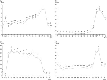
The mean plasma leptin concentrations over the period of study are shown in Figure 1a. At 7 days before AI, mean (±s.d.) plasma values were 1.54±0.36 ng/ml, whereas at insemination (day 0) leptin concentration significantly increased (3.73±0.98 ng/ml; P<0.05) to return approximately to starter levels up to 30 days of pregnancy (P>0.05). From day 30 to day 65, the plasma levels of leptin ranged from 2.5 to 3 ng/ml. At the end of mid gestation (day 72), leptin concentration significantly increased (P<0.05): the highest concentrations of leptin were reached at day 107 (5.91±0.69 ng/ml) of pregnancy and at farrowing (5.84±1.72 ng/ml). At 10 and 20 days after farrowing, plasma leptin levels drastically decreased (P<0.05) to 2.20±0.34 and 1.72±0.57 ng/ml, respectively.
Plasma leptin (a), prolactin (PRL) (b), progesterone (c) and cortisol (d) concentrations in sows (n=15). Blood sampling period started at day −7 before AI (0) and ended at day 20 after farrowing (f). Error bars indicate standard deviation. Significant differences (P<0.05) among samplings are labeled with different letters.

PRL
As shown in Figure 1b, plasma PRL levels were 4.16±0.87 ng/ml at day −7 and 2.40±1.09 at AI (P>0.05). Until late gestation (day 93), its concentration did not change (P>0.05). The PRL concentration increased 2 weeks before farrowing to reach the peak at farrowing (67.83±4.91 ng/ml; P<0.05). A positive correlation (P<0.01) was observed between changes in plasma PRL and leptin levels during mid and late pregnancy (Table 2). After farrowing, PRL concentrations were 55.39±3.12 ng/ml (day 10) and 39.53±1.62 ng/ml (day 20) (P<0.05).
Correlations between leptin and lactogenic hormone levels in relation to pregnancy periods

*P<0.05; **P<0.001.
Progesterone
Plasma progesterone level (Figure 1c) at day −7 was 6.07±0.34 ng/ml, and then it decreased at AI (0.28±0.05 ng/ml; P<0.05). From the beginning of early gestation (day 15), the levels increased 10-fold to reach the plateau (mean 32 ng/ml). The plateau was maintained up to the start of late gestation (day 86). In the following weeks, progesterone levels dropped, and at delivery they were 7.38±0.54 ng/ml (P<0.05). As shown in Table 2, in early pregnancy, a positive and moderate correlation (P<0.05) was observed between progesterone and leptin. At 10 and 20 days after farrowing, plasma progesterone levels were 0.98±0.03 and 0.87±0.05 ng/ml, respectively (P<0.05).
Cortisol
Plasma cortisol concentration (Figure 1d) dropped from 17.62±2.7 ng/ml at day −7 to 11.94±1.88 ng/ml at AI (P<0.05). During early and mid pregnancy, mean cortisol levels remained around 10 ng/ml (P>0.05). A significant increase (P<0.05) was observed close to farrowing (107 days: 32.92±2.54 ng/ml) and at farrowing (37.67±4.10 ng/ml). A significant correlation (P<0.01) between changes in plasma cortisol and leptin levels was observed in late pregnancy (Table 2). At 10 and 20 days after farrowing, cortisol plasma decreased (P<0.05), reaching values of 15.78±1.59 and of 14.26±0.94 ng/ml, respectively.
Placental expression of leptin and its receptors
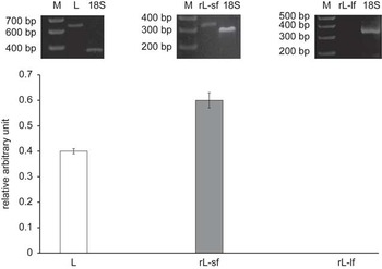
The expression analysis showed the presence of leptin and its short-form receptor in all the samples of placenta (Figure 2). The long-form receptor was not expressed.
Sow placenta (n=160) expression of leptin and short and long forms of leptin receptors. M=marker; L=leptin; rL sf=leptin receptor short-form; rL lf=leptin receptor long-form; 18S=internal positive control. Error bars indicate standard deviation.

Discussion
In pregnancy, placenta is the major source of leptin, which is an important factor linking metabolic status to reproduction (Schneider, Reference Schneider2004). Leptin is recognized as an important placental growth factor and mitogen, regulating peripherally the balance of energy reserves by the facilitation of endocrine responses typical of pregnancy (Hauguel-de Mouzon et al., Reference Hauguel-de Mouzon, Lepercq and Catalano2006; Henson and Castracane, Reference Henson and Castracane2006). In humans and mice, maternal leptin levels reach the peak between mid and late pregnancy, but hyperleptinemia in this period is not associated with a decrease of feed intake (Trujillo et al., Reference Trujillo, Spuch, Carro and Señarís2011). The key metabolic feature observed during pregnancy is the development of leptin resistance (Nagaishi et al., Reference Nagaishi, Cardinali, Zampieri, Furigo, Metzger and Donato2014). Our results indicate that in breeding sows, where hyperphagia of pregnancy is not satisfied, leptin increases from the end of the 2nd stage of pregnancy to reach the highest levels at the 3rd stage and at delivery. The high levels of the hormone are compatible with a mechanism of resistance to central leptin action. The limited access to food in our animals does not seem to affect the presence of leptin resistance. These results underline the functional importance of the central leptin resistance to allow increased nutrient availability for the fetus, as hypothesized by Tessier et al. (Reference Tessier, Ferraro and Gruslin2013) and Ladyman (Reference Ladyman2008). Leptin receptors are expressed in a variety of isoforms, the major form involved in BW regulation being the long form (Vaisse et al., Reference Vaisse, Halaas, Horvath, Darnell, Stoffel and Friedman1996). In rats, hyperleptinemia, during healthy pregnancy, leads to central leptin resistance by downregulation of the full-length leptin receptor in the hypothalamus and increased circulation of soluble receptor isoforms (Brunton and Russell, Reference Brunton and Russell2008). The widespread expression of short leptin receptor isoforms in tissues throughout the mammalian body points out that leptin also acts at periphery as a factor able to modify tissue metabolism (Harris, Reference Harris2000). We observed the expression of the short leptin receptor form in the placental tissue at delivery, confirming a direct involvement of leptin on fetal metabolism. The presence of leptin receptors in placenta suggests autocrine or paracrine effects of leptin on processes essential to ensure an adequate nutrient supply for fetal growth and development, that is, angiogenesis (Grattan et al., Reference Grattan, Ladyman and Augustine2007; Anagnostoulis et al., Reference Anagnostoulis, Karayiannakis, Lambropoulou, Efthimiadou, Polychronidis and Simopoulos2008; Ladyman, Reference Ladyman2008; Tessier et al., Reference Tessier, Ferraro and Gruslin2013). We did not find the presence of the long form of leptin receptor, but this result was not unexpected for two reasons. First, the long-form receptor is expressed in high levels in the hypothalamus and is well characterized in central lepin action, that is, regulating appetite and metabolic rate. Second, our data concerned only the end of pregnancy; leptin receptors have been found to be regulated and to change under certain circumstances such as pregnancy. In several species, the expression of multiple forms of the leptin receptor switch in placenta during the course of pregnancy (Edwards et al., Reference Edwards, Bohm, Purcell, Ratterree, Swan, Castracane and Henson2004; Smolinska et al., Reference Smolinska, Kaminski, Siawrys and Przala2009).
Pregnancy-induced changes in BW regulation are normal physiological parts of maternal adaptation, and they are likely to be induced by the hormonal changes that accompany pregnancy. Lactogenic hormones such as progesterone, PRL and cortisol are involved (Grattan et al., Reference Grattan, Ladyman and Augustine2007; Augustine and Grattan, Reference Augustine and Grattan2008; Ladyman et al., Reference Ladyman, Augustine and Grattan2010). Progesterone, a powerful orexigenic hormone, induces weight gain and greater fat depots to promote a positive energy balance. In pregnant females, progesterone hampered lipostatic control, inhibiting the concurrent enhancement in plasma and cerebrospinal fluid leptin levels expected from the increased fat mass (Grueso et al., Reference Grueso, Rocha and Puerta2001). During early pregnancy, when increase in fat stores is limited in restrictively fed pregnant sows, a positive correlation between leptin and progesterone was observed.
A similar distribution of leptin and PRL receptors in the rodent hypothalamus and brainstem was observed, and this has allowed us to hypothesize a cross talk between leptin and PRL signaling (Nagaishi et al., Reference Nagaishi, Cardinali, Zampieri, Furigo, Metzger and Donato2014). Recent findings suggest a potential role for PRL signaling in the induction of leptin resistance in pseudopregnant rats (Augustine and Grattan, Reference Augustine and Grattan2008). Our data have shown a positive correlation between PRL and leptin in mid and late pregnancy. The exact neuroendocrine changes are currently unknown; however, they may involve a loss in functional leptin receptors in key hypothalamic nuclei or a loss in leptin signaling via intracellular signaling cascades. The leptin and PRL crosstalk is more evident during lactation. In sows, hypophagia commonly occurs during the 1st week of lactation, (Cools et al., Reference Cools, Maes, Decaluwé, Buyse, van Kempen and Janssens2013) and in this period leptin levels were low (Summer et al., Reference Summer, Saleri, Malacarne, Bussolati, Beretti, Sabbioni and Superchi2009). Low leptin and high PRL concentrations could trigger at brain level the mechanism increasing food intake. In this regard, Roy et al. (Reference Roy, Benomar, Bailleux, Vacher, Aubourg, Gertler, Djiane and Taouis2007) reported a direct effect of PRL on orexigenic neuropeptide Y expression to satisfy the high energy demand for milk production. Glucocorticoids, whose main representative is cortisol, are potent regulators of leptin expression (Houseknecht and Portocarrero, Reference Houseknecht and Portocarrero1998). In mice, hyperleptinemia during pregnancy results from corticosterone-dependent upregulation of leptin secretion. In late pregnancy, we observed a positive correlation between leptin and cortisol. During pregnancy, glucocorticoids belong to factors that reduce insulin sensitivity in the dam, activate adipogenesis by differentiation of pre-adipocytes (Breton, Reference Breton2013) and promote, in sheep, the synthesis and secretion of leptin in the fetus (Forhead and Fowden, Reference Forhead and Fowden2009). It has been suggested that the glucocorticoid surge that is characteristic of late gestation may also downregulate placental leptin production (Sugden et al., Reference Sugden, Langdown, Munns and Holness2001). Changes in either fetal or maternal leptin levels may be important for parturition or late fetal development.
In summary, these findings suggest that lactogenic hormones are involved in altering leptin signal during different stages of pregnancy in sows. In the restrictively fed pregnant sow model, leptin resistance contributes mainly to allow increased nutrient availability for the fetus. The expression of the short leptin receptor in the placenta strengthens this hypothesis.
Acknowledgments
All the authors equally contributed to the paper.