INTRODUCTION
Mites are classified in the subclass Acari (class Arachnida) of the phylum Arthropoda. Although approximately 48 200 species have been described (Halliday et al. Reference Halliday, O Connor, Baker, Raven and Williams2000), a further half-million species are believed to exist worldwide (Kettle, Reference Kettle1984). More so than any other arthropod group, mites are found in highly diverse habitats: terrestrial, marine, freshwater and even in the upper atmosphere due to dispersal through aerial currents (Krantz and Walter, Reference Krantz and Walter2009). Whereas most mite species live freely in the environment, some species have evolved to be parasitic on other animals or on plants and are therefore of great agricultural and veterinary importance, although their medical impact is generally more modest. Some species are significant destructive pests of stored food products; while others (such as house dust mites) produce faecal allergens, inducing asthma. Mites can also produce serious skin conditions by feeding on the skin of domestic animals (mange) and can cause dermatitis in humans. Finally, some species act as important vectors of pathogenic microorganisms of medical and veterinary importance (Arlian et al. Reference Arlian, Vyszenski-Moher and Morgan2003; Brouqui and Raoult, Reference Brouqui and Raoult2006; Valiente-Moro et al. Reference Valiente-Moro, De Luna, Tod, Guy, Sparagano and Zenner2009a ).
The relationship between bacteria and arthropods can be divided into 2 main aspects, which are not mutually exclusive: (1) bacteria recognized as pathogens transmitted by an arthropod vector, and (2) bacteria residing as symbionts within their arthropod host. The study of the first aspect usually concerns surveillance for emerging or re-emerging diseases and interactions between the arthropod vector, environment, wildlife, domestic animals and humans. In contrast, the second research area concerns other bacteria that may influence the physical, ecological and evolutionary traits of their arthropod host, usually without transmission of these organisms to a second host in which disease may occur. These studies are often designed to characterize and define symbiont–arthropod interactions. For example, the nutritional mutualist, Buchnera aphidicola, synthesizes essential amino acids for its aphid host (Acyrthosiphon pisum) that feeds on plant phloem, which has a very low essential amino-acid content (Gunduz and Douglas, Reference Gunduz and Douglas2009). Pea aphids also harbour defensive mutualists such as Regiella insecticola, which protects the host population from a natural enemy (the pathogenic fungus, Pandora neoaphidis) by reducing the sporulation rate in aphid cadavers, thus reducing the probability of pathogen transmission to other aphids (Scarborough et al. Reference Scarborough, Ferrari and Godfray2005). Other bacterial genera are capable of manipulating their hosts’ reproduction: Wolbachia, Cardinium, Spiroplasma and Rickettsia induce detrimental phenotypes in their arthropod hosts such as cytoplasmic incompatibility, parthenogenesis induction, feminization and male killing (Stouthamer et al. Reference Stouthamer, Breeuwer and Hurst1999; Tinsley and Majerus Reference Tinsley and Majerus2006; Enigl and Schausberger, Reference Enigl and Schausberger2007; Giorgini et al. Reference Giorgini, Monti, Caprio, Stouthamer and Hunter2009). These findings may be utilized to enhance prospects for biological control since there is the potential to manipulate arthropod populations of agricultural, medical or veterinary importance.
Recently, the number of publications on arthropod-associated bacteria has substantially increased, particularly for the Diptera, Hemiptera and Hymenoptera (Baumann, Reference Baumann2005; Crotti et al. Reference Crotti, Rizzi, Chouaia, Ricci, Favia, Alma, Sacchi, Bourtzis, Mandrioli, Cherif, Bandi and Daffonchio2010; Martinson et al. Reference Martinson, Danforth, Minckley, Rueppell, Tingek and Moran2011; Taylor et al. Reference Taylor, Coghlin, Floate and Perlman2011; Martin et al. Reference Martin, Gubler, Wimmer, Germann and Bernasconi2012; Zucchi et al. Reference Zucchi, Prado and Consoli2012; Skaljac et al. Reference Skaljac, Zanic, Hrncic, Radonjic, Perovic and Ghanim2013). In parallel, studies on mite-bacterial relationships have also increased, but to a lesser extent compared with the insect orders above. Moreover, sources of information with respect to mites and their pathogens and symbionts are widely distributed in the literature, and the compilation of data in terms of review publications is still very limited. Accordingly, the aims of this literature review are (1) to obtain an overview of bacterial diversity in mites and its potential applications, and (2) to provide comparative data for mite-associated bacteria of agricultural, veterinary and medical importance to stimulate hypothesis-driven research.
MATERIALS AND METHODS
Literature search
This review focuses on reports of bacteria found in mites across the world. The publications were extracted by searching from 2 major scientific literature databases, PubMed (www.ncbi.nlm.nih.gov/pubmed) and Web of Knowledge (www.webofknowledge.com). Three main mite orders (Mesostigmata, Sarcoptiformes, Trombidiformes), some common names of mites (e.g. gamasid mite, dust mite, itch mite, spider mite, harvest mite, free-living mite, chigger etc.) or scientific names (e.g. Dermanyssus, Dermatophagoides, Leptotrombidium or Tyrophagus), were used in combination with the term ‘bacteria’ or ‘bacterium’ as the keywords and applied to the title field or abstracts in those databases. From the obtained literature, the number of bacterial taxa (genus and species level) was recorded for each mite species. Only the publications reporting a minimum of genus-level identifications of bacteria were included in the database for statistical analyses. Bacterial scientific names obtained from the literature were checked for taxonomic assignment following the NCBI Taxonomy Browser (http://www.ncbi.nlm.nih.gov/Taxonomy/Browser/wwwtax.cgi), while for the latest mite taxonomic classifications, Krantz and Walter (Reference Krantz and Walter2009) was consulted.
Mite classification
Taxonomically, the mites were classified into 3 orders and 14 superfamilies. In addition, for the purposes of the present investigation, mites were also grouped into 4 types based on life history (Krantz and Walter, Reference Krantz and Walter2009): (1) ‘Vertebrate parasite’ was defined as a mite species which at some lifecycle stage feeds on vertebrate animals or are confirmed as disease vectors; (2) ‘Invertebrate predator’ was defined as a mite species which at some lifecycle stage hunts or feeds upon other invertebrates (some of which are used as biological pest control in agricultural practice); (3) ‘Plant parasite’ was defined as a mite species that feeds on live plant tissues (with some species responsible for economic losses in agricultural products); and (4) ‘House pest and allergen’ are those mites which spoil stored foodstuffs or contain powerful allergens that induce detrimental immune responses in humans and/or animals (Table S1).
Bacterial classification
For bacteria, apart from taxonomic classification, 4 bacterial groups were categorized due to their biological characteristics. Following the scheme of Valiente-Moro et al. (Reference Valiente-Moro, Thioulouse, Chauve, Normand and Zenner2009b ), the different categories were defined as: (1) ‘Saprophyte’ – examples are bacteria which have not been described as being pathogenic; (2) ‘Opportunistic pathogen’ – species in this category cause disease in compromised vertebrate hosts but not in healthy hosts; (3) ‘Pathogen’ – most species in the genus are pathogens of vertebrates; and (4) ‘Symbiont’ – bacteria that strictly live in association with an arthropod host.
Statistical analysis
In order to visualize the distribution of the bacteria found in each mite superfamily, a principal component analysis (PCA) was performed using R freeware (R Development Core Team, 2008) with the ade4 package (Dray and Dufour, Reference Dray and Dufour2007). The PCA was calculated by counting the number of bacterial genera positively reported in each mite taxa. Before starting the analysis, data from 4 mite superfamilies (Oppioidea, Rhodacaroidea, Erythraeoidea and Eviphidoidea) were removed due to only one record of bacteria each that could cause analysis bias (outliers).
To investigate the difference of the 4 biological types of mite on bacterial diversity, the species number of bacteria (species richness) in each order was recorded across the 4 mite categories. This was analysed using the non-parametric Kruskal–Wallis test, and multiple pairwise comparison tests were performed with SPSS version 21.0 software (IBM Corporation, Armonk, NY, USA), applying 95% confidence intervals. P-values were adjusted for multiple comparisons.
RESULTS & DISCUSSION
Methodological approaches to the identification of bacteria in mites
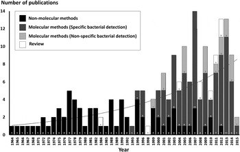
A total of 193 publications were included in this systematic review from 1964 until January 2015. The number of publications reporting mite-associated bacteria was found to increase gradually over this period (Fig. 1). However, we estimated that the total volume of literature describing mite-bacteria associations is 5 times smaller than that for ticks and 20 times smaller than that for insects (data not shown). The eligible publications covered 143 mite species belonging to 3 orders and 14 superfamilies (Table S1). The most studied mite taxon was Dermanyssoidea (46 species), followed by Trombiculoidea (34 species), Tetranychoidea (23 species) and Phytoseioidea (10 species).
The number of publications reporting mite-associated bacteria. Numbers within bars refer to a breakdown of publications by methodology. Curve represents an exponential line of best fit.

Clearly, prior to the early 1990s (Fig. 1), analyses of bacteria in mites were restricted to non-molecular methods such as conventional bacterial cultures with biochemical characterization, inoculations of laboratory animals and serological tests. For example, Mycoplasma spp. were isolated from goats’ ear mites, Psoroptes cuniculi and Raillietia caprae, by culturing the crushed mites in PPLO agar supplemented with pig serum, and then the bacteria were identified by biochemical characteristics (Cottew and Yeats, Reference Cottew and Yeats1982). Similarly, the red poultry mite, Dermanyssus gallinae, was studied for its potential vectorial role for Salmonella gallinarum and Erysipelothrix rhusiopathiae transmission in the poultry industry by culturing mite extracts in selective enrichment media, selenite broth (Zeman et al. Reference Zeman, Stika, Skalka, Bartik, Dusbabek and Lavickova1982) and crystal-violet sodium-azide broth (Chirico et al. Reference Chirico, Eriksson, Fossum and Jansson2003), respectively. In the scrub typhus research field, a number of studies have used mouse passages to amplify Orientia tsutsugamushi from wild chigger mites fed on the rodents, and different strains of the bacterium were indirectly detected by various serological methods (e.g. fluorescence antibody assays, immunoperoxidase staining and complement fixation tests; Kitaoka et al. Reference Kitaoka, Asanuma and Otsuji1974; Roberts et al. Reference Roberts, Muul and Robinson1977; Dohany et al. Reference Dohany, Shirai, Robinson, Ram and Huxsoll1978; Shirai et al. Reference Shirai, Huxsoll, Dohany, Montrey, Werner and Gan1982; Ree et al. Reference Ree, Lee and Cho1992; Frances et al. Reference Frances, Watcharapichat and Phulsuksombati2001; Lerdthusnee et al. Reference Lerdthusnee, Khlaimanee, Monkanna, Sangjun, Mungviriya, Linthicum, Frances, Kollars and Coleman2002; Phasomkusolsil et al. Reference Phasomkusolsil, Tanskul, Ratanatham, Watcharapichat, Phulsuksombati, Frances, Lerdthusnee and Linthicum2009). Of course, such specific methods allow the identification of the target organism only, and unculturable bacteria would not be detected.
With the advent of the molecular era, the development of specific PCR assays and conventional and next-generation sequencing techniques revealed a significantly higher microbial diversity than was previously estimated by culture-dependent approaches (Hugenholtz et al. Reference Hugenholtz, Goebel and Pace1998; Hubert et al. Reference Hubert, Nesvorna, Kopecky, Sagova-Mareckova and Poltronieri2014; Yun et al. Reference Yun, Roh, Whon, Jung, Kim, Park, Yoon, Nam, Kim, Choi, Kim, Shin, Kim, Lee and Baea2014). In particular, use of 16S rDNA PCR with bacterial species-specific primers has been widely used for bacterial taxonomic studies in mites (Fig. 1). Additionally, several publications used specific PCR to amplify other bacterial genes of interest. For example, the protein-coding genes: ftsZ, groEL, wsp and citrate synthase (gltA) were used in Wolbachia studies (Hong et al. Reference Hong, Gotoh and Nagata2002; Gotoh et al. Reference Gotoh, Noda, Fujita, Iwadate, Higo, Saito and Ohtsuka2005; Yu et al. Reference Yu, Zhang, Xue and Hong2011; Lu et al. Reference Lu, Zhang and Hong2012; Ros et al. Reference Ros, Fleming, Feil and Breeuwer2012; Suh et al. Reference Suh, Sim, Park and Cho2014; Glowska et al. Reference Glowska, Dragun-Damian, Dabert and Gerth2015; Zhang et al. Reference Zhang, Ding, Rong and Hong2015); outer membrane protein B gene, 17 kD antigenic gene and gltA were used for Rickettsia spp. (Reeves et al. Reference Reeves, Dowling and Dasch2006, Reference Reeves, Loftis, Szumlas, Abbassy, Helmy, Hanafi and Dasch2007; Choi et al. Reference Choi, Lee, Park, Lee, Han, Kim, Lee, Song, Choi, Kim, Park and Jang2007; Tsui et al. Reference Tsui, Tsai, Weng, Hung, Liu, Hu, Lien, Lin, Shaio, Wang and Ji2007); the 16S–23S intergenic spacer (ITS) and gltA were used for Bartonella spp. (Kabeya et al. Reference Kabeya, Colborn, Bai, Lerdthusnee, Richardson, Maruyama and Kosoy2010; Kamani et al. Reference Kamani, Morick, Mumcuoglu and Harrus2013); gyrB was used for Cardinium (Ros et al. Reference Ros, Fleming, Feil and Breeuwer2012; Zhu et al. Reference Zhu, Zhang, Zhang, Ge, Gotoh and Hong2012); the 5S–23S ITS was used for Borrelia spp. (Literak et al. Reference Literak, Stekolnikov, Sychra, Dubska and Taragelova2008); the epank1 gene was used for Anaplasma phagocytophilum (Literak et al. Reference Literak, Stekolnikov, Sychra, Dubska and Taragelova2008); and the 56-kD type-specific antigen gene was used extensively for O. tsutsugamushi (Tamura et al. Reference Tamura, Makisaka, Kadosaka, Enatsu, Okubo, Koyama, Yu and Urakami2000; Pham et al. Reference Pham, Otsuka, Suzuki and Takaoka2001; Khuntirat et al. Reference Khuntirat, Lerdthusnee, Leepitakrat, Kengluecha, Wongkalasin, Monkanna, Mungviriya, Jones and Coleman2003; Lee et al. Reference Lee, Shim, Song, Choi, Hwang, Park, Park and Shin2011; Liu et al. Reference Liu, Jia, Xing, Suo, Du, Jia, Gao, Xie, Liu and Ren2013; Seto et al. Reference Seto, Suzuki, Otani, Qiu, Nakao, Sugimoto and Abiko2013; Shin et al. Reference Shin, Roh, Park, Song, Chang, Lee, Lee, Park, Park and Shin2014; Takhampunya et al. Reference Takhampunya, Tippayachai, Promsathaporn, Leepitakrat, Monkanna, Schuster, Melendrez, Paris, Richards and Richardson2014).
An alternative approach has been the use of conserved primers to amplify 16S rRNA products in an unbiased fashion (Fig. 1), followed by cloning and sequencing of selected clones for taxonomic assignment (Hogg and Lehane, Reference Hogg and Lehane1999, Reference Hogg and Lehane2001; Hoy and Jeyaprakash, Reference Hoy and Jeyaprakash2005; Hubert et al. Reference Hubert, Kopecky, Perotti, Nesvorna, Braig, Sagova-Mareckova, Macovei and Zurek2012, Reference Hubert, Nesvorna, Kopecky, Sagova-Mareckova and Poltronieri2014; Tang et al. Reference Tang, Chang, Srinivasan, Mathaba, Harnett and Stewart2013; Murillo et al. Reference Murillo, Aubert and Raoult2014). However, to the best of our knowledge, only one publication has used the Roche 454 pyrosequencing platform targeting 16S rRNA amplicons to reveal the bacterial community of a mite species (in this case, the bulb mite, Rhizoglyphus robini; Zindel et al. Reference Zindel, Ofek, Minz, Palevsky, Zchori-Fein and Aebi2013). In a more recent study, bacterial genomic sequences from 100 species (predominantly enterobacteria) were identified during assembly of the Dermatophagoides farinae (dust mite) genome (Chan et al. Reference Chan, Ji, Yim, Liu, Zhou, Li, Yang, Li, Li, Law, Wu, Cai, Qin, Bao, Leung, Ng, Zou, Zhong, Ran, Zhong, Liu and Tsui2015).
Bacterial diversity in mites
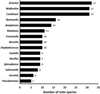
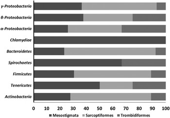
Mite species were found to be associated with 85 bacterial genera (approximately 150 identified species) belong to 7 phyla (plus 3 classes of Proteobacteria) and 25 orders (Table S2 and S3). Cardinium (in 31 mite species), Wolbachia (31 hosts) and Orientia (32 hosts) were the most prevalent bacteria; followed by Bartonella, Anaplasma and Rickettsia, with 16, 14 and 11 mite species reported, respectively (Fig. 2). Among the 7 bacterial phyla and the 3 classes of Proteobacteria (α, β and γ), Bacteriodetes, Firmicutes, Tenericutes and Actinobacteria were reported in all 3 mite orders (Fig. 3). However, Chlamydiae were reported only in mites from the order Mesostigmata, and Spirochaetes were found in the Mesostigmata and Trombidiformes, but not in the Sarcoptiformes (Fig. 3).
The top-ranked 15 bacterial genera detected in mites.

Proportions (%) of the most abundant bacterial groups in the 3 principal mite orders.

Symbionts can be obligatory or facultative, live inside or outside host cells, and can affect their host negatively, positively, or have no discernible phenotype. Some symbiotic bacteria may provide benefits to the host in particular environments, but can be disadvantageous under different circumstances (Hoy and Jeyaprakash, Reference Hoy and Jeyaprakash2008). A number of bacteria were reported as potential mite symbionts in this literature survey, including Wolbachia, Cardinium, Acaricomes, Spiroplasma, Snodgrassella, Serratia, Rickettsiella and Schineria. Wolbachia and Cardinium have been relatively well studied in terms of effects on their mite hosts, which manifest as reproductive alterations. However, the phenotypes (if any) induced by the other potential symbionts remain unknown. Wolbachia and Cardinium manipulate mite reproduction by inducing cytoplasmic incompatibility, parthenogenesis, sex-ratio distortion (e.g. male-killing and feminization), and an increase in female fecundity (Breeuwer and Jacobs, Reference Breeuwer and Jacobs1996; Weeks and Breeuwer, Reference Weeks and Breeuwer2001; Chigira and Miura, Reference Chigira and Miura2005; Gotoh et al. Reference Gotoh, Noda, Fujita, Iwadate, Higo, Saito and Ohtsuka2005; Groot and Breeuwer, Reference Groot and Breeuwer2006; Gotoh et al. Reference Gotoh, Noda and Ito2007; Novelli et al. Reference Novelli, Freitas-Astua, Segatti, Mineiro, Arthur, Bastianel, Hilf, Gottwald and Machado2008; Zhu et al. Reference Zhu, Zhang, Zhang, Ge, Gotoh and Hong2012; Zhao et al. Reference Zhao, Chen, Ge, Gotoh and Hong2013a ; Suh et al. Reference Suh, Sim, Park and Cho2014; Zhang et al. Reference Zhang, Ding, Rong and Hong2015). These reproductive manipulation strategies facilitate vertical transmission through the female line and drive the spread of the symbionts into mite populations (Zhao et al. Reference Zhao, Zhang and Hong2013b ).
Wolbachia is the most prevalent arthropod symbiont (infecting approximately 40% of terrestrial species; Zug & Hammerstein, Reference Zug and Hammerstein2012) and is also found in some species of filarial nematodes (Ferri et al. Reference Ferri, Barbuto, Martin, Lo, Uni, Landmann, Baccei, Guerrero, De Souza, Bandi, Wanji, Diagne and Casiraghi2011). In mites, although 31 species were positively reported for Wolbachia infection, the bacteria occurred only in 5 of 14 studied superfamilies: the Dermanyssoidea (various parasitic mites of vertebrates), Phytoseioidea (fungivorous, pollenophagous and predatory mites), Oppioidea (in an oribatid free-living mite, Oppiella nova), Cheyletoidea (parasitic mites of birds, but not in Demodex spp.), and Tetranychoidea (phytophagous mites). Interestingly, Cardinium was also found in 31 mite species but these were distributed across 8 superfamilies, representing a much broader host range than Wolbachia (Table S2). According to these findings, Cardinium appears to be a more important symbiont for mites than it is for other arthropods (Zug & Hammerstein, Reference Zug and Hammerstein2012).
Apart from these reproductive symbionts, another symbiotic bacterium, Acaricomes phytoseiuli, has been isolated from the predatory species, Phytoseilus persimilis, which is widely used for biological control of spider mites (major agricultural pests) (Pukall et al. Reference Pukall, Schumann, Schutte, Gols and Dicke2006). Plants damaged by feeding spider mites release volatiles to attract predacious mites when hunting their prey. Schütte et al. (Reference Schütte, Gols, Kleespies, Poitevin and Dicke2008) reported that A. phytoseiuli caused P. persimilis to become refractory to plant volatile attraction, leading to a high tendency to miss their prey (the so-called ‘non-responding syndrome’). Moreover, infected mites developed symptoms such as body shrinkage, cessation of oviposition and even death. Accordingly, the bacterium was realized as a potential pathogen of predatory mites (Schütte and Dicke, Reference Schütte and Dicke2008; Schütte et al. Reference Schütte, Gols, Kleespies, Poitevin and Dicke2008).
Mites are often overlooked as vectors of diseases when compared with ticks or haematophagous insects, but a number of pathogenic bacteria have been reported in the vertebrate-parasitic mite superfamilies Dermanyssoidea, Acaroidea, Cheyletoidea and Trombiculoidea. In terms of veterinary importance, mites have been reported as potential vectors and reservoirs of several pathogenic bacteria of livestock. E. rhusiopathiae, the causative agent of erysipelas, and S. gallinarum, causing fowl typhoid, were reported in the poultry red mite, D. gallinae (Zeman et al. Reference Zeman, Stika, Skalka, Bartik, Dusbabek and Lavickova1982; Chirico et al. Reference Chirico, Eriksson, Fossum and Jansson2003; Wales et al. Reference Wales, Carrique-Mas, Rankin, Bell, Thind and Davies2010; Brännström et al. Reference Brännström, Hansson and Chirico2010; Valiente-Moro et al. Reference Valiente-Moro, Thioulouse, Chauve and Zenner2011). These diseases rapidly spread in infected flocks with moderate to high morbidity, resulting in significant economic damage (Takahashi et al. Reference Takahashi, Takagi, Yamamoto and Nakamura2000; Shah et al. Reference Shah, Lee, Park, Lee, Eo, Kwon and Chae2005). With respect to mammalian livestock, Anaplasma spp., such as A. phagocytophilum (causing tick-borne fever in ruminants) were found in various mite species of the superfamily Dermanyssoidea (Fernandez-Soto et al. Reference Fernandez-Soto, Perez-Sanchez and Encinas-Grandes2001; Reeves et al. Reference Reeves, Dowling and Dasch2006); whereas the opportunistic pathogen Serratia marcescens was found in the scab mites, Psoroptes ovis and P. cuniculi, although a role for this bacterium in the pathogenesis of psoroptic mange has not been demonstrated (Mathieson and Lehane, Reference Mathieson and Lehane1996; Hogg and Lehane, Reference Hogg and Lehane1999; Perrucci et al. Reference Perrucci, Rossi, Fichi and O'Brien2005).
For human public health, the most researched mite-associated bacterium is the scrub typhus agent, O. tsutsugamushi. This Rickettsia-like bacterium has been mainly found in chiggers (the larval stage of trombiculid mites), with more than 30 species reported as hosts (Kitaoka et al. Reference Kitaoka, Asanuma and Otsuji1974; Shirai et al. Reference Shirai, Huxsoll, Dohany, Montrey, Werner and Gan1982; Ree et al. Reference Ree, Lee and Cho1992; Kelly et al. Reference Kelly, Dasch, Chan and Ho1994; Urakami et al. Reference Urakami, Tamura, Tarasevich, Kadosaka and Shubin1999; Frances et al. Reference Frances, Watcharapichat and Phulsuksombati2001; Jensenius et al. Reference Jensenius, Fournier and Raoult2004; Tilak et al. Reference Tilak, Kunwar, Wankhade and Tilak2011; Phasomkusolsil et al. Reference Phasomkusolsil, Tanskul, Ratanatham, Watcharapichat, Phulsuksombati, Frances, Lerdthusnee and Linthicum2012; Seto et al. Reference Seto, Suzuki, Otani, Qiu, Nakao, Sugimoto and Abiko2013). Thus, in contrast with Cardinium and Wolbachia (Zug and Hammerstein, Reference Zug and Hammerstein2012), Orientia appears to be a highly specialized symbiont of a single mite superfamily. Three genera of chiggers, Leptotrombidium, Schoengastia and Blankaartia, were also implicated in having a vectorial role for Bartonella tamiae, one of several Bartonella spp. that cause illness in Asian populations (Kosoy et al. Reference Kosoy, Morway, Sheff, Bai, Colborn, Chalcraft, Dowell, Peruski, Maloney, Baggett, Sutthirattana, Sidhirat, Maruyama, Kabeya, Chomel, Kasten, Popov, Robinson, Kruglov and Petersen2008; Kabeya et al. Reference Kabeya, Colborn, Bai, Lerdthusnee, Richardson, Maruyama and Kosoy2010). Moreover, Bartonella spp. have been detected in other mite taxa, the Dermanyssoidea, Acaroidea, Glycyphagoidea and Cheyletoidea (Reeves et al. Reference Reeves, Dowling and Dasch2006; Kopecky et al. Reference Kopecky, Nesvorna and Hubert2014; Murillo et al. Reference Murillo, Aubert and Raoult2014), suggesting that several mites could play an important role as vectors or reservoirs of human bartonellosis.
In addition to the poultry pest D. gallinae, members of the superfamily Dermanyssoidea that may feed on humans have been found to be infected with pathogenic bacteria of medical importance. For example, the intracellular pathogens, Rickettsia akari (causing rickettsialpox) was isolated from the mouse and rat mites, Liponyssoides sanguineus and Ornithonyssus bacoti (Jensenius et al. Reference Jensenius, Fournier and Raoult2004; Brouqui and Raoult, Reference Brouqui and Raoult2006; Reeves et al. Reference Reeves, Loftis, Szumlas, Abbassy, Helmy, Hanafi and Dasch2007); Rickettsia typhi (causing murine typhus) was also found in O. bacoti (Grabarev et al. Reference Grabarev, Suroviatkin, Tikhonova, Mishchenko and Potapenko2009); and Rickettsia prowazekii (the causative agent of epidemic typhus) was isolated from Androlaelaps fahrenholzi and Haemogamasus reidi in addition to the main louse vector of the disease (Kettle, Reference Kettle1984; Jensenius et al. Reference Jensenius, Fournier and Raoult2004; Bitam, Reference Bitam2012). Moreover, another intracellular pathogen, Coxiella burnetii (the causative agent of Q-fever) was detected in L. sanguineus, O. bacoti, D. gallinae, Eulaelaps stabularis, Androlaelaps spp. and Haemogamasus spp. (Zemskaya and Pchelnika, Reference Zemskaya and Pchelnika1968; Kettle, Reference Kettle1984; Kocianova, Reference Kocianova1989; Reeves et al. Reference Reeves, Loftis, Szumlas, Abbassy, Helmy, Hanafi and Dasch2007); the spirochete, Borrelia burgdorferi (a causative agent of Lyme disease) was found in O. bacoti, Myonyssus gigas, Laelaps agilis, E. stabularis, Euryparasitus emarginatus, Eugamasus sp. and Haemogamasus spp. (Lopatina et al. Reference Lopatina, Vasileva, Gutova, Ershova, Burakova, Naumov and Petrova1999; Netusil et al. Reference Netusil, Zakovska, Horvath, Dendis and Janouskovcova2005, Reference Netusil, Zakovska, Vostal, Norek and Stanko2013); and a further highly-virulent pathogenic bacterium in humans and other mammals, Francisella tularensis (causative agent of tularaemia), was isolated from O. bacoti, Hirstionyssus spp., Haemogamasus spp. and Laelaps spp. (Timofeeva, Reference Timofeeva1964; Petrov, Reference Petrov1971; Zuevskii, Reference Zuevskii1976; Lysy et al. Reference Lysy, Nosek, Vyrostekova and Kovacik1979).
Patterns of mite-bacterial association
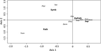
The PCA revealed well-separated associations between the mite taxa (10 superfamilies) and bacterial types, with the first and second dimensions explaining 96% of the total variance (Fig. 4). Trombiculoidea were reported to be strongly associated with pathogenic bacteria, whereas symbiotic bacteria were clustered with mites from the Phytoseioidea and Tetranychoidea. However, the remainder of mite taxa (Dermanyssoidea, Acaroidea, Analgoidea, Glycyphagoidea, Hemisarcoptoidea, Sarcoptoidea and Cheyletoidea) were less strongly associated with opportunistic pathogens and saprophytes. Undoubtedly, in part these data reflect important biases related to bacteria-specific studies, such as the exclusive focus on the Trombiculoidea as vectors of O. tsutsugamushi; or the fact that the relationship between symbiotic bacteria and mites has been better studied in the Phytoseioidea and Tetranychoidea than in the other mite taxa.
Principal Component Analysis of 10 mite superfamilies (Acar, Acaroidea; Anal, Analgoidea; Chey, Cheyletoidea; Derm, Dermanyssoidea; Glyc, Glycyphagoidea; Phyt, Phytoseioidea; Hemi, Hemisarcoptoidea; Sarc, Sarcoptoidea; Tetr, Tetranychoidea and Trom, Trombiculoidea) associated with the categorized bacterial groups (Sap, Saprophytes; OpPath, Opportunistic Pathogens; Path, Pathogens; Symb, Symbionts).

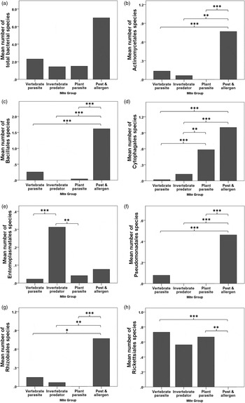
Of 25 bacterial orders, only 7 taxa showed significant differences in bacterial species richness among the 4 biological mite groups: Actinomycetales (χ2 = 24·97, p < 0·0001), Bacillales (χ2 = 30·64, p < 0·0001), Cytophagales (χ2 = 79·21, p < 0·0001), Entomoplasmatales (χ2 = 19·33, p < 0·0001), Pseudomonadales (χ2 = 24·77, p < 0·0001), Rhizobiales (χ2 = 17·69, p = 0·001) and Rickettsiales (χ2 = 21·82, p < 0·0001) (Fig. 5). However, there were no significant differences in total bacterial species richness among the 4 mite groups (χ2 = 5·72, p = 0·126). Mites in the ‘pest and allergen’ group showed higher total bacterial richness than mites in the other groups (significantly so for Actinomycetales, Bacillales, Cytophagales, Pseudomonadales and Rhizobiales); however, Rickettsiales were completely absent therein (Fig. 5H). Mite species in this category are well recognized as generators of allergens and carriers of some pathogenic fungi in human stored food products (Franzolin et al. Reference Franzolin, Gambale, Cuero and Correa1999; Hubert et al. Reference Hubert, Stejskal, Munzbergova, Kubatova, Vanova and Zdarkova2004, Reference Hubert, Kopecky, Perotti, Nesvorna, Braig, Sagova-Mareckova, Macovei and Zurek2012). Invertebrate-predator mites harboured a high number of Entomoplasmatales (Fig. 5E), although these were exclusively derived from the genus Spiroplasma. The bacteria in this genus are known as reproductive manipulators of insect predators and plant pests (Enigl and Schausberger, Reference Enigl and Schausberger2007; Di Blasi et al. Reference Di Blasi, Morse, Mayberry, Avila, Morando and Dittmar2011; Rivera et al. Reference Rivera, Cedillo, Hernandez, Romero and Hernandez2013), demonstrating that predacious and phytophagous mites and their plant hosts form an important habitat for maintaining Spiroplasma in nature.
Analysis of differences in bacterial species richness among mite taxa with multiple pairwise comparisons after Kruskal–Wallis test (*p < 0·05, **p < 0·01, ***p < 0·001) for all bacterial species combined (A), Actinomycetales (B), Bacillales (C), Cytophagales (D), Entomoplasmatales (E), Pseudomonadales (F), Rhizobiales (G) and Rickettsiales (H).

Outcomes and perspectives
This systematic review of the literature suggests that important differences in bacterial flora may exist between mites with different lifestyles, since ‘house pests and allergens’ displayed a particularly diverse bacteriome enriched for several of the bacterial orders included in the analysis (with the notable exceptions of the Entomoplasmatales and Rickettsiales). A key priority for allergy research will be to determine whether these apparent associations are confirmed by further unbiased, high-throughput sequencing methods; and if so, the extent to which the bacterial flora of mite pests may modulate conditions such as atopic dermatitis (Sonesson et al. Reference Sonesson, Bartosik, Christiansen, Roscher, Nilsson, Schmidtchen and Back2013). It would also be interesting to investigate the putative absence of Rickettsiales from this group of mites to reveal any potential barriers to colonization, especially as the Rickettsiales are clearly widespread in other mite categories.
On the basis of the mite literature published to date, very few bacterial species have become uniquely adapted to mites, with only Orientia spp., R. akari and A. phytoseiuli contending as mite-specific symbionts. For the former 2 species, the possibility that they are not restricted to mites with a vertebrate-parasite lifestyle should be considered. Indeed, other arthropod-transmitted human pathogens, such as Rickettsia felis, have been detected in non-biting arthropods (Thepparit et al. Reference Thepparit, Sunyakumthorn, Guillotte, Popov, Foil and Macaluso2011). Our review of the literature also raises the hypothesis that Cardinium is so widely distributed in mites (Weeks et al. Reference Weeks, Velten and Stouthamer2003) that it may be better adapted to this taxon [and perhaps other arachnids, Duron et al. (Reference Duron, Hurst, Hornett, Josling and Engelstadter2008)] than it is to insects.
In conclusion, this review provides useful reference data of mite-associated bacteria for further research, with the intention to increase awareness of the potential for mites to transmit disease. A deeper understanding of the impact of symbionts on their arthropod hosts may also facilitate the development of intervention tools for vector and pest control, for which precedents for insects already exist (Jeffery et al. Reference Jeffery, Thi, Nam, Nghia, Hoffmann, Kay and Ryan2009; Iturbe-Ormaetxe et al. Reference Iturbe-Ormaetxe, Walker and O’ Neill2011). Manipulation of the bacteriome could lead to future opportunities to decrease the medical, veterinary and agricultural impact of mites, although major challenges in the handling and colonization of many species lay ahead.
Supplementary material
To view supplementary material for this article, please visit http://dx.doi.org/10.1017/S0031182015000530
FINANCIAL SUPPORT
We gratefully acknowledge the Mahidol-Liverpool Chamlong Harinasuta Scholarship scheme for financial support of KC.