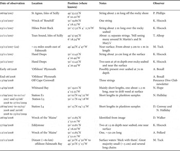
Published records of the occurrence of the physonect siphonophore Apolemia uvaria (Lesueur, 1815) in British waters are sporadic and widely distributed, from the Celtic Sea and the south-west continental shelf edge to the northern North Sea (Fraser, Reference Fraser1961; Kirkpatrick & Pugh, Reference Kirkpatrick and Pugh1984; Mapstone, Reference Mapstone2003). The species has not previously been recorded from Cornish waters in the Environmental Records Centre for Cornwall and the Isles of Scilly (ERCCIS) database (P. Gainey, personal communication), and is not mentioned in either the Plymouth Marine Fauna (Marine Biological Association, 1957) or in correspondence about unusual sightings held by the Marine Biological Association (MBA). Although the first MBA records are from 2007, A. uvaria has been seen regularly in the western part of south Cornwall for many years, with 2003 and 2008 being years of particularly numerous sightings (D. Roberts, Kennack Diving, personal communication). Offshore of Falmouth, A. uvaria has been seen since 2004, particularly during autumn and winter months (M. Tuck, personal communication). Furthermore, boatmen in the Isles of Scilly indicated, late in 2007, sightings of A. uvaria over many years (A. Gall, Isles of Scilly Wildlife Trust, personal communication). Sampling off Plymouth at the long-term monitoring stations L4, L5 and E1 has predominantly caught A. uvaria in coarse mesh plankton tows taken throughout the water column. Table 1 summarizes observations since 2007 made by or forwarded to the authors and which are considered reliable (they were sampled or good quality images were taken). In summer 2009, we received several records including some images of siphonophores from the Isles of Scilly to off Plymouth but they were almost certainly all of Agalma elegans.
Sightings of Apolemia uvaria reported in 2007 and 2008.

Apolemia uvaria is prone to fragmentation (Mapstone, Reference Mapstone2009, p. 21) and the records in Table 1 probably represent fragments rather than complete colonies. Physonect siphonophores, often called ‘string jellies’, are long, complex polymorphic cnidarians. They are divided into two principal parts: a nectosome comprising a small float (pneumatophore) and numerous swimming bells (nectophores) for propulsion, and an attached siphosome of serially repeated units (cormidia) for feeding and reproduction. Although whole colonies have been collected from the Mediterranean Sea, there are no reliable records yet for the nectosome portion of A. uvaria from the Atlantic Ocean (Mapstone Reference Mapstone2003, p. 194). This nectosome portion often breaks free from the siphosome in A. uvaria and swims away independently, leaving lengths of siphosomal stem that drift around in the water for some time, but cannot swim.
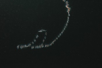
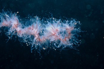
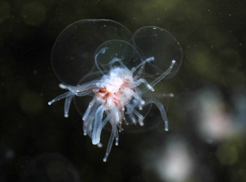
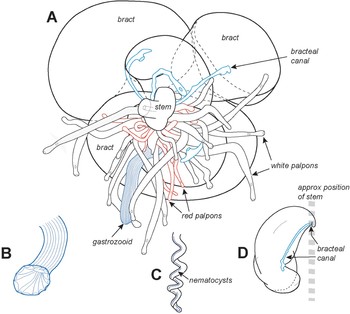
Although apolemiids are most easily identified from their nectosomal nectophores, the siphosomal stem fragments shown in Figures 1–3 can be positively identified as A. uvaria for several reasons, which are worth describing here to facilitate future identifications. Each fragment is long (Figure 1) and consists of groups of cormidia separated by lengths of bare stem (internodes). Cormidia of A. uvaria are distinctive because in addition to one or more mouths (gastrozooids), they contain two types of palpons (white and red; Figures 3–4), whereas in other apolemiid species only one palpon type is present. In addition, each cormidium bears several bracts, and these help to buoy up the heavy stem as well as protect the mouths and palpons from damage. In A. uvaria the canals of these bracts have a characteristic tip that fuses with the lower surface of the bract and then extend a short distance beyond this point (Figure 4); in the bracts of other apolemiids there is no such extension (see Mapstone Reference Mapstone2003, figure 13a–f). Also, in the present siphosomal stem lengths of A. uvaria (Figures 2–4) no reproductive structures (gonophores and gonodendra) are observed, which is to be expected because in A. uvaria gonophores only form at the extreme posterior (distal) end of the stem (Mapstone Reference Mapstone2003, p. 200). In at least one other apolemiid species, gonophores are known to be distributed along most of the siphosome (Mapstone, personal observation).
Siphosomal fragment of Apolemia uvaria bearing many cormidia. Hilsea Point Rock, South Devon. Length of stem fragment ~1–2 m.

Part of a siphosomal fragment of Apolemia uvaria showing several cormidia with gastrozooids, many translucent palpons and some shorter red palpons. Hand Deeps, offshore south Cornwall. Diameter of cormidial clusters <5 mm.

Detached single cormidium of Apolemia uvaria (see Figure 4 below for interpretive drawing) Stoke Point, South Devon. Diameter of cormidial cluster <5 mm.

Schematic interpretation of Figure 3. (A) A cormidium of Apolemia uvaria with 5 bracts attached to the central siphosomal stem with stem canal (out of page and to left), a gastrozooid, and many white and red palpons. Each bract has a bracteal canal (blue); the muscular gastrozooid ingests prey and the thinner white and red palpons assist with digestion and waste removal; (B) sketch of gastrozooid (mouth) in open feeding mode; (C) example of coiled tentacle with simple row of nematocysts (not shown in A above); (D) side view of jelly-filled bract showing approximate life position of stem. Diameter of cormidial cluster in Figure 4A <5 mm.

In whole unfragmented apolemiid colonies, many elongate tentacles emerge from the siphosomal stem, one from each gastrozooid or palpon. When feeding these hang down some distance (Mapstone Reference Mapstone2003, figure 14), exposing a row of stinging cells (nematocysts), which inflict a painful and often lethal sting to the prey. In the single cormidium shown in Figure 3, the tentacles are withdrawn between the palpons (so not visible), but in other cormidia (images available from the authors) tentacles like that illustrated in Figure 4C are visible.
Occurrences of A. uvaria become particularly newsworthy when the species causes mortality in farmed salmon, as happened in south-west Ireland, Shetland and Norway in the late 1990s. In autumn 1998, ‘string jellies’ were present in samples from the Sea of the Hebrides and The Minch (S. Hay, personal communication). Fraser (Reference Fraser1961) observed that A. uvaria had become established in the northern North Sea and was not limited to the Mediterranean as previously thought.
The occurrence of Apolemia uvaria as far north as Norway (Båmstedt et al., Reference Båmstedt, Fosså, Martinussen and Fosshagen1998) and, at least from the 1960s in the northern North Sea, makes it unlikely that seawater warming is a reason for its recent presence off Plymouth. It seems more probable that some variation in ocean currents or upwelling of deeper waters has caused more specimens to be brought into British waters, as suggested by Båmstedt et al. (Reference Båmstedt, Fosså, Martinussen and Fosshagen1998) for the Norwegian increase.
ACKNOWLEDGEMENTS
Thanks to all of the observers who have provided records and especially to Michael Tuck for keeping us regularly informed of occurrences out of Falmouth; also to Barry Mapstone for Figure 4.