

Introduction
Carbon monoxide (CO) poisoning is the most common cause of death and injury among all poisonings, and its incidence is 16/100,000.Reference Hampson and Weaver1 Myocardial injury is detected in one-third of CO poisonings.Reference Lippi, Rastelli, Meschi, Borghi and Cervellin2 It has been found that the risk of acute myocardial infarction (MI) is 1.45-times higher in CO poisoning compared to that in normal population.Reference Huang, Ho and Chen3 Despite its common occurrence, it is still unclear as to which diagnostic and treatment algorithms must be used while approaching CO poisoning cases with myocardial injury. This Case Report presents the diagnosis and treatment approach to a case of CO poisoning in whom transient ST segment elevation was detected by electrocardiography.
Case Report
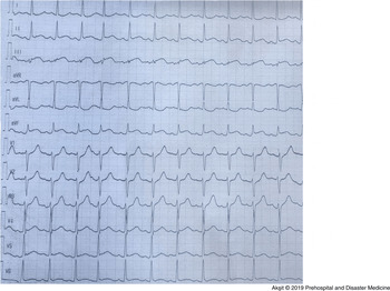
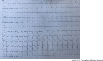
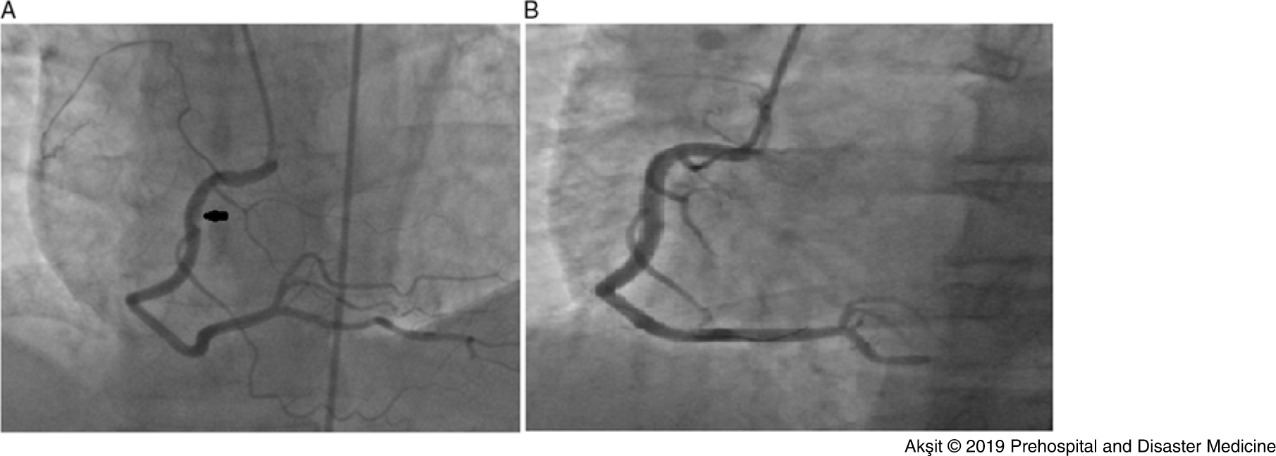
A 41-year-old male patient was found unconscious at home by his relative. The relatives suspected that the patient was poisoned by the smoke that came from the heating stove. An ambulance was called and the patient was brought to the emergency department. It was unknown for how long the patient remained unconscious at home and how many hours he had been exposed to smoke. The patient was treated with high-dose oxygen (12–15L/min) in the ambulance. The patient was conscious with a Glasgow Coma Score of 13 upon admission to the emergency room. The patient did not report any symptoms, except mild headache. Apart from smoking history, the patient’s past history was not remarkable for coronary artery disease. When the patient was admitted to the emergency department, his arterial blood pressure was 135/72mmHg, pulse rate was 90 beats/minute, and respiratory rate was 25 breaths/minute. Carboxyhemoglobin (HbCo) level was 12.2% (n: 0.5%–1.5%) and partial pressure of oxygen (PO2) was 85% (n: 83–108) in the initial blood gas analysis. The initial electrocardiogram (ECG) showed 1mm ST segment elevation in leads DII, DIII, and aVF (Figure 1). As the patient did not describe chest pain and had no cardiac symptoms, ECG was repeated 10 minutes later, and it was seen that ST segment elevation disappeared (Figure 2). From that point on, ST segment elevation was not observed in any of the patient’s control ECGs. When laboratory parameters were examined, high-sensitive Tn-T (HsTn-T) was 62.32ng/L (n: 0–14), aspartate aminotransferase (AST) was 56U/L (n: 5–40), alanine transaminase (ALT) was 23 (n: 5–41), creatinine was 1.89mg/dl (n: 0.7–1.2), urea was 47mg/dl (n: 5–50), and potassium was 4.76mmol/L (n: 3.5–5.1). Cranial computed tomography and cranial diffusion magnetic resonance imaging of the patient after neurology consultation in the emergency department revealed no significant pathology other than small lacunar infarcts. Left ventricular ejection fraction was measured as 60% in echocardiography, and there was no wall motion abnormality. The patient had no ECG change or cardiac complaints in the following day, but the control HsTn-T values increased up to 609ng/dl. As the patient had a transient ST segment elevation and a high HsTn-T, the patient was informed and transferred to the coronary angiography laboratory after an informed consent was obtained. Acetylsalicylic acid 300mg, clopidogrel 600mg, and intravenous 7500 units of unfractionated heparin were administered to the patient. Coronary angiography was performed from the right femoral artery. The patient’s left coronary system was normal, but a thrombus image narrowing the lumen by approximately 60% was observed in the right coronary artery (Figure 3A). Thrombus aspiration could not be performed because there was no appropriate catheter. Tirofiban, an antiaggregant drug, was administered at a bolus dose of 25microgram/kg within three minutes, followed by a maintenance dose of 0.15microgram/kg/minute infused over 48 hours. Control coronary angiography showed continuing thrombus formation and a 4.0 × 22mm bare metal stent was deployed at 12 atmosphere (atm; Figure 3B). The patient had no complaints and was discharged one week after admission. Follow-up examination was performed at the cardiology out-patient clinics two weeks after discharge. Patient did not have angina and there was no change in ECG. Cardiac markers and renal and liver functions that were found to be impaired at the time of admission were within normal limits. The patient was advised against smoking and second-hand smoking. The patient was recommended to continue acetylsalicylic acid 100mg, clopidogrel 75mg, and atorvastatin 40mg tablets, and to attend another follow-up examination one month after in an out-patient clinic. Patient’s informed consent was obtained before writing this Case Report.
The Patient’s First Electrocardiography Shows ST Segment Elevation in Leads DII, DIII, and aVF.

Control Electrocardiography Obtained after 10 Minutes Shows No ST Segment Elevation.

(A). Coronary Angiography Showed a Thrombus Image in the Right Coronary Artery, Causing Approximately 60% Stenosis in the Lumen. (B). Thrombus Formation Persisted in Control Coronary Angiography Performed after Tirofiban Infusion, and a 4.0 x 22mm Bare Metal Stent was Implanted to the Patient.

Discussion
Carbon monoxide poisoning causes serious damage to organs that are highly dependent on oxygen, such as the heart, brain, and kidneys. Depending on cardiac and neurological effects, symptoms such as chest pain, syncope, shortness of breath, palpitation, dizziness, or blurred vision can occur.Reference Reumuth, Alharbi and Houschyar4 In this case, although the HbCO level was 12.2%, there were no other symptoms except mild headache. In their study, Kalay, et al showed in patients with CO poisoning that coronary arteries may appear normal, even in the presence of elevated levels of cardiac markers and myocardial dysfunction ameliorated in most of the cases they followed in the first 24 hours.Reference Kalay, Ozdogru and Cetinkaya5 In the same study, it was reported that the most important determinants of myocardial injury were CO level and time of exposure to CO. Therefore, early treatment is very important. Under normal conditions, the half-life of CO is 320 minutes; this is reduced to 80 minutes with 100% oxygen therapy, and hyperbaric oxygen therapy further reduces the half-life to 23 minutes.Reference Genç, Baydın, Aygün and İncealtın6 One of the indications for hyperbaric oxygen therapy is the presence of findings suggesting myocardial injury.Reference Gülbay and Önen7 In the present case, 100% oxygen therapy was administered because hyperbaric oxygen therapy was not available.
The main causes of myocardial injury in CO poisoning are tissue hypoxia, mitochondrial injury, and reactive oxygen radicals. The cause of acute MI in CO poisoning is the rupture of atheroma plaque, and less frequently, acute thrombosis.Reference Rose, Wang and Xu8 Unlu, et al have successfully applied thrombolytic therapy in a patient with inferolateral MI induced by acute thrombosis after CO poisoning.Reference Unlu, Ozturk and Demirkol9 Hsu, et al treated thrombotic lesion causing critical left anterior descending coronary artery stenosis in a patient with acute anterior MI after CO poisoning by stent implantation.Reference Hsu, Lin and Su10 In the present case, the patient initially received intensive antiplatelet and antiaggregant treatment because the thrombus did not produce critical stenosis (stenosis was less than 70%). A stent was implanted since the thrombus image persisted in the control coronary angiography.
Environmental air pollution studies have shown that CO and other air pollutants increase the risk of arterial and venous thrombosis.Reference Lippi, Rastelli, Meschi, Borghi and Cervellin2 An increased risk of thrombosis has been reported due to increased platelet aggregation caused by the binding of CO to fibrinogen-bound haem.Reference Dziewierz, Ciszowski and Gawlikowski11 Carbon monoxide has a direct binding affinity to myoglobin that is 60-times higher than that of oxygen.Reference Nelson12 Mitochondrial injury induced by CO may cause stunned myocardium-like syndrome (the state of myocardial hypokinesia, although coronary arteries are normal).Reference Tritapepe, Macchiarelli and Rocco13 In CO poisoning, two different clinical patterns have been identified in terms of the effects of CO poisoning on the cardiovascular system. The first clinical pattern relates to young patients with low cardiovascular risk, in whom global left ventricular hypokinesia is observed in echocardiography and coronary arteries often appear normal. The second clinical pattern is observed in patients in a relatively older age group who have cardiovascular risk factors and segmental wall motion abnormality in echocardiography.Reference Satran, Henry, Adkinson, Nicholson, Bracha and Henry14 The most important problem in these patients is to determine the patients that should undergo coronary angiography. There is no consensus as to which group of patients with acute MI due to CO poisoning should undergo angiography. As a result of the literature review, it was noticed that coronary angiography was generally performed to the patients conforming to the second clinical pattern mentioned above. Recent observational studies involving a large number of patients have shown that MI may develop even months after CO poisoning.Reference Kaya, Coşkun and Beton15 As there were no cardiac symptoms in this case and echocardiography showed normal ejection fraction, and no wall motion abnormality, HsTn-T elevation could have been attributed to myocardial injury and coronary angiography could have been skipped. The reason for being insisted on coronary angiography was transient ST segment elevation, which was incidentally detected once and only for a short time.
Limitations
A major limitation of this report is that hyperbaric oxygen therapy was not administered, as it was not available. Administration of this therapy could enable the examination of its effect on coronary thrombus.
Conclusion
This is the first reported case with transient ST segment elevation associated with acute coronary thrombus caused by CO poisoning. It may be recommended that patients with CO poisoning should be followed-up with a 12-lead ECG monitor or 24-hour ECG Holter monitoring, even if they show no cardiac symptoms and echocardiography shows no wall motion abnormality. Early coronary angiography upon detection of such dynamic ECG changes in these recordings as ST segment elevation can reduce the risk of MI and mortality in these patients.
Conflicts of interest
none