INTRODUCTION
Praziquantel (PZQ) is a broad-spectrum anthelmintic used largely in the control of human schistosomiasis. Its cestocidal activity has been demonstrated in the treatment of metacestodes of Echinococcus granulosus (Thomas, Andrews and Mehlhorn, 1982; Andrews et al. 1983; Gemmell and Parmeter, 1983; Urrea-París et al. 1999), Taenia solium (Sotelo et al. 1985) and Mesocestoides corti (Novak, 1977; Hrčkova and Velebný, 1997), but higher concentrations and/ or longer treatments were necessary for PZQ to be effective in vivo. As suppression of host immune responses is a well-described phenomenon in metacestode infections, a stimulation of humoral and cellular components of the host′s immunity might serve to increase the therapeutic effect of drugs. Among immunostimulating substances, glucans represent a very promising group and belong to a class of drugs known as biological response modifiers. In the present work, we used carboxymethylated (1-3)-D-β-glucan from yeast Saccharomyces cervisie. Glucans are glucose polymers found in the cell walls of plants, fungi and bacteria, and both particulate and soluble β-glucans have been shown to activate the innate immune system (reviewed by Bohn and BeMiller, 1995). They also have anti-tumour as well as anti-infective activities including protection against bacterial, viral, fungal and parasitic infections. In our previous studies, co-administration of liposomized glucan with albendazole or fenbendazole to mice with toxocariasis significantly increased the efficacy of treatment (Hrčkova and Velebný, 2001). The most severe pathological consequence of metacestode parasitation in the liver is fibrosis and granulomatous inflammation. The key issue in pathogenesis of hepatic fibrosis is activation by hepatic stelate cells (HSC), mediated by various cytokines and reactive oxygen species released from the damaged hepatocytes and Kupffer cells. Results of activation include overproduction of extracellular matrix components collagen type I, III, fibronectin and others (see review by Wu and Zern, 2000). Various authors have proposed that periparasitic fibrosis might restrict parasite migration and development as, in resistant mice, lower intensity of infection correlated with higher collagen deposits (Guerret et al. 1998; White et al. 1988). On the other hand, the overall liver fibrosis interferes profoundly with the metabolic and functional capacity of the liver parenchyma (Burt, 1993). Thus, it would be desirable if the increase of drug effectiveness against metacestode stages of infections would not lead to the more intense fibrosis, but rather contribute to collagen degradation and resorption. In murine Schistosoma mansoni infection, PZQ treatment resulted in the slow resorption of liver fibrous tissue, and decreased size of fibrotic granulomata was observed several months post-treatment (Stone, 2000; Singh et al. 2004). Although PZQ is also a cestocidal drug, its effect on fibrogenesis during larval cestode infections has not been examined to date.
The larval stage (tetrathyridium) of the cestode M. vogae (syn. M. corti) (Etges, 1991) multiplies asexually by anterio-posterior fission in the liver and peritoneal cavity of several hosts (Specht and Vogae, 1965; Hrčkova and Velebný, 1994; Toplu, Yildiz and Tunay, 2004). In our previous study on M. vogae infection in mice, administration of glucan or liposomized glucan resulted in lower parasite numbers and moderately increased collagen content in the whole liver (Ditteová, Velebný and Hrčkova, 2003a,b). Similarly, White et al. (1988) found stimulation of liver fibrosis and reduced larval burdens in the livers and peritoneal cavities during the course of M. vogae infection treated with a glucan of fungal origin – lentinan. There is growing evidence that mast cells (MC) have the potential directly to influence fibroblasts, the main ‘players’ of fibrosis, and/or indirectly other cells leading to a pro-fibrotic response (see review Puxeddu et al. 2003). Moreover, mast cells function in the host defence against parasites (Lee, Swieter and Befus, 1986), and take part in the pathophysiological conditions such as wound healing and tissue reconstruction (Garbuzenko et al. 2002). They have been found in a high numbers in M. vogae-infected livers and peritoneal cavity of rats (Chernin et al. 1988), but it is not known if mast cells are involved in glucan-stimulated fibrogenesis and liver tissue reconstruction during cestode infections.
Our aims were to investigate the impact of LG co-administration on the larvicidal effect of PZQ in M. vogae infected mice, modulation of liver fibrogenesis following therapy and the involvement of mast cells in this process in relation to the efficacy was also studied. Finally, a preliminary insight into the cellular basis of liver tissue re-modelling after PZQ and/or LG treatment was achieved by means of nuclear DNA markers.
MATERIALS AND METHODS
Infection
Tetrathyridia of Mesocestoides vogae (M. corti) were recovered from the peritoneal cavity of ICR strain mice 9 weeks post-infection (p.i.). Larvae were washed several times and placed into Hanks' balanced salt solution until used for infection. Male mice of the same strain, aged 5–6 weeks, were orally infected with 55–60 tetrathyridia in 0·5 ml of warm saline.
Drugs and liposome preparation
Praziquantel was purchased from Sigma (Germany) and suspended in 0·4% cremophor oil in distilled water with concentration of the drug 7 mg.ml−1. Carboxymethylated 1,3-D-β-glucan from Saccharomyces cerevisiae (a kind gift from Dr Kogan, Institute of Chemistry, SAS, Slovakia) was entrapped into negatively charged liposomes as described previously (Velebný et al. 1997). The phospholipid mixture consisted of 0·056 mmol Phospholipon 100H (donated by Phospholipid com., Germany), 0·016 mmol cholesterol and 0·008 mmol dicetylphosphate (both from Serva, Germany). One dose of liposomized glucan (LG) (5 mg.kg−1) contained approximately 1·35 mg of liposomal lipids.
Liposomes containing 2 cellular DNA markers: [methyl-3H]-thymidine and its non-radioactive analogue 5-bromo-2′-deoxyuridine (BrdU) (ICN, USA), were also prepared. Briefly, a chloroform[ratio ]methanol (4[ratio ]1, v/v) mixture consisting of 0·170 mmol Phospholipon 100H (Phospholipid com., Germany), 0·049 mmol cholesterol and 0·024 mmol dicetylphosphate (Serva, Germany) in a molar ratio of 7[ratio ]2[ratio ]1 was dried under reduced pressure on a rotary evaporator. The thin lipid film formed on the round flask's walls was hydrated with 12·5 ml of saline containing 0·163 mmol BrdU and 60 μCi [methyl-3H]-thymidine by gentle shaking giving rise the liposome suspension with entrapped markers. Non-entrapped markers were removed during overnight dialysis against saline. The final concentration of liposomized BrdU was 4 mg.ml−1 and that of [methyl-3H]-thymidine was 4·8 μCi.ml−1.
Experimental design
Infected mice were divided into 4 groups, each comprising 27 animals, and treatment started on day 14 p.i. PZQ was administered to mice orally twice a day for 3 consecutive days (total 6 doses) with one dose containing 35 mg PZQ.kg−1 of body weight (Group 1). Liposomized glucan was administered intramuscularly (hind leg) in 3 daily doses on the same days as PZQ in combination with PZQ (Group 2) or alone (Group 3). Infected untreated mice (Group 4) served as the control. One dose of LG contained approximately 3 mg.kg−1 of body weight of glucan. Group 5 comprised 12 healthy uninfected mice that were treated with LG at the same dose and livers were examined for collagen content and mast cell counts before and after treatment on the same days.
Examination of parasite burden in the livers and peritoneal cavities was carried out on days 1, 7 and 14 post-therapy (p.t.) and 8 mice/group/day were killed by cervical dislocation.
Tetrathyridia from the peritoneal cavity of mice were collected and counted. Livers were cut into very small pieces and samples for determination of larval numbers were digested in 0·25% trypsin solution (pH 7·8–8·0) at 37 °C for 4×30 min. The total number of tetrathyridia in 1 g was calculated as the average from 3 counts/mouse and then the absolute number in the whole liver was determined. Finally, the mean larval number±S.D. in each group/day was calculated.
The effect of therapy was expressed as the percentage reduction of worm numbers and calculated using the formula:

The efficacy of drugs was calculated for larvae isolated from the total livers, whereas further tests were performed on 2 liver parts (A and B), which differed in intensity of infection within the experimental period. The reason for this was following: after the oral infection, parasitization in individual liver lobes is not homogenous and larvae migrate primarily into the central part of the large lobe, then gradually accummulate also in the smaller lobes. Some larvae probably also penetrate from the peritoneal cavity (personal observations; Specht and Voge, 1965). The pharmacokinetic properties of PZQ indicate that availability of PZQ is the same for the whole liver (Steiner et al. 1976), and that is why we decided to calculate efficacy from total larval counts. Based on published data cited in this study, indicating a very close relation between parasitic antigens (secreted and somatic) and fibrogenesis/mastocytosis, these host-parasite interactions were evaluated in 2 separated liver lobes, differing in intensity of infection. From total larval numbers, the ratio 1[ratio ]4 for intensity of infection between A and B parts was determined in random samples before treatment (personal observations).
Cell proliferation and DNA synthesis in the livers
For determination of cell proliferation following therapy, the incorporation of 2 nuclear markers BrdU and 3H-thymidine into DNA of cells undergoing mitosis was monitored. Three mice from each experimental group received single dose of liposomized (BrdU/3H-thymidine) formulation on day 13 p.t. (28 p.i.) corresponding to the regenerative phase (Specht and Widmer, 1972) intraperitoneally. The dose contained approximately 2 mg of BrdU and 2·4 μCi of -[methyl-3H]-thymidine/0·5 ml per mouse weighing 40 mg. Mice were killed 24 h later and samples from 2 liver parts differing in the intensity of infection and fibrosis were collected in triplicate from each mouse. These represent: part A – the peripheral part of the smaller lobe (weak infection) and part B – central parts of large lobes (the heavy infection). Samples were further processed for immunohistological localization of BrdU-labelled cell nuclei and radiometric measurements of DNA-incorporated 3H-thymidine.
Radiometric determination of 3H-thymidine incorporation in the liver
Samples of the livers (100 mg) were hydrolysed in H2O2 and HClO4 after a modified method of Mahin and Lofberg (1966) and then mixed with 5 ml of liquid scintillation cocktail (ICN, USA). Radioactivity in samples was measured on a liquid scintillation counter, Rackbeta 1217 (LKB Wallace, Finland). Results were expressed as CPM per gram of the tissue (mean±S.D., n=9).
Hydroxyproline content in the liver
Total collagen content was examined by means of biochemical determination of hydroxyproline concentration (the most abundant amino acid of collagen) obtained from 3 livers/group/day. Three samples of liver homogenates from liver parts A and B (3 from each homogenate, n=9) weighing 100 mg each, were hydrolysed in 4 ml of 6 M HCl for 20 h at 110 °C, then the acid was evaporated to dryness and re-suspended in 1 ml of distilled H20. Hydroxyproline concentration was determined after a modified colorimetric method of Woessner (1961) in 96-well flat-bottomed plates. The OD of samples was measured at 550 nm and expressed as μg.g−1 of wet weight of the liver tissue.
Histochemistry
Liver samples were fixed in Histochoice Fixative solution (Amresco, USA) for 20 h at 4 °C, dehydrated and embedded in paraffin wax. Sections (6 μm in thickness) were cut and mounted on glass slides. Haematoxylin/eosin staining was used to evaluate the overall liver pathology and the collagen fibrils were visualized after combined blue trichrome (Mallory). The liver mast cells were stained with toluidine blue at pH 0·5. Stained mast cells were counted in the whole area of 80 independent ocular grids at magnification 600×(area of the circle=0·076 μm2) on the LEICA microscope and mean mast cell numbers±S.D./field were calculated. Finally, the mean numbers for 1 mm2 of section area for each group of mice were calculated. In addition, non-serial sections taken from a single tissue block were used for counts in order to avoid counting the same mast cells profile.
Immunohistochemistry
Liver samples from treated and control mice, which also received liposomes with entrapped BrdU and 3H-thymidine, were processed as described above, cut and mounted on glass slides. Sections were de-paraffinized, washed in PBS (7·2) and endogenous peroxidase was blocked by exposure to 0·03% H2O2 in methanol for 30 min. The nuclear DNA was denaturated during incubation of slides with 4 M HCl/PBS for 20 min at 36 °C, then neutralized with 0·1 M borate buffer for 5 min and washed several times with PBS. After blocking of non-specific binding sites with 4% Top-block solution (Sigma) for 30 min at 37 °C, sections were incubated with monoclonal anti-BrdU primary antibody (Sigma) for 2 h at 37 °C to visualize BrdU incorporated in the cell nuclei. Rinsed sections were exposed to the polyclonal rabbit anti-mouse IgG-Px (Sigma) for 1 h at 37 °C, then rinsed and incubated with DAB solution (Vector, USA). Finally, counter-staining of the liver tissue was performed with Gill′s haematoxylin and the colour reaction stopped with Scott water (pH 8·0).
Statistics
Statistical differences within all 4 groups were assessed using one-way ANOVA, followed by post-hoc Tukey′s test (P level indicated in the text). This allowed comparison between groups at each day of examination. Analysis was performed using the Statistica 6.0 (Stat Soft, Tulsa, USA) statistical package.
RESULTS
Efficacy of treatment
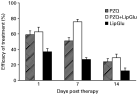
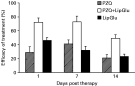
The larvicidal activity of praziquantel after co-administration of liposomized glucan (LG) was examined in the livers and peritoneal cavities of M. vogae-infected mice. In the livers (Fig. 1) administration of LG alone resulted in a small reduction of the larval counts. Simultaneous administration of LG and PZQ significantly enhanced larvicidal effect of the drug on day 7 p.t. (75·5±4·4%) in comparison with PZQ-treatment (51·0±3·1%) (P<0·05). However, as unaffected larvae continued to proliferate, efficacy was only 24·3±4% after PZQ and 29·0±4·9% in the group treated with PZQ+LG, on day 14 p.t. The larvicidal effect of PZQ was lower on the larvae in the peritoneal cavity (Fig. 2), in contrast on the larvae in liver. In the peritoneal cavity, efficacy of combined treatment with PZQ and LG (72·2±6·4%) was increased in comparison with PZQ alone (29·5±8%) (P<0·01) and LG-alone (46±4·2%) already on day 1 p.t. and lasted up to the end of the experiment.

Fig. 1. Efficacy of 6 doses of praziquantel (given orally twice a day), 3 doses of liposomized glucan (given once a day s.c.) administered alone or in combination, on Mesocestoides vogae tetrathyridia in the liver. Mice were infected with 60 larvae and treatment commenced on day 14 p.i. for 3 consecutive days. Larval numbers were counted within 2 weeks post-therapy (p.t.).

Fig. 2. Efficacy of 6 doses of praziquantel (given orally twice a day), 3 doses of liposomized glucan (given once a day s.c.) administered alone or in combination, on Mesocestoides vogae tetrathyridia in the peritoneal cavity. Experimental design as in Fig. 1.
Mast cells counts and their histochemical localization
Mast cell counts in the A and B parts of the control livers, and following treatment, are summarized in Table 1. In all groups of mice, the numbers of mast cells were higher in the B-liver parts, in which more intense fibrosis was also found. Kinetics of mastocytosis was profoundly modulated by praziquantel and the immunodulator glucan. After administration of LG the highest stimulation of mastocytosis was seen already on next day after the therapy (B-part, 7·26±8·8), administration of PZQ alone was followed by an increase of mast cell counts between days 1–7 p.t. (B-part, 4·62±4·2). Regardless of the day of examination, administration of PZQ partially abolished LG-stimulated mastocytosis in both liver parts. On day 14 p.t. mastocytosis was profoundly lowered in PZQ- treated groups, but only moderately in LG-treated mice compared with the control.

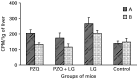
Fig. 3. 3H-thymidine incorporation into nuclear DNA of proliferating cells in 2 liver parts from mice infected with tetrathyridia of Mesocestoides vogae. 3H-thymidine entrapped in liposomes was given to mice intraperitoneally on day 13 p.t. and incorporated marker was determined 24 h later (day 29 p.i.) as counts per min (CPM) in the liver parenchyma from control and treated groups of mice.
Table 1. The number of hepatic mast cells stained with toluidine blue in two liver parts (A, B) from mice infected with tetrathyridia of Mesocestoides vogae (M. corti) and treated with praziquantel alone or in combination with liposomized glucan (LG) and LG alone (Data represent mean numbers of mast cells±S.D. counted on 80 non-overlaping fields/group/day at magnification 600×(area of the circle=0·076 μm2). Mean mast cells numbers at area of 1 mm2 were then calculated.)

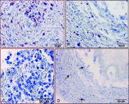
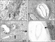
In histological examinations of liver sections from all groups, mast cells were localized predominantly in the fibrotic granulomatous lesions (Fig. 4A) and collagenous capsules containing larvae (Fig. 4B) in both liver parts. Mastocytosis was more developed in livers of mice given LG. In PZQ-treated groups, mast cells were scarce in the fresh inflammatory infiltrates (Fig. 4C), where their numbers increased along with progressing fibrosis. No mast cells were seen in intact areas of liver parenchyma. In the vicinity of larvae, in which extensive destruction of the larval surface was observed, most notably after PZQ+LG treatment on day 7 and 14 p.t., mast cells were present in a greater numbers (Fig. 4D).

Fig. 4. Histochemical localization of mast cells (purple, arrows) on the liver sections from Mesocestoides vogae-infected control and treated mice (Toluidine Blue staining). The high numbers of mast cells were in the fibrotic foci (A) and periparasitic capsules (B), control B-part, day 14 p.t. (C) Detail of parenchymal cell infiltration near larval migratory track from A-liver part of PZQ-treated mice. (D) Thick fibrous capsule with scattered mast cells (arrows) surrounding the apparently damaged larva, B-liver part after PZQ+lip.glucan treatment on day 14 p.t.
Determination of the hydroxyproline concentration
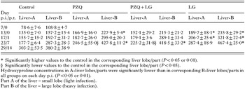
Hydroxyproline concentration data are summarized in Table 2. In all groups of mice (treated and control), hydroxyproline content was higher in more heavily infected central areas of large lobes (B) in contrast to the smaller liver lobes (A) (P<0·05) and gradually increased. In comparison with the control, PZQ-alone-treatment caused gradual lowering of hydroxyproline concentration in A-liver parts, whereas in B-liver parts, rate of hydroxyproline accumulation was higher than in control, most notably on day 14 p.t. Administration of glucan in liposomes resulted in higher hydroxyproline values than in PZQ-treated group and control. This effect of LG was inhibited after simultaneous administration of PZQ. We also monitored the effect of liposomized glucan on liver collagen content in healthy uninfected mice using the same treatment/examination regime. There was no significant change in hydroxyproline concentration and values were 83·4±5·6 μg.g−1 tissue (before administration) and between 83·6±12·2 and 71·5±8·6 (within 2 weeks p.t.).
Table 2. Hydroxyproline concentration (μg.g−1 of tissue) in the livers of mice infected with Mesocestoides vogae (M. corti), untreated and treated with praziquantel (PZQ) alone, liposomized glucan (LG) and after combined therapy with both compounds (Data represent mean values ±S.D. (n=9).)

Histochemical localization of total collagen on the liver sections
The overall pathological appearance in liver sections was similar in treated and control groups. Inflammatory cells comprising neutrophils, eosinophils and macrophages were the dominant cells in the fresh inflammatory foci surrounding larvae and also filled their migratory tracts. Collagen fibres were seen as a diffuse fibrosis in the paranchymal tissue in B-liver parts by day 17 p.i. (Fig. 5A ) and from this period, the collagenous capsules started to form around the larvae. At the end of the period some of the larvae were surrounded by thick capsules (Fig. 5B) but also new (unencapsulated) larvae penetrating, probably from the peritoneal cavity, were seen in livers from all groups (Fig. 5C). On this day, fibrosis was very widespread and intense and collagen fibres lined the penetration tracks. Many of larvae in livers from PZQ and PZQ+LG-treated mice appeared to have a damaged tegument (Fig. 5D) and were already necrotic.

Fig. 5. Histology and collagen staining (Blue Trichrome-Mallory) of the livers from mice infected with tetrathyridia Mesocestoides vogae. (A–D) Histopathology was observed in the control and treated groups. (A) Parenchymal accummulation of collagen fibres (arrow) associated with a diffuse leucocyte infiltration, control B-part, day 17 p.i. (B) Intact larva inside well-developed collagenous capsule, control B-part, day 29 p.i. (C) Collagen fibres at the border of larval migratory track (arrow) filled with the erythrocytes. Note 2 penetrating larvae surrounded by a dense layer of inflammatory cells. (D) Larva with damaged tegument (arrow) surrounded by the inflammatory cells inside thick capsule, PZQ+lip.glucan B-part, day 14 p.t.
Quantitative analysis of cell proliferation
Synthesis of nuclear DNA was monitored by means of incorporation of 3H-thymidine and its non-radioactive analogue BrdU during S-phase. Radiometrical data (expressed as CPM) obtained on the next day after administration (14 p.t.) and summarizing the total incorporated 3H-thymidine in both selected liver parts, are shown in Fig. 3. The most interesting observation was the different kinetics of thymidine incorporation into DNA between two liver lobes in control and all treated groups of mice. Thus, in the infected untreated group, a similar amount of marker was determined in part (B) with a higher infection (147·7±12·3) as in the much less infected lobe (A, 139·0±8·1). Either treatment significantly elevated (P<0·05) 3H-thymidine uptake in A-liver parts in comparison with B-parts. LG given alone exerted the highest stimulatory effect on DNA synthesis recorded (A, 267·4±38; B, 204·9±17) whereas PZQ had the lower effect (A, 204·9±19; B, 133·4±9). After simultaneous administration of both compounds 3H-thymidine incorporation in both liver lobes was slightly lower (A, 173·9±32; B, 114·8±21).
Immunocytochemical examination of BrdU-positive cells
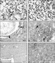
Several kinds of cells residing in the livers (hepatocytes, hepatic stelate cells – HSC, fibroblasts or even Kupffer cells) have the ability to divide or regenerate after various stimuli. To evaluate the impact of treatment on mitotic activity of the above-mentioned cells, incorporation of BrdU into replicating nuclear DNA was investigated immunocytochemically. The various labelling patterns of the nuclei, and their localization in treated and control groups, indicated the presence of a few distinct populations of BrdU-labelled cells. Very intense BrdU nuclear immunostaining was associated with the nuclei of round cells and stelate-like cells localized inside fibrotic granulomatous foci, in which collagen fibres prevailed over the inflammatory cells (Fig. 6A). BrdU staining was seen already in ‘younger’ inflammatory infiltrates where collagen fibres were rare (Fig. 6B). The migratory tracts of larvae also contained BrdU-positive nuclei of different staining patterns (not shown). Labelled flattened cells, apparently fibroblasts, formed the inner layer around the larvae (Fig. 6C). BrdU labelling was seen also in another kind of cell, probably hepatocytes, foci of which were more abundant in the A-liver parts from treated groups (Fig. 6D) often localized next to the paths after larval migration. The least frequent round cells, displaying very intense BrdU labeling, possibly Kupffer cells, were usually seen scattered among hepatocytes. The most abundant was this population of cells in the livers from groups that received liposomized glucan, more in A-liver parts (Fig. 6E) than in B-parts (Fig. 6F). Apart from the last staining pattern, distribution of other BrdU-labelled cells was similar in the livers from all groups of mice.

Fig. 6. Immunostaining of BrdU incorporated into nuclear DNA of proliferating cells in the liver. (A) In the mature fibrotic foci BrdU labelling was associated with nuclei of round-shapes and stelate-shaped cells (arrow). (B) Young cell infiltration contained small labelled nuclei (black). (C) BrdU incorporated into flat cells, probably fibroblasts (arrows), forming the innner border of developing larval capsule. (D) Area of necrotic hepatocytes with intense BrdU-labelling indicates (arrow) on ongoing cell regeneration/proliferation. BrdU-positive cells, possibly Kupffer cells, were scattered in the liver parenchyma from liposomized glucan-treated mice (black nuclei). A-part (E) and B-part (F). This pattern of cell labelling was more widespread in A-parts than in B-parts.
DISCUSSION
There is still no consensus regarding the treatment regimes and PZQ doses for the treatment of larval stages of cestode infections in humans and animals (WHO, 1995) and usually treatment lasting several weeks is required to inhibit larval multiplication or to eliminate infection (Novak, 1977; Thomas and Gönnert, 1978). The doses used in our study were not designed completely to eliminate this infection, but rather to evaluate the dynamic relationship between efficacy of drug and pathological processes (fibrosis, mastocytosis, tissue re-modelling) in the liver.
Macrophages play a crucial role in defence against helminth infections, and M. vogae metacestode infection appears directly to reduce effector macrophage functions and unresponsiveness to IFN-γ activation (Jenkins et al. 1990). Beta-glucans are a broad group of molecules in which an immunostimulating effect is manifested by activation of leukocyte effector functions, for example phagocytic ability, production of reactive oxygen radicals (Wakshull et al. 1999), cytokines and release of inflammatory mediators (Williams, Mueller and Browder, 1996; Kogan, 2000). Therefore, in order to activate larvicidal capacity of macrophages, polysaccharide β-glucan entrapped in liposomes (LG) was co-administered with PZQ in our study. Addition of LG resulted in a significantly higher efficacy of treatment in comparison with PZQ alone in the peritoneal cavity within the whole examined period and on day 7 p.t. in the liver. The observed reduction of larval numbers after LG treatment might indicate the activation of cytotoxic functions of effector immune cells.
Among the disadvantages of injecting soluble forms of immunomodulators into the body is that only a small fraction can reach macrophages in order to be activated. In this respect, conventional liposomes are suitable cariers for site-specific delivery of drugs to macrophages (Gregoriadis, 1988). Liposomized glucan could be internalized by macrophages via complement receptors after opsonization of liposome particles or after their phagocytosis or endocytosis, which led to cell activation (Szebeni, 1998). Thus, we assume that in our in vivo studies, glucan liberated from taken-up liposomes affected activity of mononuclear phagocytic cells directly without binding to the glucan receptors present on the cells (Brown and Gordon, 2003).
Mastocytosis and eosinophilia are prominent host immunopathological responses to the helminth infections, occuring in the liver, gastrointestinal tract and other tissues. Very little information is available about modulation of mastocytosis after PZQ treatment of cestode infections (Starke and Oaks, 1999). This is the first study, in this respect, on mice with the metacestode stage of M. vogae infection. Cook et al. (1988) demonstrated that in rats infected with M. corti (M. vogae) liver mast cells did not seem to play any role in resistance to infection. No adherence capacity was seen for these cells to undamaged larvae but this increased on larvae with a partially damaged surface. In our study, mast cells numbers were low between days 14 and 17 p.i. in the livers from control mice and progressively increased within 1 month p.i., more in a heavily infected (B) lobules. Treatment with PZQ and/or with LG in our work markedly modulated mastocytosis. In the case of treatment with PZQ-alone, elevation of mast cells numbers within 7 days p.t., was more intense in highly infected liver parts, and corresponded to the highest reduction of larvae during this period. We believe that antigens released from PZQ-damaged larvae could either directly stimulate early recruitment of mast cells from the circulation and their activation or, indirectly, via other immune mechanisms.
Parasite antigens initiate a series of MC-membrane and cytoplasmic events which lead to cell activation and degranulation (Lee et al. 1986). Activated mast cells can kill the worms indirectly by means of the array of released cytotoxic molecules or via cytokines and other molecules (Foreman, 1994). Taking all our data together, we assume that MC contributed to the increased PZQ efficacy, but their role was only partial in comparison with that of other effector cells. In contrast, much higher mast cell numbers in both liver parts, peaking on day 1 p.t., were found after administration of LG-alone. Precursors of MC leave the bone marrow via the blood and complete their differentiation in the tissues, but MC can also proliferate in the tissues (Foreman, 1994). Histologically, we could hardly see any mast cells in the livers of uninfected mice treated with LG. Therefore, we hypothesize that at least two mechanisms could be responsible for the early onset of a high mastocytosis following administration of liposomized glucan. One could be the chemotactic factors released from the activated mononuclear phagocytic cells but LG might also directly stimulate mast cells proliferation in the liver. Regarding the direct stimulation, it is possible that a part of injected LG was taken up by mast cells as their ability to phagocytose via opsonin-dependent (Fc and C3 receptors) and opsonin-independent receptors (Toll-like receptors, dextran receptors) was recently confirmed (Henz et al. 2001; Feger et al. 2002). Interestingly, when LG was co-administered with PZQ, efficacy was elevated, but PZQ nearly completely suppressed LG-induced mastocytosis. The direct cytotoxic effect of PZQ on MC is hardly the reason for this as PZQ was shown to be non-toxic (Cioli and Pica-Mattoccia, 2003; Montero and Ostrosky, 1997). In addition, apoptotic down-regulation of mucosal mastocytosis, following PZQ therapy of infection with adult Hymenolepis diminuta, started after 4 weeks post-treatment when worms were already removed from the gut (Starke and Oaks, 1999). At present, with limited information, we can only assume that PZQ-induced mast cell downregulation was initiated by the particular antigens released or revealed from damaged larvae, which probably modulated production of IL-3 and IL-4 cytokines which are essential for mast cells survival (Foreman, 1994). Moreover, we assume that LG-activated macrophages/Kupffer cells have accounted for the overall higher reduction of larvae after combined treatment and that the role of mast cells was more regulatory than the larvicidal.
There are many reports indicating the important pathogenic role of MC in chronic inflammation and fibrosis and there is limited information how anthelmintic therapy modulates these pathological responses. In the present study, the decline of MC numbers within the experimental period was accompanied by a significant elevation of the collagen content in all treated groups, the most in LG-treated mice.
Chernin et al. (1988) found that livers of M. corti-infected rats contained mast cells with histochemical properties of both mucosal (MMC) and connective mast cells (CTMC), which differ in granule content (Puxeddu et al. 2003). Fibroblasts are an important source of growth factors only for connective MC and can also regulate their effector functions (Levi-Schaffer, Kelav-Appelbaum and Rubinchik, 1995; Sellge et al. 2004). On the other hand, CTMC have the potential directly to influence fibroblasts, the main components of fibrosis, and/or indirectly other cells, leading to a pro-fibrotic response. They can release cytokines with pro-fibrogenic activities such as TNF-α, TGF-β, and others.
From the second week post-infection, larvae were surrounded by an increasing amount of collagen fibres forming the capsule, together with the formation of perivascular and diffuse parenchymal fibrosis. It seems that encapsulation of larvae in the liver plays a role in the defence against parasite development/migration, as the higher hydroxyproline concentrations were found in treated groups, mainly in B-parts. In E. multilocularis-resistant NMRI mice, the higher collagen content was inversely correlated with the size of the parasitic lesion (Guerret et al. 1998). The same was observed in mice with M. corti infection (Pollacco et al. 1978), indicating that collagenous capsules around parasites probably restrict multiplication of larvae. So we assume that fibrosis after PZQ-treatment was, in B-parts (high larval numbers), stimulated by antigens released from the damaged larvae, which triggered a cascade of pro-fibrotic reactions. Mast cell-produced factors probably also contributed. Fibrosis in A-parts was localized mainly near larval migratory tracts and in inflammatory infiltrates, free from larvae. Here the decline of collagen content after PZQ therapy in comparison with controls, could be the result of release of antifibrotic humoral factor IFN-γ from local macrophages/mast cells due to the limited antigen stimulation. This was suggested by Lortat-Jacob et al. (1997) in the study on S. mansoni-infected mice, where administration of IFN-γ reduced liver parenchymal collagen in egg-free lobes but induced a small increase in the collagen concentration of the granulomas surrounding eggs. Information on the effect of PZQ on liver fibrosis during metacestode infections is lacking and most data come from the studies on S. mansoni-infected animals. Here the greatest stimulation of fibrogenesis was seen after administration of LG alone. In our previous studies we have described the elevation of hydroxyproline content after β-glucan-treatment in the livers of mice with M. vogae infection along with the decrease in parasite burdens (Ditteová et al. 2003a,b). A similar reduction in M. corti larval numbers in the peritoneal cavity and liver, along with increased collagen deposition/fibrosis after administration of a T-cell-dependent immunomodulator β-(1-3) glucan – lentinan, was observed by White et al. (1988). It was believed that glucan stimulates collagen deposition in the wounds indirectly via release of macrophage pro-fibrotic factors, but recent in vitro studies revealed that glucan can directly activate collagen synthesis in human skin fibroblasts in a time-dependent manner (Kougias et al. 2001; Wei et al. 2002). It is possible that the much higher hydroxyproline content found in LG-treated groups than in PZQ-treated, or even in control (A-parts), could partially be the result of a direct stimulation of liver fibroblasts. Similarly, as in the case of mastocytosis, PZQ suppressed LG-stimulated collagen synthesis. As not only collagenesis, but also mastocytosis, were more intense after administration of LG during the experimental period, than in other groups, a role of mast cells in the regulation of collagen synthesis by fibroblasts is plausible. Thus it appears that LG triggered early and very intense mastocytosis, and these activated MC, at least partly, stimulated fibrosis by releasing pro-fibrotic cytokines. Among them, tumor necrosis factor-alfa (TNF-α) plays an essential role as it is pro-inflammatory cytokine directly involved in activation of HSC, which are then transformed onto fibroblasts (Wu and Zern, 2000). In E. multilocularis metacestode infections TNF-α was shown to stimulate liver periparasitic fibrosis (Amiot et al. 1999). Elevation of MC numbers preceeded an increase of fibrosis, which might provide indirect evidence about the activating role of MC for fibroblasts during M. vogae infection. However, the liver injury due to parasitization and larval somatic antigens seem to be the initiating factors as, in uninfected mice, no elevation of hydroxyproline concentration and mast cells was observed.
Fibrogenesis is a dynamic process, which generally forms part of the tissue repair response to hepatocyte injury, which results in accumulation of extracellular matrix proteins (ECM). The liver has a remarkable reparative capacity and both Kupffer cells and mast cells were postulated to play a key role in liver regeneration/remodelling (Takeishi et al. 1999). In the present work we examined the impact of treatments on cell proliferation using DNA nuclear markers 3H-thymidine and BrdU, which have been succesfully employed as the mitotic S-phase markers of hepatocyte proliferation (Miyagawa et al. 1996). Immunohisto-chemically, BrdU incorporation was observed in several morphologically different cell types. In general, the most intense labelling was in cells localized in advanced fibrotic foci, collagenous capsules and in collagen fibres lining migratory tracts in all groups, which could be HSC and fibroblasts, but incorporation to MC can not be excluded. Areas of regenerating hepatocytes were also observed. Cell proliferative capacity is attributed not only to the parenchymal liver cells, but also to Kupffer cells, HSC, fibroblasts, mast cells and pit cells (Takeishi et al. 1999 and others). In the control groups, 3H-thymidine values were similar in both liver parts as were also hydroxyproline concentrations, but mast cell counts differed. Therefore, we believe that HSC and fibroblasts are the main proliferating cells in the control mice and that doubling of MC numbers in the B-part compared with the A-part was not due to their in situ proliferation, but probably to their recruitment from blood MC.
Interestingly, in all treated mice 3H-thymidine incorporation in the liver tissues obtained from A and B-liver lobes showed marked differences, being higher in A-parts than in B-parts.
Discrete BrdU-positive round-shaped nuclei were found scattered among intact hepatocytes in the A-parts (Fig. 6D) of LG-treated groups, but not after treatment with PZQ-alone. No similar staining pattern for mast cells was seen in intact parenchyma in A-lobes from treated and control groups, free from fibrosis. As in this liver part from LG-treated mice a maximal level of 3H-thymidine was also determined, we suppose that these BrdU-labelled cells were proliferating Kupffer cells rather than mast cells or possibly HSC, which were associated with the fibrous tissues. Direct mitotic effects of glucan on other liver cells is also possible, but cannot be confirmed in the light of present data. Many studies have confirmed the primary role of Kupffer cell-produced factors in hepatocyte proliferation (for example, Matsumoto and Nakamura, 1997; Takeishi et al. 1999). Thus, in the LG-treated group, an elevated population of Kupffer cells might produce stronger signals for regeneration of parenchymal tissue seen as areas of BrdU-positive hepatocytes that probably also accounted for the highest recorded 3H-thymidine levels. In this group, fibroblast/HSC proliferation in B-parts seems to be dominant, but mast cell/Kupffer cell proliferation could also have contributed to the high 3H-thymidine radioactivity observed here. Furthermore, in the A-liver lobes from mice treated with PZQ-alone and in combination with LG, the increased radioactivity was inversely correlated with mast cell counts and fibrosis/collagen content. It might suggest, that after PZQ treatment, cell division in A-parts took place predominantly in the regenerating hepatocytes that were also localized after BrdU immunostaining. In the B-liver parts, 3H-thymidine levels were comparable with the control, in contrast to the diminished population of MC and elevated amount of hydroxyproline. In addition, BrdU immunostaining of cells resembling the Kupffer cells was scarce here. Therefore, it seems that in heavily infected lobes (B), after PZQ treatment, the proliferating fibroblasts and HSC, in accordance with the higher collagen deposit found here, were the main proliferating cells and that parenchymal regeneration was slowed down here.
In conclusion, the anthelmintic effect of PZQ could be enhanced after co-administration of the immunomodulator β-glucan entrapped in the liposomal carrier. Intense mastocytosis seen after treatment with LG can be abolished after simultaneous administration of PZQ in a time-dependent manner. The reparative processes of liver parenchyma were enhanced in an inverse correlation with the intensity of infection after treatment with PZQ, as indicated by the pattern of cell proliferation.
This work was supported by grant ‘VEGA’ no. 2/4180/04 of the Slovak Grant Agency and by The State programmes of the research and development šPVV-2003 SP 51/028-09-08.