Introduction
Trehalose (α-d-glucopyranosyl α-d-glucopyranoside) is composed of two glucose subunits linked by an α,1 → 1-glycosidic bond. This is a non-reducing disaccharide, more stable than other disaccharides. It goes through slow hydrolysis, and does not undergo the Maillard reaction. The arrangement of hydroxyl groups in trehalose facilitates the formation of hydrogen bonds between these groups of the sugar and the phosphate of head groups of the membrane phospholipids (Portmann & Bircht, 1995; Temesvari et al., Reference Temesvari, Klein and Cotter1996; Behm, Reference Behm1997; Sato et al., Reference Sato, Okamoto, Minami, Kohri and Yamamoto1999; Pereira et al., Reference Pereira, Lins, Chandrasekhar, Freitas and Hürenberger2004). Physical and chemical properties make trehalose not only an energy source, but also a compound assisting in maintaining the integrity of membranes and other cell structures during environmental stresses such as dehydration, high osmotic pressure, freezing and high-temperature shock (Crowe & Crowe, Reference Crowe and Crowe1982; Lee et al., Reference Lee, Waugh and Griffin1986; Takahashi et al., Reference Takahashi, Ohmae and Hatta1997; Oliver et al., Reference Oliver, Leprince, Wolkers, Hincha, Heyer and Crowe2001). Trehalose is present in members of many systematic groups, such as bacteria, fungi, plants, insects, nematodes and other invertebrates (Wyatt & Kalf, Reference Wyatt and Kalf1957; Kaazen et al., Reference Kaazen, McDougall and Strřm1994; Behm, Reference Behm1997; Nambu et al., Reference Nambu, Nambu and Tanaka1997; Nwaka & Holzer, Reference Nwaka and Holzer1998; Müller et al., Reference Müller, Aeschbacher, Winkler, Boller and Wiemken2001; Lapinski & Tunnacliffe, Reference Lapinski and Tunnacliffe2003; Dmitryjuk et al., Reference Dmitryjuk, Łopieńska-Biernat and Farjan2009). This sugar is not present in the blood of mammals, but its hydrolysis from the diet has been observed (Ishihara et al., Reference Ishihara, Taketani, Sasai-Takedatsu, Kino, Tokunaga and Kobayashi1997; Oesterreicher et al., Reference Oesterreicher, Marcesich and Henning2001).
There is only one trehalose biosynthetic route in fungi, plants and animals, where two enzymes participate in trehalose synthesis, trehalose 6-phosphate (T6P) synthase (TPS; EC 2.4.1.15) and T6P phosphatase (TPP; EC 3.1.3.12). The first one catalyses the transfer of the glucose unit from uridinediphosphate glucose (UDPG) on to glucose 6-phosphate (G6P) with T6P formation; the second one is responsible for intermediate dephosphorylation and formation of trehalose and the inorganic phosphate (Pi) molecule. However, in bacteria there are five different trehalose biosynthesis pathways: (1) TPS/TPP; (2) trehalose synthase; (3) maltooligosyl-trehalose synthase/maltooligozyl-trehalose trehalohydrolase; (4) trehalose phosphorylase; and (5) trehalose glycosyltransferring synthase (Avonce et al., Reference Avonce, Mendoza-Vargas, Morett and Ituriaga2006). It is known that in nematodes, as in other animals, trehalose is synthesized in the classic way, with the participation of TPS and TPP (Behm, Reference Behm1997). The activity of both enzymes participating in trehalose synthesis has been confirmed in the muscles, haemolymph and reproductive system of Ascaris suum (Dmitryjuk et al., Reference Dmitryjuk, Łopieńska-Biernat and Farjan2009). Other possibilities in the synthesis of this sugar in representatives of nematodes have not been demonstrated so far.
Properties of the participating enzymes in trehalose synthesis have been studied in Pseudomonas (Ohguchi et al., Reference Ohguchi, Kubota, Wada, Yoshinaga, Uritani, Yagisawa, Ohishi, Yamagishi, Ohta and Ishikawa1997; Ma et al., Reference Ma, Xue and Sun2006), Mycobacterium (Klutts et al., Reference Klutts, Pastuszak, Edavana, Thampi, Pan, Abraham, Carroll and Elbein2003; Pan et al., Reference Pan, Edavana, Jourdian, Edmondson, Carroll, Pastuszak and Elbein2004), archaebacteria (Nakada et al., Reference Nakada, Ikegami, Chan, Kubota, Fukusa, Sugimoto, Kurimoto and Tsujisaka1996), Saccharomycopsis fibuligera (Liang et al., Reference Liang, Wang, Zhu and Chi2006), Selaginella (Valenzuela-Soto et al., Reference Valenzuela-Soto, Marquez-Escalante, Iturriaga and Figueroa-Soto2004; Márquez-Escalante et al., Reference Márquez-Escalante, Figueroa-Soto and Valenzuela-Soto2006) and the amoeba Dictyostelium discoideum (Killick, Reference Killick1979). Genes of TPS have been characterized in a number of organisms representing many different systematic groups, from bacteria, yeasts, insects, nematodes and crabs, to plants (De Virgilio et al., Reference De Virgilio, Bürckert, Bell, Jenö, Boller and Wiemken1993; Kaazen et al., Reference Kaazen, McDougall and Strřm1994; Vogel et al., Reference Vogel, Fiehn, Jean-Richard-dit-Bressel, Boller, Wiemken, Aeschbacher and Wingler2001; Chen et al., Reference Chen, Ma, Behar, Xu and Haddad2002, Pellerone et al., Reference Pellerone, Archer, Behm, Grant, Lacey and Somerville2003; Chung, Reference Chung2008). TPS activity and the presence of tps genes have not been observed in mammals. Additionally, TPP has not been found in mammals. Therefore, the synthesis pathway occurring in parasites may play an important role for therapeutic aims. This pathway can be a target for antinematode drugs.
Considering the above, research involving the purification of TPS and determination of enzyme properties or tps gene sequencing from the muscles of Ascaris is a good start for these studies.
Materials and methods
Purification of trehalose 6-phosphate synthase from the muscles of Ascaris suum
The research material consisted of muscles of adult females of A. suum. Prepared muscle tissue was frozen in liquid nitrogen and stored at − 70°C. Samples were then homogenized in a DI 18 basic type Yellow line homogenizer (IKA-Werke, Staufen, Germany) in a 1:4 (w/v) ratio in 0.9% NaCl. The homogenate was centrifuged at 1500 g for 15 min at 4°C. The supernatant was a crude extract, which then served for TPS purification.
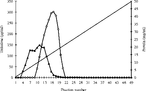
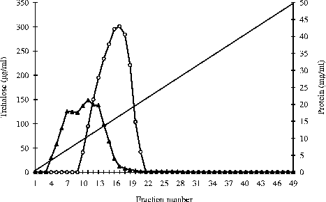
The enzyme was purified according to a three-step procedure, namely with ammonium sulphate precipitation, ion exchange chromatography and molecular filtration. In the first place, the enzymatic protein was precipitated from the crude extract using ammonium sulphate within the saturation range of 0.6–0.8. The sediment was collected by centrifugation at 2000 g for 20 min at 4°C. It was then dissolved in 0.9% NaCl and dialysed overnight against deionized water at 4°C. In the second step the dialysate was lyophilized (Martin Christ freeze dryer Alpha 1-4 LD, Osterode am Harz, Germany) and dissolved in 5 ml of 0.9% NaCl, and then centrifuged for 10 min at 1500 g. The supernatant was applied to a column (3.5 × 24.5 cm; Pharmacia Biotech, Piscataway, New Jersey, USA) of DEAE SEPHACELTM ionite (Diethylaminoethyl-Sephacel, Sigma-Aldrich, St. Louis, Missouri, USA). Proteins were eluted from the column with a linear gradient of 0.1–0.5 M NaCl. Protein elution was monitored spectrophotometrically at 280 nm using a Nano Drop ND-1000 spectrophotometer (Thermo Fisher Scientific, Waltham, Massachusetts, USA). The fractions were collected with a flow rate of 1 ml/min (fig. 1). Then, the active fractions (nos 13–17; fig. 1) were pooled and dialysed overnight (33 × 21 mm dialysis tubes, Sigma) against deionized water at 4°C and lyophilized. As a further step the lyophilizate was dissolved in 2.5 ml of 0.9% NaCl, and centrifuged at 1500 g for 15 min. The supernatant was applied to a column (3.5 × 27 cm) of Sepharose 6B (Sigma), equilibrated with 0.1 M acetic acid–ammonia buffer (pH 4.2). Proteins were eluted from the gel with the same buffer. Protein elution was monitored spectrophotometrically. Fractions were collected with a flow rate of 1 ml/min. The active fractions were pooled, dialysed and then lyophilized.
DEAE SEPHACELTM column chromatography. The protein bound to the column was eluted using a linear sodium chloride gradient between 0 and 0.5 m in 0.1 m acetic acid–ammonia buffer pH 4.2 (—). The protein present in each fraction (solid triangles) was monitored spectrophotometrically at 280 nm. The activities of trehalose 6-phosphate synthase (TPS) fractions (numbers 13–17) were pooled (open circles).
Characteristics of the enzyme
The activity of TPS was determined using a method described by Giaever et al. (Reference Giaever, Styrvold, Kaasen and Strøm1988). The end-product of the reaction (trehalose) was assayed by high-performance liquid chromatography using a method described by Dmitryjuk et al. (Reference Dmitryjuk, Łopieńska-Biernat and Farjan2009). The activity of TPS was expressed in units (u) per mg protein measured with the Bradford (Reference Bradford1976) method. One unit defines the quantity of trehalose (μmol) synthesized during 1 min at 37°C. The effect of pH on the enzyme was determined in 0.1 M acetic acid–ammonia buffer within the range of pH 3.0–8.6. Study of the optimum temperature of the enzyme was carried out in the temperature range of 20–90°C in 0.1 M acetic acid–ammonia buffer at pH 4.2. For each subsequent sample, the temperature was increased by 5°C. To study the thermal stability of the enzyme, test samples containing buffered enzymatic protein were preincubated for 15 min at temperatures from 20 to 90°C. Next, the samples were cooled or warmed to 37°C and a enzymatic reaction was started by introduction of substrates: UDPG and G6P. For assaying the influence of chemical compounds, 10 mM solutions of the compounds CaCl2, NaCl, ZnCl2, FeCl3, MgCl2, KCl and ethylenediaminetetra-acetic acid (EDTA) were used. The activity of the control sample, which contained water instead of the effector, was assumed at 100%. The substrate specificity of TPS was studied against 10 mM solutions of glucose, fructose, G6P and fructose 6-phosphate (F6P) (Sigma). The molecular weight of the denatured enzymatic protein was established using the sodium dodecyl sulphate–polyacrylamide gel electrophoresis (SDS-PAGE) non-continuous system of Laemmli (Reference Laemmli1970). Protein separation was conducted using EV243 apparatus (Sigma) and SigmaMarkerTM Low Range (molecular weight 29–200 kDa). After the electrophoresis, the gel was stained with the method described by Neuhoff et al. (Reference Neuhoff, Stamm and Eibl1985) using Coomassie brilliant blue G-250 colloidal dye.
An aliquot of 10 μg of TPS protein was used to designate the isoelectric point (pI). The protein was dissolved in 400 μl of denaturation buffer (7 M urea, 2 M thiourea, 2% CHAPS (3-[(3-cholamidopropyl)dimethylammonio]-1-propanesulphonate) buffer, 0.5% immobilized pH gradient (IPG) buffer, 0.002% bromophenol blue). Then, 2.5 μl of ampholytes (pH 3.0–10.0; Sigma) were added. At the same time, a second sample with isoelectric focusing (IEF) protein markers (amyloglucosidase from Aspergillus niger pI 3.6, trypsin inhibitor from Glycine max pI 4.6 and carbonate anhydrase isoenzyme from human erythrocytes pI 6.6; Sigma) was prepared. The pI of the protein was determined using gel strips (24 cm long) and EtanTMIPGphorTM 3 IEF System apparatus (GE Healthcare, Piscataway, New Jersey, USA). The separation was conducted according to the following parameters: 500 V/1.00 h/0.5 kVh; 1000 V/1.00 h/0.8 kVh; 8000 V/3.00 h/13.5 kVh; 8000 V/3.00 h/30 kVh; 10,000 V/3.00 h/16.5 kVh; 10,000 V/2.75 h/27.2 kVh. After removing the gel strips, they were immersed in a fixative fluid (10% acetic acid, 40% ethanol) for 1 h. Next, they were stained using a modified Neuhoff et al. (Reference Neuhoff, Stamm and Eibl1985) method. According to the localization of marker bands, the pI of the studied enzymatic protein was determined.
Sequencing of trehalose 6-phosphate synthase genes from Ascaris suum muscles
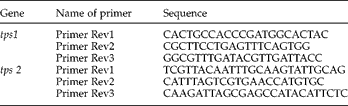
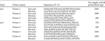
The research material was isolated from live worms brought in a warm Ringer solution from a meat-processing plant. Samples of 50 mg muscle were separated under sterile conditions, frozen in liquid nitrogen and stored at − 70°C until later analysis. Total RNA extraction was performed using a Total RNA Kit (A&A Biotechnology, Gdynia, Poland). RNA quality was confirmed electrophoretically. A TransScriba Kit was used (A&A Biotechnology) for the synthesis of cDNA. In the first stage, 5 μl of total RNA were preincubated with 0.2 μl of appropriate reverse primer (table 1) in a total volume of 11 μl at 70°C for 5 min and then cooled at 4°C for 6 min. In the next stage, 4 μl of 5 × reverse transcriptase (RT) reaction buffer, 2 μl of 10 mM mix of triphosphate deoxyribonucleotides (dNTPs) and 1 μl of Riboblock were added to the mixture and incubated for 5 min at 37°C. In the third stage, 2 μl of TransScriba Reverse Transcriptase (20 U/μl) were added to the mixture to a total volume of 20 μl and incubated for 90 min at 37°C, and for the next 10 min at 70°C. In the polymerase chain reaction (PCR) primers were used with sequences shown in table 2. The reaction was carried out in a volume of 50 μl. The reaction mixture consisted of 5 μl cDNA, 25 μl Mix Plus High GC (A&A Biotechnology), 0.1 μl appropriate tps forward primer concentration of 100 mM and 0.1 μl appropriate tps reverse primer at the same concentration, and 23.8 μl of nuclease-free water. The PCR was performed in a Gradient thermocycler (Eppendorf, Hamburg, Germany) using the following temperature–time profile: 94°C, 2 min; (94°C, 30 s, 65°C, 45 s, 72°C, 90 s) × 40 cycles; 72°C, 5 min. PCR products were applied to electrophoresis on 2% agarose gel containing 0.01% ethidium bromide. During the cloning into the pJET1 vector (3.1 kb; Fermentas, Burlington, Ontario, Canada), a non-purified DNA fragment was reacted with 0.5 μl of polymerase WALK (thermostable polymerase Pwo; A&A Biotechnology) within 5 min at 70°C using a Gradient thermocycler (Eppendorf). After electrophoresis, products were isolated from the 2% gel and purified using a Gel-Out Kit (A&A Biotechnology). PCR products were thus obtained with blunt ends suitable for cloning into the vector. The ligation reaction was adjusted to 20 μl. The ligation mixture consisted of: 1 μl of pJET1 DNA, 9 μl of the corresponding tps gene fragment, 1 μl of DNA T4 ligase (A&A Biotechnology) and 9 μl of water. Ligation was performed for 18 min at room temperature. The ligation mixture was transformed into competent cells of Escherichia coli TOP10F' (Novagen, Madison, Wisconsin, USA). Competent cells were prepared using an E. coli Transformer Kit (A&A Biotechnology). To transform, 100 μl of competent cells were used, to which was added the entire volume of the ligation mixture. Then the sample was placed on ice for 60 min and sonicated in Sonic3 (Polsonic, Poznań, Poland) for 3 s. The prepared samples were distributed on plates with LA (Luria agar) solid medium with ampicillin (50 μl/ml). Then the plates were incubated for 16 h at 37°C. The potential presence of clones in the colonies was checked for tps gene fragments by PCR with primers and reaction conditions as above. Next, transformant colonies with confirmed presence of the gene were transferred into 5 ml of liquid LB (Luria broth) medium with ampicillin (50 μl/ml) and incubated for 17 h at 37°C. Recombinant plasmids were isolated from cultures using a Plasmid Mini AX Kit (A&A Biotechnology). The resulting plasmids were investigated for the presence of gene fragments by PCR as above. PCR product sequencing was performed by a 3730xI DNA Analyzer (Applied Biosystems, Carlsbad, California, USA). The obtained sequences submitted in whole contigs and phylogenetic trees were constructed using Vector NTI® software (Invitrogen, Carlsbad, California, USA).
Sequences of the primers for reverse transcription.
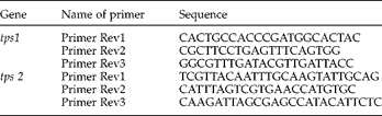
Sequences of the primers for polymerase chain reaction.

Results
Characteristics of the purified enzyme
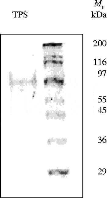
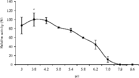
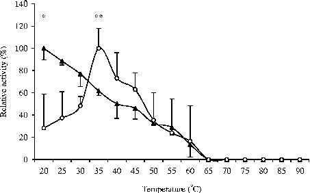
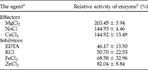
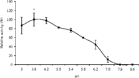
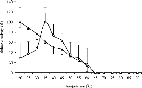
TPS was purified 265-fold by ammonium sulphate fractionation, ion-exchange chromatography on DEAETM (fig. 1) and molecular sifting on Sepharose 6B (table 3). The molecular weight of TPS (66 kDa) was determined based on SDS-PAGE (fig. 2). TPS is active in a range of pH 3.0–7.0. It was established that pH 3.8–4.2 is the optimum pH for the enzyme activity. The lowest TPS activity was observed at neutral pH. At a basic pH, the enzyme was inactive (fig. 3). The pI of TPS from A. suum muscles was 5.4. The optimum temperature for maximum activity of TPS was 35°C (fig. 4). At 40°C, the enzyme demonstrated 87.04% of its maximum activity. A rapid decrease in the enzyme activity occurred at 50°C, when TPS demonstrated only 35.27% of its maximum activity determined for the optimum temperature. The enzyme completely lost its activity above 60°C. TPS from the muscles of Ascaris is not a thermostable enzyme. The enzyme demonstrated activity in a range of 20–60°C during incubation without substrates. Maximum activity (100%) was assumed at a temperature of 20°C. With the increase in temperature, the activity of the enzyme gradually decreased and 35.37% of initial activity was achieved at 50°C. This enzyme was inactive at higher temperatures (fig. 4). The strongest activator of TPS was 10 mM MgCl2, followed by CaCl2 and NaCl. The presence of MgCl2 in the reaction mixture elevated TPS activity almost twofold (203%). With both NaCl and CaCl2 in the reaction mixture, the enzyme demonstrated 144% of control activity. EDTA, KCl, FeCl3 and ZnCl2 were inhibitors of TPS. Activity at the level of 45% was recorded for the control trial in the presence of 10 mM EDTA. A significant decrease in the enzyme activity (up to 50%) was observed in the presence of KCl. Activity at the level of 68% was recorded in the presence of FeCl3 (table 4). TPS from A. suum muscles was not fully specific towards G6P, which acts as a glucose acceptor in the reaction of synthesis of T6P. It was demonstrated that after the introduction of F6P into the reaction mixture, 60.83% (115.4 ± 61.81 u/mg) of control activity (189.7 ± 10.54 u/mg) was obtained for G6P. The enzyme was inactive in the presence of glucose and fructose.
Purification of trehalose 6-phosphate synthase.

a μmol of trehalose min− 1 mg− 1 protein; mean ± SD; n= 3.
Sodium dodecyl sulphate–polyacrylamide gel electrophoresis (SDS-PAGE) of trehalose 6-phosphate synthase (TPS) from Ascaris suum muscle. Right-hand lane: marker proteins – myosin from rabbit muscle (molecular weight (M r) = 200 kDa), α-galactosidase from Escherichia coli (M r = 116 kDa), phosphorylase b from rabbit muscle (M r = 97 kDa), albumin from bovine serum (M r = 66 kDa), glutamic dehydrogenase from bovine liver (M r = 55 kDa), ovoalbumin from chicken egg (M r = 45 kDa), glyceraldehyde 3-phosphate dehydrogenase from rabbit muscle (M r = 36 kDa) and carbonic anhydrase from bovine erythrocytes (M r = 29 kDa). Left-hand lane: TPS from A. suum muscle (7.6 μg).

Dependence of activity of trehalose 6-phosphate synthase (TPS) from Ascaris suum muscle on pH. The activities of samples at pH 3.8 (179.94 ± 26.04 u/mg of protein) were taken as 100%.
Thermostability and optimum of temperature of trehalose 6-phosphate synthase (TPS) from Ascaris suum muscle. Thermostability (solid triangles) and optimum temperature (open circles) of enzyme activities of samples at 20°C (*, 116.94 ± 11.96 u/mg of protein) and at 35°C (**, 136.15 ± 9.72 u/mg of protein) were taken as 100%.
Influence of different effectors and inhibitors on the activity of trehalose 6-phosphate synthase.
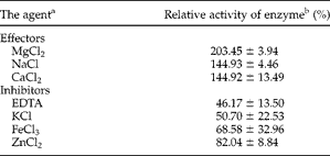
a The agent concentration was 10 mmol.
b The activities of control sample without effectors (126.4 ± 26.5 u/mg of protein) were taken as 100%. Mean ± SD; n= 3.
Sequencing of trehalose 6-phosphate synthase genes
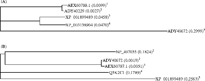
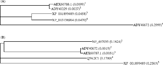
We isolated and sequenced two whole genes encoding TPS from A. suum muscle. Complete coding sequences for tps1 and tps2 were 3917 bp and 3976 bp, respectively. These are deposited in GenBank (GenBank accession numbers JF412033.2 and JF412034.2). Translation products were 1265 amino acids for AEX60788.1 and 1298 amino acids for AEX60787.1. Phylogenetic trees were constructed using Vector NTI showing that the product of tps1 from A. suum muscle exhibits high sequence similarity with analogous proteins of parasitic nematodes, such as Brugia malayi and Loa loa (fig. 5A). However, the tps2 gene product showed high sequence similarity with proteins derived from the free-living nematodes Caenorhabditis elegans and Aphelenchus avenae (fig. 5B).
Phylogenetic tree analysis of deduced amino acids of trehalose 6-phosphate synthase (TPS). (A) Product of tps1. 1Ascaris suum, tps1 (JF412033.2); 2Ascaris suum, α,α-trehalose phosphate synthase uridine diphosphate (UDP)-forming 1; 3Brugia malayi trehalose-phosphate synthase protein 1; 4Loa loa trehalose-phosphate synthase protein 1; 5Ascaris suum α,α-trehalose phosphate synthase UDP-forming 2. (B) Product of tps2. 1Caenorhabditis elegans, trehalose 6-phosphate synthase family member (tps2); 2Ascaris suum, α,α-trehalose phosphate synthase UDP-forming 2; 3Ascaris suum, tps2 (JF412034.2); 4Aphelenchus avenae, α,α-trehalose phosphate synthase UDP-forming 2; 5Brugia malayi, trehalose 6-phosphate synthase protein 1.
Discussion
Many researchers consider trehalose and T6P as regulators of carbohydrate metabolism in members of various systematic groups. These groups are mainly plants, fungi and invertebrates. In plants, T6P plays the role of a signal sugar in the regulation of carbohydrate utilization and starch metabolism, as well as in other pathways of carbohydrate metabolism (Lunn et al., Reference Lunn, Feil, Hendriks, Gibon, Morcuende, Osuna, Scheible, Carillo, Hajirezaei and Stitt2006; Paul, Reference Paul2007). In higher plants, trehalose is a potentially toxic compound; accumulation of T6P is harmful for pathogenic fungi as well (Veluthambi et al., Reference Veluthambi, Mahadevan and Maheshwari1981; Petzold et al., Reference Petzold, Himmelreich, Mylonakis, Rude, Toffaletti, Cox, Miller and Perfect2006). In Arabidopsis, the level of T6P is strongly correlated with TPS, TPP and trehalase activities (Winkler, Reference Winkler2002; Eastmond & Graham, Reference Eastmond and Graham2003). Trehalose is the main sugar occurring in the haemolymph of insects. In the insect fat body, synthesis of trehalose is stimulated by hypertrehalosaemic hormones and is used mainly for flying (Becker et al., Reference Becker, Schloder, Steele and Wegener1996; Thompson, Reference Thompson2003). The TRET1 gene codes for a trehalose transporter responsible for the release of trehalose from the insect fat body. Other tissues use the released trehalose as a carbon source, which might influence the regulation of trehalose level in insect haemolymph (Kanamori et al., Reference Kanamori, Saito, Hagiwara-Komoda, Tanaka, Mitsumasu, Kikuta, Watanabe, Cornette, Kikawada and Okuda2010). Moreover, trehalose plays a significant role in nematode metabolism. It is an energy reserve for adult nematodes and nematode larvae developing in eggs. It protects organisms from environmental stresses such as drying or freezing, improves thermostability and extends nematodes' lifespan (Elbein et al, Reference Elbein, Pan, Pastuszak and Carroll2003; Honda et al., Reference Honda, Tanaka and Honda2010).
The current state of knowledge about the metabolism and role of trehalose and T6P in members of various systematic groups within Nematoda is incomplete. Thus far, properties of TPS have been described only in one nematode, A. avenae (Loomis et al., Reference Loomis, Madin and Crowe1980); because of this, the discussion will largely rest on representatives of different systematic groups. Genetic aspects of trehalose metabolism have been studied more often. The tps genes of C. elegans and filarial nematodes have been determined (Pellerone et al., Reference Pellerone, Archer, Behm, Grant, Lacey and Somerville2003). Occurrence of tps genes has also been shown in A. avenae (Goyal et al., Reference Goyal, Browne, Burnell and Tunnacliffe2005). For the first time in nematodes, Kormish & McGhee (Reference Kormish and McGhee2005) found the existence of the gene encoding TPP. Besides the encoding of TPP by the gob-1 gene in C. elegans, it was observed that the concurrent inhibition of tps1 and tps2 genes abolishes the lethal phenotype of gob-1. This has been interpreted to mean that gob-1 lethality is not due to low trehalose levels but rather to the build-up of the toxic intermediate T6P.
Some aspects concerning the synthesis of trehalose in A. suum have been presented in studies by Feist et al. (Reference Feist, Read and Fisher1965). In our previous study, the activity of TPS and TPP was demonstrated in muscles and individual parts of the female reproductive system as well as in the haemolymph of A. suum. No activity of TPS and TPP has been observed in parasite intestine and cuticle (Dmitryjuk et al., Reference Dmitryjuk, Łopieńska-Biernat and Farjan2009). Results demonstrated in the present study expand our knowledge about the synthesis of trehalose in nematodes by providing detailed information about biochemical and genetic properties of TPS isolated from A. suum muscles. Because there is no synthesis pathway of trehalose in parasite hosts, these studies have practical aspects in searching for nematotoxic drugs that will be safe and effective for animals infected with Ascaris.
The yield of TPS purification from A. suum muscles was low (10.6%). Nishimoto et al. (Reference Nishimoto, Nakano, Nakada, Chan, Fukusa, Sugimoto, Kurimoto and Tsujisaka1996) also obtained low purification efficiency for trehalose synthase (11.25%) isolated from Pimelobacter sp. A very low yield was obtained by Vandercammen et al. (Reference Vandercammen, Francois and Hers1989) in the purification of TPS from Saccharomyces cerevisiae. This low yield of the purified enzyme from A. suum muscle may be due to the fact that this protein is strongly associated with the muscle cell membrane structures, as was the case for acid trehalase from the same source (Dmitryjuk & Żółtowska, Reference Dmitryjuk and Żółtowska2003). Others, using different chromatographic procedures, have obtained better results. Slightly higher purification efficiencies (36 and 60%) have been obtained for trehalose synthase isolated from Grifola frondosa and Saccharomycopsis fibuligera, respectively (Saito et al., Reference Saito, Kase, Takahashi, Takahashi and Horinouchi1998; Liang et al., 2006). A very high purification yield (89%) was obtained for TPS isolated from Selaginella lepidophylla using a Ni-dipicolylamine (DPA) matrix (immobilized metal affinity chromatography) (Valenzuela-Soto et al., Reference Valenzuela-Soto, Marquez-Escalante, Iturriaga and Figueroa-Soto2004).
The purified enzyme demonstrated the highest activity at pH 3.8–4.2. TPS isolated from Ascaris muscles is surprisingly very acidic, similar to trehalase from the same source (Dmitryjuk & Żółtowska, Reference Dmitryjuk and Żółtowska2003). The enzyme isolated from A. avenae demonstrated the highest activity at basic pH 8.0 (Loomis et al., Reference Loomis, Madin and Crowe1980), whereas TPS from A. suum muscles at this pH is already inactive. TPS from the thermophile bacterium, Thermus thermophilus, demonstrated the highest activity at pH 6.0 (Silva et al., Reference Silva, Alarico and Da Costa2005). Moreover, the enzyme isolated from G. frondosa had optimum activity at a pH range of 6.5–6.8 (Saito et al., Reference Saito, Kase, Takahashi, Takahashi and Horinouchi1998). The optimum pH of trehalose synthase activity from Thermobifida fusca was 6.5 (Wei et al., Reference Wei, Zhu, Luo, Lu, Chen, Wang, Huang, Meng, Wang and Huang2004). As compared with TPS from A. suum, a higher optimum pH (pH 7.0) was also recorded in S. lepidophylla and Mycobacterium smegmatis (Pan et al., Reference Pan, Edavana, Jourdian, Edmondson, Carroll, Pastuszak and Elbein2004; Valenzuela-Soto et al., Reference Valenzuela-Soto, Marquez-Escalante, Iturriaga and Figueroa-Soto2004). Trehalose synthase originating from Pimelobacter sp. demonstrated a similar optimum pH (pH 7.5) (Nishimoto et al., Reference Nishimoto, Nakano, Nakada, Chan, Fukusa, Sugimoto, Kurimoto and Tsujisaka1996). Trehalose synthase from Pseudomonas stutzeri (pH 8.5–9.0) represented an alkaline enzyme as well (Lee et al., Reference Lee, Lee, Kim, Lee, Kim, Park and Chung2005).
The pI of TPS from A. suum was 5.4. Similarly, trehalose synthase from Pimelobacter sp. has a lower pI (4.6) than the studied enzyme (Nishimoto et al., Reference Nishimoto, Nakano, Nakada, Chan, Fukusa, Sugimoto, Kurimoto and Tsujisaka1996). However, pIs obtained for trehalose synthases isolated from Pseudomonas sp. (pI 5.8) and Ginkgo biloba (pI 5.83) were higher than recorded in A. suum (Ohguchi et al., Reference Ohguchi, Kubota, Wada, Yoshinaga, Uritani, Yagisawa, Ohishi, Yamagishi, Ohta and Ishikawa1997; Wu et al., Reference Wu, Pang, Shen, Lu, Lin, Wang, Sun and Tang2006).
The optimum temperature for the activity of synthase from A. suum muscles was 35°C, close to temperatures recorded for Ascaris hosts. A similar optimum temperature of activity was found for trehalose synthases isolated from Pseudomonas putida, P. stutzeri and G. frondosa (Saito et al., Reference Saito, Kase, Takahashi, Takahashi and Horinouchi1998; Lee et al., Reference Lee, Lee, Kim, Lee, Kim, Park and Chung2005). A lower optimum temperature (20°C) was recorded for trehalose synthase isolated from Pimelobacter sp. (Nishimoto et al., Reference Nishimoto, Nakano, Nakada, Chan, Fukusa, Sugimoto, Kurimoto and Tsujisaka1996). The enzyme originating from T. thermophilus had a significantly higher temperature of activity and its optimum action was established at 98°C; such a high temperature resembles the living environmental conditions for these bacteria (Silva et al., Reference Silva, Alarico and Da Costa2005). An opposite result was obtained by Wei et al. (Reference Wei, Zhu, Luo, Lu, Chen, Wang, Huang, Meng, Wang and Huang2004) for TPS from T. fusca. Here, the optimum temperature for enzyme action was 25°C. A higher temperature was recorded in S. lepidophylla. The optimum temperature for the fraction eluted with 1 M KCl was 55°C (Márquez-Escalante et al., Reference Márquez-Escalante, Figueroa-Soto and Valenzuela-Soto2006).
TPS represents a non-thermostable enzyme. The purified enzyme had only 14% of its maximum activity at 60°C. Like TPS from A. suum muscle, that from P. putida does not have thermostable properties (Ma et al., Reference Ma, Xue and Sun2006); however, the enzyme from T. thermophilus was extremely thermostable and it maintained 50% of its activity at 100°C (Silva et al., Reference Silva, Alarico and Da Costa2005). In addition, trehalose synthase isolated from Pimelobacter sp. also demonstrated higher stability than TPS from A. suum (Nishimoto et al., Reference Nishimoto, Nakano, Nakada, Chan, Fukusa, Sugimoto, Kurimoto and Tsujisaka1996).
Two reagents, 10 mM EDTA and 10 mM KCl, were shown as inhibitors of TPS; however, the strongest activator of TPS was MgCl2, followed by NaCl and CaCl2. An increase in the enzyme activity in P. putida and T. thermophilus in the presence of Mg2+ was demonstrated by Ma et al. (Reference Ma, Xue and Sun2006) and Silva et al. (Reference Silva, Alarico and Da Costa2005). TPS from S. cerevisiae was activated by 5 mM Mg2+, but higher concentrations of Mg2+ may inhibit the enzyme (Vandercammen et al., Reference Vandercammen, Francois and Hers1989). K+ ions have a proactive effect on trehalose synthase isolated from P. putida and an inhibiting effect on TPS from A. suum, whereas Zn2+ ions inhibit the enzyme in both species. Contrary to the enzyme studied here, 10 mM Ca2+ is an inhibitor of trehalose synthase isolated from P. stutzeri (Lee et al., Reference Lee, Lee, Kim, Lee, Kim, Park and Chung2005). Activation of the enzyme isolated from A. suum by Na+ ions is consistent with the results obtained for trehalose synthase from E. coli (Giaever et al., Reference Giaever, Styrvold, Kaasen and Strøm1988). Zn2+ ions were shown to inhibit the enzyme isolated from Pimelobacter sp. (Nishimoto et al., Reference Nishimoto, Nakano, Nakada, Chan, Fukusa, Sugimoto, Kurimoto and Tsujisaka1996) and TPS from A. suum. Moreover, Zn2+ ions act as an inhibitor of synthase from T. fusca (Wei et al., Reference Wei, Zhu, Luo, Lu, Chen, Wang, Huang, Meng, Wang and Huang2004). As well as A. suum, Mg2+, Ca2+ and Na+ ions have an impact on the activation of enzymes from S. lepidophylla, whereas K+ ions activate only TPS from S. lepidophylla (Valenzuela-Soto et al., Reference Valenzuela-Soto, Marquez-Escalante, Iturriaga and Figueroa-Soto2004).
The molecular weight of TPS isolated from A. suum was 66 kDa on SDS-PAGE. Based on BLASTP (http://blast.ncbi.nlm.nih.gov), the protein of TPS is a homotetramer. There was only a slight difference in the molecular weight of trehalose synthase (67 kDa) from Pseudomonas sp. The molecular weight was determined with gel filtration and suggested that the native enzyme from Pseudomonas was composed of four units (Ohguchi et al., Reference Ohguchi, Kubota, Wada, Yoshinaga, Uritani, Yagisawa, Ohishi, Yamagishi, Ohta and Ishikawa1997). A lower weight (52 kDa) determined by SDS-PAGE electrophoresis was reported for the enzyme isolated from T. thermophilus (Silva et al., Reference Silva, Alarico and Da Costa2005). The enzyme isolated from Mycobacterium tuberculosis demonstrated a lower molecular weight (58 kDa) than the enzyme isolated from A. suum and also represented a tetramer (Pan et al., Reference Pan, Carroll and Elbein2002). Synthase from M. smegmatis had a similar molecular weight (68 kDa) as the TPS studied here; however, the active enzyme was probably a hexamer (Pan et al., Reference Pan, Edavana, Jourdian, Edmondson, Carroll, Pastuszak and Elbein2004). Trehalose synthase from Pimelobacter sp. (Nishimoto et al., Reference Nishimoto, Nakano, Nakada, Chan, Fukusa, Sugimoto, Kurimoto and Tsujisaka1996) had a slightly lower weight (62 kDa). The active enzyme from G. frondosa was a dimer (120 kDa; Saito et al., Reference Saito, Kase, Takahashi, Takahashi and Horinouchi1998). A higher molecular weight (97.9 kDa) was recorded for TPS from G. biloba (Wu et al., Reference Wu, Pang, Shen, Lu, Lin, Wang, Sun and Tang2006).
The enzyme isolated in the present study did not demonstrate complete specificity towards the glucose acceptor, G6P. In the presence of F6P, TPS from A. suum demonstrated more than half of its maximum activity recorded for G6P. On the contrary, T6P synthases from T. thermophilus, M. tuberculosis and S. lepidophylla were completely specific towards G6P (Pan et al., Reference Pan, Edavana, Jourdian, Edmondson, Carroll, Pastuszak and Elbein2004; Valenzuela-Soto et al., Reference Valenzuela-Soto, Marquez-Escalante, Iturriaga and Figueroa-Soto2004; Silva et al., Reference Silva, Alarico and Da Costa2005). Similar to E. coli, TPS from A. suum did not show activity after the replacement of G6P with glucose (Giaever et al., Reference Giaever, Styrvold, Kaasen and Strøm1988). TPS from S. lepidophylla demonstrated around 30% activity in the presence of glucose as an acceptor; however, both enzymes from A. suum and S. lepidophylla did not show activity against fructose (Valenzuela-Soto et al., Reference Valenzuela-Soto, Marquez-Escalante, Iturriaga and Figueroa-Soto2004).
In our studies, we isolated and sequenced two whole genes encoding the TPS from A. suum muscle. Sequences of these genes exhibit high sequence similarity with analogous genes from different developmental stages of A. suum (JI164351.1 and JI164934.1; Wang et al., Reference Wang, Czech, Crunk, Wallace, Mitreva, Hannon and Davis2011) and also in further stages from other parasitic and free-living nematodes. BLASTP analysis showed that the investigated enzyme belongs to the blood type B glucosyltransferase (GTB)-type superfamily (multi-domains of trehalose-OtsA; specific hits of GT1-TPS). Earlier overall phylogenetic analysis carried out by Avonce et al. (Reference Avonce, Mendoza-Vargas, Morett and Ituriaga2006) showed that the nematode TPS sequences were found within the cluster comprising the eubacterial and archaeal proteins, suggesting a possible lateral gene transfer event. We carried out cluster analysis of Nematoda and constructed phylogenetic trees showing that products of tps1 from A. suum muscle exhibit high sequence similarity with analogous proteins of parasitic nematodes (B. malayi and L. loa), and the tps2 gene product showed a high sequence similarity with proteins derived from free-living nematodes (C. elegans and A. avenae). The phylogenetic analysis of Goyal et al. (Reference Goyal, Browne, Burnell and Tunnacliffe2005) showed the close relationship between nematode TPS sequences (to 70% identity, from A. avenae, C. elegans and C. briggsae). According to the authors, this supports the hypothesis that two C. elegans genes diverged before the evolutionary separation from A. avenae, after which a reduplication event occurred in A. avenae to give rise to tps1 and tps2. It is unknown if this was the case with parasitic nematodes. Possibly a similar situation occurred with TPS genes in A. suum.
Only one protein having TPS activity was isolated from Ascaris muscles; however, the presence of both genes was observed. Therefore, it is highly probable that two forms of this enzyme can exist in muscle cells, like trehalase from Ascaris (Dmitryjuk & Żółtowska, Reference Dmitryjuk and Żółtowska2003). However, further studies are required to demonstrate the intracellular localization of this enzyme. The necessity for further studies on the intracellular level as well as tissue localization of the enzyme in Caenorhabditis was suggested by Pellerone et al. (Reference Pellerone, Archer, Behm, Grant, Lacey and Somerville2003).
Summing up, TPS isolated from A. suum muscles is mostly activated by Mg2+ and strongly inhibited by EDTA, is a non-active enzyme in alkaline pH, is a non-specific enzyme for G6P and is also a non-thermostable protein. Two genes (tps1 – JF 412033.2 and tps2 – JF412034.2), similar to those of other nematodes, encode TPS in the muscles of the parasite.
Acknowledgements
This research was supported financially by The Ministry of Science and Higher Education (grant no. 2PO4C 124 29, 2005–2009). We would like to thank Anita Franczak, PhD, and research scientists at the Department of Animal Physiology of the Biology Faculty at the University of Warmia and Mazury, Olsztyn (Poland) for their invaluable help in molecular studies.