Implications
Blood cortisol is a component that indicates stress condition in livestock. However, blood cortisol concentrations vary because of different factors including environmental temperature or humidity, management and physiological conditions. Measuring cortisol in the hair of animals is a new technique that precisely reveals stress condition in livestock. Water restriction can increase stress in animals, especially during hot and humid weather. This study shows that cortisol concentration in wool is a better indicator than blood cortisol in sheep exposed to heat stress, receiving water restriction.
Introduction
In any type of grazing system, water is often a limiting factor for sheep (Casamassima et al., Reference Casamassima, Pizzo, Palazzo, DʼAlessandro and Martemucci2008). Water restriction may exacerbate the impact of heat stress, especially in hot and humid climates (Marai et al., Reference Maria, El-Darawany, Fadiel and Abdel-Hafez2007). Water restriction for 4 days in sheep increased the blood cortisol concentrations (Li et al., Reference Li, Christopherson and Cosgrove2000). Cortisol is a biomarker of the physiological response to stress, including heat stress. This hormone is usually measured in blood serum or plasma, requiring stressful handling procedures such as capturing, restraining animals, and venipuncture or blood collection from catheter. These procedures can potentially confound the reliability of the assessment (Creel et al., Reference Creel, Creel, Wildt and Monfort1992; Davenport et al., Reference Davenport, Tiefenbacher, Lutz, Novak and Meyer2006) and provide highly variable data in blood. Some researchers (Boandl et al., Reference Boandl, Wohlt and Carsia1989; Creel et al., Reference Creel, Creel, Wildt and Monfort1992) found increased blood cortisol concentration in response to handling, capturing or restraining animals. Concentration of cortisol in the animal’s hair may provide precise and reliable data because the concentration changes with a positive correlation with stressors such as environment or temperature (Kalra et al., Reference Kalra, Einarson, Karaskov, Van Uum and Koren2007; Yamada et al., Reference Yamada, Stevens, de Silva, Gibbins, Beyene, Taddio, Newman and Koren2007). Coping with this issue, stressful sample collection can be avoided by using alternative sample matrices such as urine, feces or saliva (Accorsi et al., Reference Accorsi, Carloni, Valsecchi, Viggiani, Gamberoni, Tamanini and Seren2008). Each has clear limitations as explained by Davenport et al. (Reference Davenport, Tiefenbacher, Lutz, Novak and Meyer2006). Samples derived from restrained animals are problematic because stress may alter blood and urine hormonal levels (Creel et al., Reference Creel, Creel, Wildt and Monfort1992). One technique to minimize these limitations is to measure cortisol in the matrix of hair, suggested by Yamada et al. (Reference Yamada, Stevens, de Silva, Gibbins, Beyene, Taddio, Newman and Koren2007). Cortisol concentrations in the biological matrices may be affected by stress-induced changes in the hypothalamus–pituitary axis over extended periods of time (Comin et al., 2011). As cortisol in the hair is not affected by short-term environmental factors such as handling, it may be a reliable approach to measure endogenous cortisol. To the best of the author’s knowledge, there are no reports measuring cortisol in the wool of sheep, especially during short periods of heat stress. Water restriction could intensify the effect of heat stress or reduce excessive stress if available. Therefore, this study was conducted to examine wool and blood cortisol concentrations, and water consumption patterns of sheep exposed to heat stress receiving water restriction.
Material and methods
Nine 3-year-old Corriedale female sheep (average BW=43±6.5 kg), individually fed diets based on maintenance requirement in metabolic crates, were assigned to treatments according to a Latin square design for three periods of 21 days each. Treatments included free access to water (FAW), 2 h water restriction (2hWR) and 3 h water restriction (3hWR) after feeding. Following the water restriction time, ewes were given FAW. Water was provided in 5 l buckets for each sheep individually and residual water every day was collected twice and weighed, and fresh water was offered every time. Feed was provided as a TMR (70% concentrate, 30% forage) having 60% DM. Analyzed composition of TMR (DM basis) was CP: 16.1%; NDF: 39.9%; and TDN: 69.1%. Feed was weighed and offered twice daily at 0900 and 1800 h. Water was available free of choice to the FAW group; however, it was provided after feeding time and at 1100 h and 2000 h for the 2hWR group, and at 1200 h and 2100 h for the 3hWR group, respectively. Blood was collected by jugular venipuncture in two vacutainer tubes (one tube containing EDTA for measuring neutrophils and another tube with no additive for collecting serum) at 1300 h on day 21 of each period. After collection, the serum was obtained by centrifugation (1200×g for 20 min) and then placed in storage tubes and were used for cortisol analysis (RIA method). Blood in vacutainers containing EDTA was used for neutrophil analysis by laser flow cytometry (Hemo Vet 950, HemoVet. Co., San Antonio, USA). Experimental room temperature and relative humidity were monitored at hourly intervals throughout the trial by using a temperature–humidity data logger device (CEM-DT-172, No. 11048007, Shenzhen, China). Average temperature–humidity index (THI) in the experimental room was 27.9 throughout the experiment calculated using the equation of Marai et al. (Reference Maria, El-Darawany, Fadiel and Abdel-Hafez2007), which defines heat stress condition
 $${\rm THI}=db\,\left(\!{{}{\deg}\!{\rm C}} \right){\minus}\left\{ {\left( {0.31{\minus}0.31{\rm RH}} \right)\left( {db\,\left(\!{{}{\deg}\!{\rm C}} \right){\minus}14.4} \right)} \right\}$$
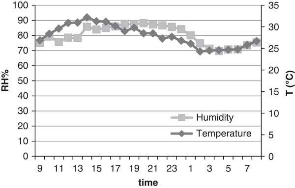
where db is the dry bulb temperature (°C) and RH is the relative humidity. The THI values obtained indicate the following: <22.2=absence of heat stress; 22.2 to <23.3=moderate heat stress: 23.3 to <25.6=severe heat stress, and 25.6 and more=extreme severe heat stress. Average daily mean temperature (°C) and relative humidity (RH%) were presented in Figure 1 where the minimum and maximum temperature and relative humidity were 25.1±0.09°C, 30.0±0.12°C, 75.5±4.7%, and 88.2±2.3%, respectively.
Average daily pattern of temperature (°C) and relative humidity (RH%) in experimental room (0900 to 0800 h).

Wool cortisol analysis
At the end of each period (21 days), wool was carefully shaved at 1400 h without damaging the skin or contaminating the sample using commercially available pet grooming clippers. Then, the wool samples were wrapped in aluminum foil as described by Davenport et al. (Reference Davenport, Tiefenbacher, Lutz, Novak and Meyer2006). Preparing wool for cortisol analysis included shaving (from the posterior vertex region of the neck between the cisterna magna and scapular bones), wrapping in aluminum foil, unwrapping and washing with isopropanol and methanol, drying, grinding and then applying hair cortisol assay using the kit, according to the manufacturer’s recommendation (Salimetrics, high sensitivity salivary cortisol, enzyme immuneassay kit, No. 1-3002, State College, Pennsylvania, USA). Washing procedure using isopropanol and methanol was performed as described by Davenport et al. (Reference Davenport, Tiefenbacher, Lutz, Novak and Meyer2006) followed by drying (Paulsen et al., Reference Paulsen, Wilkins, Slawson, Shaw and Rollins2001). The dried wool samples were reduced to fine particles using scissors. Davenport et al. (Reference Davenport, Tiefenbacher, Lutz, Novak and Meyer2006) used a Retsch ball mill for grinding long hair from monkeys. This may induce errors as steroids may stick to the surface of the ball mill during the grinding process, resulting in lower values. However, in the present study, we measured cortisol in wool that was short in length. Pooled wool samples (250 mg) were placed in 9 ml polypropylene tubes. Then 5 ml of isopropanol was added to the samples twice. Samples were then gently mixed on a rotator at room temperature for 3 min per wash, and 2 ml of the solvent from each wash was collected in a separate tube to determine the concentration of overall cortisol lost during the washing procedure and mentioned as washed wool cortisol. The wool samples were then allowed to dry for 7 days in a clean protected hood (Paulsen et al., Reference Paulsen, Wilkins, Slawson, Shaw and Rollins2001). After drying, ∼50 mg of finely reduced wool samples (by scissors) was weighed and placed into a 2 ml microcentrifuge tube. One milliliter of methanol was added to each microcentrifuge tube, and the tubes were incubated at room temperature for 24 h with slow rotation to extract steroid hormones. After extraction, samples were spun for 30 s in a microcentrifuge tube and a 0.6 ml aliquot of methanolic extract was added to a new tube and dried at 38°C under a stream of N gas. This volume was chosen to avoid contamination of the supernatant with wool particles. The dried extracts were reconstituted with 0.4 ml of phosphate buffer provided in the assay kit as described by Davenport et al. (Reference Davenport, Tiefenbacher, Lutz, Novak and Meyer2006) for analyzing cortisol concentration and mentioned as dry wool cortisol. The remaining fraction of finely reduced wool dissolved in methanol (included wool and methanol after excluding 0.6 ml of methanol) was analyzed for cortisol concentration and mentioned as residual extract.
Wool cortisol concentrations were analyzed using two methods. First, by using a human cortisol test method using chemiluminescence immunoassay (Kohen et al., Reference Kohen, Pazzagli, Kim and Lindner1980), the test sensitivity was 0.2 µg/dl. Second, by using salivary ELISA test method (Bennett and Hayssen, Reference Bennett and Hayssen2010), the sensitivity was 0.003 µg/dl. All standards and reagents were provided in the kit. Data from the first method were not accepted and shown as results that were inconsistent from period to period, and sometimes values were not detected. The salivary ELISA test method provided very precise and consistent data with the lowest standard deviation. The intra- and inter-assay coefficients of variations were 1.11 and 10.61, respectively.
Water consumption and pattern
Water consumption was recorded two times daily for the 5 final days of each period. Duration of water consumption defines the duration (seconds) that sheep in the 2hWR and 3hWR groups spent drinking water after 2 and 3 h restriction, respectively. A timer was used to measure the duration of water consumption for each sheep individually, immediately after offering water and stoppage of drinking water by ewes of the 2hWR and 3hWR groups. Water consumption rate (WCR) was calculated in the following manner: When sheep in the 2hWR and 3hWR groups stopped water intake (time recorded), the remaining water was weighed and WCR was calculated by dividing the volume of water consumed to the duration spent on water consumption. For the FAW group water was always available, and thus the rate of water consumption was calculated based on the mean of 2 h (7200 s) and 3 h (10 800 s). The experimental procedure and methods were approved by the animal welfare and ethics authority of Kangwon National University, Chuncheon, Korea.
Statistical analysis was carried out using GLM procedure of SAS (version 9.0; SAS institute Inc., Cary, NC, USA) for a Latin square (3×3) design. The model included effects of sheep, treatment and period as follows:
 $$Y_{{ijkl}} =\mu {\plus}\alpha _{i} {\plus}S_{j} {\plus}P_{k} {\plus}{\varepsilon}_{{ijkl}} $$
where Y ijkl is each observation, μ the total mean, α i the effects of treatment, S j the effects of sheep, P k the effects of period and ε ijkl the residual effect. Experimental room environment was controlled throughout the experiment and the effects of sheep and period was not significant. Duncan multiple range tests were used for ranking treatment means and statistical differences were considered significant at P<0.05.
Results and discussion
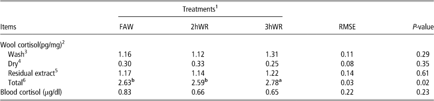
No differences (P>0.05) were found in wool cortisol concentration in each fragment of wool; however, total wool cortisol concentration was the highest (P<0.05) in the 3hWR group compared with other groups (Table 1). This suggests that the effect of stress was exacerbated by increasing the time of water restriction after feeding. Effect of higher (P<0.05) wool cortisol concentration in the 3hWR group might be the reason for suppressing neutrophil concentration (%) in blood compared with 2hWR group (36.82±1.84 and 32.42±2.72 for 2hWR v. 3hWR, respectively). The reason why neutrophil concentration in FAW group (29.67±3.35) was lower than two other groups remained unknown. Neutrophil/lymphocyte ratio showed the highest value in the 2hWR group (0.62±2.05) than other treatments (0.464±2.76, and 0.518±2.71 for FAW and 3hWR, respectively). Neutrophils are considered as the first cellular defense line against infections (Craven and Williams, Reference Craven and Williams1985) and could be suppressed by a high level of cortisol concentration. Suppressing neutrophil concentration in the 3hWR group compared with the 2hWR group might explain the more precise and reliable data in wool cortisol compared with blood cortisol, as blood cortisol did not show any difference between treatments. No difference was observed in the wool cortisol concentration between the FAW and 2hWR group, suggesting that the sheep receiving 2hWR were not stressed. However, no differences were found in the blood cortisol concentrations among treatments. This may be explained by the fact that the blood cortisol concentrations vary because of environmental stressors such as handling procedure (Boandl et al., Reference Boandl, Wohlt and Carsia1989; Creel et al., Reference Creel, Creel, Wildt and Monfort1992). Thus, individual variation among sheep resulted in large standard errors.
Effects of water restriction on wool and blood cortisol concentration in sheep exposed to heat stress

FAW=free access to water; 2hWR=2 h water restriction; 3hWR= 3 h water restriction.
1 Treatments included FAW, 2hWR and 3hWR after feeding.
2 All samples were derived from the same large pool of wool shaved from the posterior vertex region of the neck between the cisterna magna and scapular bones.
3 Wash wool cortisol represents the concentration of cortisol in the wash buffer (isopropanol); the dried wool samples were reduced to fine particles using scissors.
4 Dry wool cortisol represents the concentration of cortisol in fine wool particles after washing with methanol. Exactly 0.6 ml of the supernatant of this methanolic extract was dried and reconstituted with 0.4 ml of phosphate buffer.
5 Residual extract represents the concentration of cortisol in methanolic extract with fine wool particles remained in tube after removing 0.6 ml supernatant for dry wool cortisol analysis.
6 Total cortisol represents sum of concentrations of cortisol in wash, dry and residual extract.
a,bValues within a row with different superscripts differ significantly at P<0.05.
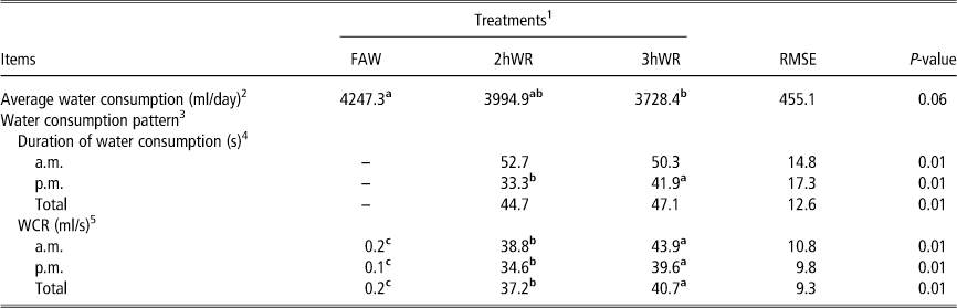
Water is the most important nutrient and should be available to livestock in adequate amounts during heat stress. In grazing systems, it is not unusual for sheep to have limited access to water at times. This can negatively affect their well-being (Silanikov, Reference Silanikov1987; Casamassima et al., Reference Casamassima, Pizzo, Palazzo, DʼAlessandro and Martemucci2008). However, water restriction for any reason can act as a stressor and will intensify the severity of stress, particularly during heat stress. No differences in average water consumption (P>0.05) were observed between the FAW and 2hWR groups and between the 2hWR and 3hWR groups (Table 2). However, sheep in the FAW group had higher (P<0.01) average water consumption than the 3hWR group. This is in agreement with Casamassima et al. (Reference Casamassima, Pizzo, Palazzo, DʼAlessandro and Martemucci2008) who reported higher water consumption for sheep with FAW compared with sheep that were restricted to 60% and 80% of total water consumption. The WCT in the 3hWR group was higher (P<0.01) than the 2hWR group for the p.m. recording, but means for the a.m. and total were not different (P>0.05). The THI was always higher in the afternoon than in the morning (26.16 v. 25.54). Sheep consumed more water during the hotter afternoon (exactly after access to water not the whole water consumption), especially when restricted from water for more than 2 h. Sheep were offered diets based on maintenance requirements and changed within the treatments in different periods; thus, individual variation and feed intake had no apparent effect on water consumption. It appears that the most important factor that affected water consumption of sheep was THI. The WCR was higher (P<0.01) in 3hWR group for all times (a.m., p.m. and total) than the 2hWR group. Sheep deprived of water for 3 h had higher (P<0.01) drive to consume water than sheep deprived for 2 h. However, total water consumed by the 3hWR group was less (P<0.01) than for other groups.
Effects of water restriction on water consumption in sheep exposed to heat stress

FAW=free access to water; 2hWR=2 h water restriction; 3hWR= 3 h water restriction; WCR=water consumption rate.
1 Treatments included FAW, 2hWR and 3hWR after feeding.
2 Average water consumption is the average of daily water consumed by each sheep in the treatment group during the last 7 days of each period.
3 Water consumption pattern includes duration of water consumption and rate.
4 Duration of water consumption is the duration (seconds) that sheep in the 2hWR and 3hWR groups spent drinking water after 2 and 3 h restriction, respectively.
5 WCR is calculated by dividing the volume of water consumed by each sheep to the duration spent on water consumption.
a–cValues within a row with different superscripts differ significantly at P<0.01.
The authors conclude that wool cortisol is a better predictor of stress than blood cortisol and is suitable to evaluate stress status of sheep during heat stress. Water restriction for more than 2 h after feeding can increase stress status in sheep by increasing the concentration of wool cortisol. Therefore, water should be available to sheep within 3 h after feeding.
Acknowledgments
Jerrold S. Meyer and Amanda F. Hamel (Psychology Department at the University of Massachusetts, Amherst, USA) are greatly acknowledged for providing their detailed procedure of analyzing hair cortisol in rhesus macaques. Authors also thank Saeed Hassani (Department of Animal Science, Gorgan University of Agricultural Sciences & Natural Resources, Iran and Jeon-Koo Lee (Department of Animal Resource Science, Kangwon National University, South Korea) for their help in planning the experimental design and SAS analysis.