Introduction
The aetiology of facial nerve damage is broad, and includes tumour infiltration, inflammatory disease, mastoid surgery and trauma. Of all the cranial nerves, the facial nerve is the most susceptible to injury from skull base trauma (frequently caused by motor vehicle accidents), due to its complex course through the narrow temporal bone canal.Reference Adegbite, Khan and Tan1, Reference Brodie and Thompson2 Such injuries (especially those involving temporal bone fractures) can lead to acute or delayed traumatic facial palsy, a common clinical entity which accounts for approximately one-third of cases of peripheral facial paralysis.
The severity of facial nerve palsy can vary greatly, ranging from transient, unilateral paresis to permanent, bilateral paralysis. As the major sequelae of this disorder have a significant impact on the patient's quality of life, accurate diagnosis and appropriate, effective medical intervention are necessary.Reference Melvin and Limb3 There is a direct correlation between the degree of facial nerve damage and the prognosis. It is crucial to determine the location of facial nerve damage, in order to select appropriate treatment (surgical or otherwise).Reference Melvin and Limb3 Clinical, testing of facial nerve alignment and function is performed in order to diagnose facial nerve injury. Alignment tests include temporal high resolution computed tomography (CT), the Schirmer test, and testing of taste and stapedial reflexes. Functional testing mainly comprises electrophysiological analysis, of which electroneurography (ENoG) is the most relevant.
Electroneurography estimates the percentage of degenerating facial nerve fibres by comparing the compound action potentials of the injured and normal nerves. However, ENoG results cannot indicate the nature of nerve fibre damage. Following facial nerve trauma, most of the nerve fibres are not completely interrupted. Moreover, the conductive function in surviving fibres can be blocked by other factors, such as extrusion, traction, altered temperature or the presence of a lesion. In such a situation, compound action potentials may be undetectable within surviving fibres, even though the conduction block may be completely reversible. In addition, the existence of the facial nerve canal makes it difficult to determine the pathophysiological changes after temporal bone fracture because the facial nerve is most susceptible to the injury due to its complex course through the narrow canal.
Given the complex pathogenesis of facial nerve injury and its clinical implications, it is necessary to study appropriate animal models of traumatic facial nerve damage, in order to better understand the changes involved.
Controversy still exists over the clinical diagnosis and treatment of traumatic facial paralysis, in particular: (1) which cases should be operated upon; (2) when and how surgery should be performed; (3) whether the nerve sheath should be opened; and (4) whether full decompression is necessary.Reference Chang and Cass4–Reference Greer, Cody, Lambert and Weiland9
In an attempt to address these contentious issues, the present study created three rat models of facial nerve injury, in order to investigate changes in facial nerve function, morphology and neurotrophic factor 3 expression.
Materials and methods
Materials
Forty-five male, adult, Wistar rats (weighing approximately 250–350 g) were used. All were confirmed to have normal facial movements prior to surgery. The rats were divided stochastically into three groups of 15 rats each. Each group was subdivided into three equal subgroups, to correspond to the three post-operative days of interest.
In each rat, the left facial nerve was exposed, damaged in different ways and not repaired. All the rats' right facial nerves were exposed similarly but not damaged; these nerves were regarded as the control group.
The study was performed in the central laboratory of the Shandong Provincial Hospital (affiliated with Shandong University). The experimental protocols were approved by the animal care and use committee of Shandong University, People's Republic of China.
Operative procedure
All procedures were conducted under general anaesthesia. The following experimental injuries were inflicted on the left facial nerve, and the wound was immediately sutured without nerve anastomosis.
In group one rats, the proximal, extracranial facial nerve was exposed between the stylomastoid foramen and the branches. The nerve was partially transected with a blade in a direction perpendicular to the nerve course, to a depth of one-third of the nerve trunk diameter (Figure 1a).
Facial nerve procedures. (a) Group one: partial transection between the stylomastoid foramen and the branches. (b) Group two: partial transection with obliteration of overlying otocyst and half of the bone canal 2 mm proximal to stylomastoid foramen. (c) Group three: complete section of the facial nerve trunk. (d) Control group (right facial nerve): facial nerve exposure only, without injury. (See text for further details.)

In group two, the otocyst was obliterated by drilling and the facial canal exposed. The facial nerve was partially transected with a blade, in the same direction and to the same depth as in group one. In addition, half the bone canal was subsequently obliterated 2 mm proximal to the stylomastoid foramen (Figure 1b).
In group three, the facial nerve was exposed in the same manner as in group two, and the facial nerve trunk fully transected (Figure 1c).
All the rats' right facial nerves were treated as a control group, and were exposed but not damaged. The wound was immediately sutured (Figure 1d).
On the first (n = 15), seventh (n = 15) and 21st (n = 15) post-operative day, animals were sacrificed and the whole length of both facial nerves removed and fixed in 4 per cent formaldehyde-calcium solution for approximately 20 hours.
All the facial nerves were then divided into two segments at the site of the lesion, embedded in paraffin and sectioned into 5-μm thick slices. Sections were histologically and immunohistochemically analysed, in order to assess changes in nerve morphology and neurotrophic factor 3 expression.
Functional assessment
All rats were assessed on each post-operative day for signs of functional change, specifically as regards blink reflex, vibrissae movement and nose tip position. Briefly, at intervals after the operation, the blink reflex was elicited by a puff of air directed onto the cornea of the operated side, from a distance 2 cm away from the eye. The number of vibrissae movements in 30 minutes was counted, and the nose tip position was recorded at the same time.
Improved trichrome staining
Slides were immersed in different concentrations of alcohol to remove the paraffin. They were stained with Harris haematoxylin for 2 minutes, washed with water for 15 minutes, and then immersed into trichrome solution for 30 minutes. Subsequently, 0.3 per cent glacial acetic acid was used twice to wash away excess trichrome solution. Slides were further washed until no further decolourisation occurred. After gradient ethyl alcohol dehydration, the slides were cover-slipped with neutral gum, and then viewed with a light microscope.
Immunohistochemistry
Slides were washed with phosphate-buffered saline (PBS) for 15 minutes, followed by 3 per cent H2O2 for 10 minutes, and then incubated with normal goat blood serum for 10 minutes at 37°C. The primary antibody (rabbit anti-rat neurotrophic factor 3 antibody; 1:100) was added to the slides and incubated for 12 hours at 4°C, followed by incubation with biotinylated goat anti-rabbit antibody and streptavidin peroxidase for 30 minutes at 37°C. The slides were washed with phosphate-buffered saline for 15 minutes, cover-slipped with neutral gum, and viewed with a light microscope.
Statistics
In order to compare the number of normal and degenerative axons in the different groups, data were expressed as means ± standard deviation (SD) and examined for statistical significance using Student's t-test. A value of p < 0.05 was considered statistically significant.
Results
Functional changes
Functional changes on the operated side were assessed on the first, seventh and 21st day after surgery.
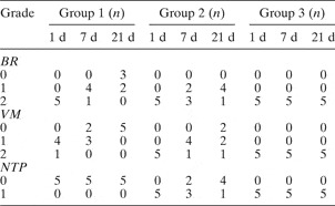
In group one, on the first post-operative day, the blink reflex disappeared and the nose tip position was normal in all rats, while little vibrissae movement was observed in 13 of the 15 rats. On the seventh post-operative day, the blink reflex recovered partially in eight of the 10 remaining rats, and vibrissae movement was observed in four of the 10 rats. On the 21st post-operative day, normal blink reflex, vibrissae movement and nose tip position were observed in only three of the five remaining rats.
In group two, on the first post-operative day, all rats showed no blink reflex, no vibrissae movement and an abnormal nose tip position. On the seventh post-operative day, the blink reflex partially recovered in four of the 10 remaining rats, while vibrissae movements were abnormal in eight of the 10 rats. On the 21st post-operative day, recovery was significantly better (compared with the seventh post-operative day): of the remaining five rats, the blink reflex was almost normal in four and vibrissae movement had recovered completely in two.
In group three, no functional recovery was observed at any stage.
In contrast, the function of the (right-side) control facial nerves was normal throughout; no facial paralysis was observed during the post-operative observation period (Table I).
Rats' function grading: first, seventh and 21st post-operative day

D = post-operative day; BR = blink reflex; VM = vibrissae movement; NTP = nose tip position
Morphological changes
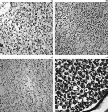
On the seventh post-operative day, in group one, the facial nerve size had altered and the nerve fibres had visibly degenerated, compared with those in control group (Figure 2a). In group two, degenerated fibres were also observed, but these were observed to be more generally dispersed throughout the nerve, both distal and proximal to the injury, and also to be more concentrated in the region of the transection, compared with group one (Figure 2b). In addition, congestion, oedema or fresh granulation tissue were observed in this work. In group three, facial nerve structure was disordered, with most nerve fibres appearing necrotic (Figure 2c). In the control group facial nerves, normal appearances were observed (i.e. improved trichrome staining showed axons as blue, myelin as red and the remaining neural components as green) (Figure 2d).
Facial nerve morphological changes in response to injury, improved trichrome staining, seventh post-operative day. (a) Group one: nerve fibre sizes differ in the facial canal segment, and some myelin sheaths and axons have degenerated (×400). (b) Group two: cross-section showing degenerated fibres present diffusely throughout the specimen, and showing substantial deterioration compared with the first post-operative day (×200). (c) Group three: most nerve fibres show necrosis and disordered organisation (×400). (d) Control group: evenly distributed fibres of uniform size, with a consistent ratio of myelin thickness to axon diameter. Normal staining is evident: axons stain blue, myelin stains dark red (surrounding the axon) and residual components stain green (×400).

On the 21st post-operative day, regeneration of nerve fibres was observed in groups one and two, whereas no obvious regeneration was seen in group three (data not shown).
Axon count
The total number of normal and degenerative axons from the intratemporal facial nerve segment was counted in the stained sections, using a 40-times counter under light microscopy. The mean number of axons ±SD was 2058 ± 485 in the control group. The mean number ±SD for normal and degenerative axons was respectively 1754 ± 312 and 256 ± 187 in group one, and 1353 ± 354 and 534 ± 257 in group two. There was a statistically significant difference between the numbers of normal and degenerative axons in those two groups (p < 0.05). We thus concluded that the degeneration caused by the intratemporal segment injury was worse than that caused by the extratemporal segment injury (Figure 3).
Numbers of normal and degenerative axons in the different groups. The comparison was made between group one and group two. The numbers of normal and degenerative axons were statistically significant (p < 0.05). Axonal degeneration was worse following intratemporal segment injury (group two) compared with extratemporal segment injury (group one).

Group three axons could not be counted at any post-operative stage, as most of the nerve fibres had disintegrated.
Neurotrophic factor 3 expression changes
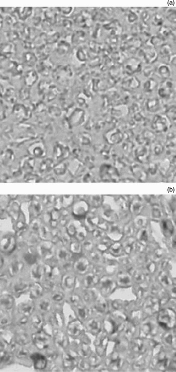
Expression of neurotrophic factor 3 was weak in the Schwann cells of the control group. However, strong positive staining for neurotrophic factor 3 was observed in the axonal facial nerve fibres of groups one and two on the seventh post-operative day. In contrast, neurotrophic factor 3 expression was only weakly positive in Schwann cells, and negative in degenerative fibres (Figure 4).
Facial nerve immunoreactivity for neurotrophic factor 3. (a) Control group, seventh post-operative day: weak immunoreactivity in Schwann cells. (b) Group two, seventh post-operative day: strongly positive immunoreactivity in axonal fibres (darker staining), with weak immunoreactivity in Schwann cells and no immunoreactivity in degenerative fibres.

Discussion
Controversy still exists regarding the necessity for facial nerve decompression after damage. The available data show that approximately 70 to 90 per cent of patients with traumatic facial paralysis recover from their condition. Some authors believe that surgery is indispensable in such patients. For instance, Darrouzet et al. state that surgery should be performed as soon as possible once immediate paralysis occurs, if high resolution CT demonstrates a clear fracture line and/or if ENoG indicates a poor prognosis.Reference Darrouzet, de Bonfils-Dindart and Bébéar10 In contrast, Chang and Cass state that facial nerve decompression should be avoided in cases in which paralysis is immediate, incomplete or no longer deteriorating, when combined with less than 95 per cent nerve fibre degeneration on ENoG.Reference Chang and Cass4
In the current study, assessment of facial nerve function (Table I) indicated that damage was more severe in group two, and that recovery was slower in group one than in group two (even though the depth of facial nerve injury was the same). These findings can be attributed to the increased nerve pressure caused by swelling of the nerve within the restrictive confines of the facial canal, which disrupts the surviving nerve fibres' axoplasmic transport and blocks their nerve conduction, which in turn results in more severe paralysis.Reference Takeda, Takeuchi, Kishimoto and Saito11, Reference Böhmer, Herzog and Dillier12 In group three, the facial nerve was paralysed completely on the injured side, although not all nerve fibres had degenerated and necrosed. The physiological block will last for a period after the damage. In the group two facial nerve sections taken on the seventh post-operative day, degenerative nerve fibres were scattered throughout the nerve, and the number of degenerative fibres was markedly increased compared with group one. This may be explained by the fact that, when oedema and haematoma occur within the facial nerve sheath, the pressure on the nerve compresses its microvasculature, restricting the blood supply, which worsens nerve fibre necrosis and functional paralysis.
Guerrissi found that extratemporal blunt trauma can produce a temporary facial palsy or paresis, due to injury of the main trunk or peripheral facial branch; however, complete recovery usually follows.Reference Guerrissi13 Intratemporal closed trauma with or without temporal bone fracture can produce a definitive facial nerve paralysis, and recovery is uncertain. These results support our findings regarding changes in facial nerve function after different types of injury. We conclude that the anatomy of the temporal bone facial canal contributes significantly to the differing results of damage to the intratemporal facial nerve versus the extratemporal facial nerve (and more peripheral nerve segments). Thus, we believe it is imperative to decompress the facial nerve canal in cases of nerve paralysis caused by temporal fracture or iatrogenic injury.
There are many differing opinions on the appropriate scope of such decompression. May proposed that, when the facial canal geniculate ganglion is damaged by fracture, the whole length of the facial nerve should be decompressed promptly, from the stylomastoid foramen to the internal auditory canal.Reference May14 Recently, other authors have stated that, if the facial nerve damage is limited, then a more limited decompression should be conducted, rather than decompression of the whole length of the nerve. In the current study, in group two facial nerve sections observed on the seventh post-operative day, congestion and oedema were seen throughout the nerve, as well as fresh granulation tissue growing into the injury. Also, it was shown in slides that degeneration of the proximal facial nerve is obviously and the same to the distal parts. The current study findings indicate that the pathological response to facial nerve injury initially occurs at the site of injury but subsequently involves the whole length of the facial nerve. This finding constitutes direct evidence which may assist the clinician in determining the scope of facial nerve decompression. Clinically, we believe the facial canal should be progressively opened up to the point at which neural oedema can be seen to resolve, on either side of the injury, rather than expose the nerve only at the site of injury. However, we emphasise the need to avoid over-extensive decompression, as decompression surgery is an invasive procedure (especially when a combined approach is used).
Schwann cells express many neurotrophic factors which support neurons, decrease their necrosis and promote their growth.Reference Nakamura, Takada, Shimabuku, Matsuo, Matsuo and Negishi15 In this study, we demonstrated that axonal expression of neurotrophic factor 3 was strongly positive at the seventh post-operative day, whereas expression by Schwann cells was only weakly positive, suggesting that expression of neurotrophic factor 3 is enhanced in surviving axonal fibres after injury. Neurotrophic factor three has been found to promote the growth of neurons and to increase the number and the length of axons in vitro.Reference Xu, Michalski, Racine and Fahnestock16 Therefore, neurotrophic factor 3 may play a positive role in facial nerve repair. Furthermore, dynamic process of degeneration, necrosis and regeneration was reflected effectively through observing activities of neurotrophic factor 3 before and after damage.Reference Xu, Michalski, Racine and Fahnestock16
• This study assessed facial nerve function, morphology and neurotrophic factor 3 expression following three types of nerve injury
• Facial nerve function and histological degeneration were worse following injury of the intratemporal segment, compared with the extratemporal segment
• Nerve fibre degeneration appeared not only at the site of injury but also throughout the length of the nerve
• The authors suggest that surgical facial nerve decompression is warranted following damage, as the restrictive effect of the temporal bone canal is a major contributor to increased nerve pressure
Conclusion
These study findings suggest that facial nerve decompression should be performed immediately after injury. This may benefit the patient by decreasing oedema pressure, preventing intensive degeneration and scar tissue formation, and promoting nerve fibre regeneration. The nerve should be progressively exposed either side of the injured segment, up to the point at which neural oedema disappears. The observed increase in neurotrophic factor 3 expression by surviving axonal fibres provides evidence that administration of neurotrophic factor 3 to the site of facial nerve injury may represent an alternative approach by which to promote nerve recovery.