Introduction
Mastoiditis is a rare complication of otitis media which can lead to the formation of a sub-periosteal abscess, also known as acute coalescent mastoiditis. The clinical presentation is one of pain and swelling of the mastoid process, pushing the pinna anterolaterally. A fluctuant collection may be felt overlying the mastoid, and the patient displays signs and symptoms of systemic infection. Treatment consists of intravenous antibiotics and, often, surgical drainage of the abscess via cortical mastoidectomy. Acute mastoiditis may be complicated by spread of the infection to the skull base or cranial cavity, or lateral or sigmoid sinus thrombosis.
We report a case of acute mastoiditis caused by metastatic deposition of non-small cell lung cancer, and we review the literature regarding this presentation.
Case report
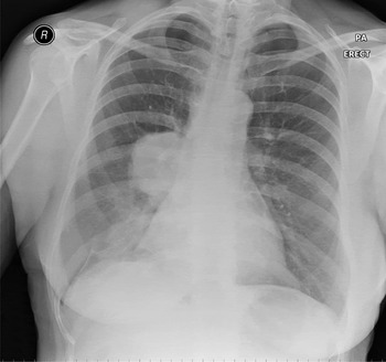
A 62-year-old woman was referred to the otolaryngology department with right-sided otalgia and decreased hearing. Her past medical history included a diagnosis of stage four, non-small cell lung cancer (see Figure 1). This had been treated with palliative chemo-radiotherapy, and was thought to be in remission.
Postero-anterior (PA) chest radiograph showing a 6 cm, right hilar carcinoma. R = right

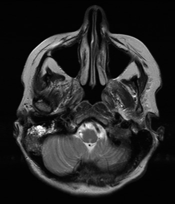
A magnetic resonance imaging scan was performed by the patient's oncology team to exclude intracranial metastases. The scan confirmed the absence of signs of intracranial disease, and demonstrated fluid within the right mastoid air cells (Figure 2).
Axial, T2-weighted magnetic resonance image showing abnormal soft tissue and fluid within the right mastoid air cells.

On otoscopy, the right tympanic membrane was noted to be erythematous, with a small degree of conductive hearing loss on free field and tuning fork testing. The rest of the ENT examination was normal.
A diagnosis of acute otitis media was made, and the patient was commenced on a course of oral clarithromycin (due to a penicillin allergy). Routine follow up was arranged for four weeks hence.
At review, the patient's pain had increased in severity and her tympanic membrane remained erythematous.
The next day, the patient underwent an examination of the ear under general anaesthesia. Myringotomy confirmed the presence of a purulent middle-ear effusion, and a ventilation tube was inserted. There was no evidence of post-auricular erythema or swelling at this time. Post-operatively, the patient's symptoms improved, and she was discharged on topical antibiotic and steroid drops.
Nine days after discharge, the patient was reviewed in clinic. Her symptoms had deteriorated once again, with increased otalgia. The patient felt systemically unwell, with pyrexia. Examination revealed mastoid tenderness and swelling, with pinna protrusion.
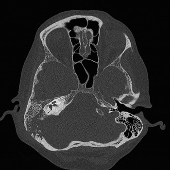
An urgent computed tomography scan (Figure 3) demonstrated opacification of the right mastoid air cells, with evidence of ill-defined bony destruction and loss of cortical definition.
Pre-operative computed tomography scan showing ill-defined bony destruction of the inner and outer cortices of the right mastoid.

The patient was admitted for intravenous antibiotics, and scheduled for an urgent right cortical mastoidectomy.
At surgery, necrotic debris was found beneath the mastoid periosteum, and was sent for microbiological and histological analysis. A cortical mastoidectomy was carried out, which revealed pus extending into the mastoid antrum. An abnormal area of necrotic tissue was noted within the posterior aspect of the mastoid cavity, arising around the sigmoid sinus. This was biopsied and sent for histological examination.
The patient's pain reduced post-operatively and the wound healed well. Four days after surgery, she developed a partial facial palsy which resolved following a course of oral steroids.
The biopsies taken from the posterior part of the mastoid showed malignant epithelial cells, with immunohistochemical staining consistent with metastatic non-small cell carcinoma. She was referred to the oncology department for consideration of further treatment.
Discussion
Metastatic malignancy is a rare differential diagnosis of acute mastoiditis. In order to identify reported cases, a systematic literature review was undertaken (on 8 June 2010), searching the Medline (1950 to present), Embase (1974 to present) and CINAHL databases. The search terms ‘mastoiditis’, ‘carcinoma’, ‘cancer’ and ‘metasta*’ were combined with Boolean operators. Duplicate results were removed and reference lists were cross-referenced for additional relevant studies. Results were limited to English-language publications.
Four case reports of mastoiditis secondary to metastatic carcinoma were identified. Two cases were caused by metastatic breast carcinoma, one by renal cell carcinoma and one by cholangiocarcinoma.Reference Johnson and Brown1–Reference Mansell4 We found no reported cases of mastoiditis caused by metastatic lung cancer.
Metastatic temporal bone tumours were initially thought to be rare, but this belief has been refuted by a number of post-mortem studies.
• Temporal bone metastasis is a rare cause of mastoiditis
• This case report highlights the potential for lung cancer metastasis to cause mastoiditis
• Post-mortem studies of cancer patients have demonstrated temporal bone metastases in 22 per cent, although significant symptoms seemed rare
The largest autopsy series was published in 2000 by Gloria-Cruz et al. Reference Gloria-Cruz, Schachern, Paparella, Adams and Fulton5 This study reviewed the temporal bone collection at the University of Minnesota. Autopsy reports from 864 patients were reviewed, and 212 patients were identified with non-systemic malignancies (excluding diseases such as lymphoma, leukaemia or multiple myeloma). Histological examination of these temporal bones demonstrated metastatic deposits in 47 patients (22.2 per cent).
The commonest primary source for metastatic spread to the temporal bone appears to be the breast, accounting for 18–29 per cent of cases.Reference Belal6–Reference Streitmann and Sismanis8 Other possible sources include lung, renal, stomach, prostate and laryngeal cancers.
In Gloria-Cruz and colleagues' series, the commonest otological feature of temporal bone metastasis was hearing loss (occurring in 40.4 per cent of cases), although this could not necessarily be attributed to the metastatic disease. In this series, 36.2 per cent of patients were asymptomatic. Otalgia was only present in four cases (8.5 per cent), suggesting that acute otitis media and mastoiditis are rare presentations of metastatic disease.
Conclusion
The presented case represents the first report of mastoiditis caused by metastatic lung cancer. Post-mortem histopathological studies suggest that metastasis to the temporal bone is not uncommon. However, it would appear to be a rare cause of mastoiditis.