Introduction
Schistosomiasis remains a neglected tropical disease (NTD) and an important public health problem in many non-industrialized countries (Utzinger et al., Reference Utzinger, N'Goran, Caffrey and Keiser2011). At least 261 million people required preventive treatment for schistosomiasis in 2013. More than 40 million people were treated for schistosomiasis in 2013 (WHO, 2015).
The disability-related outcome associated with chronic schistosomiasis is measured by disability-adjusted life-year (DALY) estimates. It was found that the disability of schistosomiasis due to the three most common infecting species – Schistosoma mansoni (S. mansoni), S. haematobium and S. japonicum – ranged from 2 to 15% (King et al., Reference King, Dickman and Tisch2005). An effective treatment of schistosomiasis was first achieved by praziquantel (PZQ) against all Schistosoma species (Cioli & Pica-Mattoccia, Reference Cioli and Pica-Mattoccia2003). However, PZQ resistance has been reported. In addition, parasites might survive treatment in immunocompromised hosts, because of the active role of the host immune system in the process of eliminating worms after PZQ administration (Brindley et al., Reference Brindley, Strand, Norden and Sher1989). Also, in vivo studies showed that PZQ is less effective in killing immature parasites (Sabah et al., Reference Sabah, Fletcher, Webbe and Doenhoff1986). Despite the prolonged drug research and control measures, this disease has not yet been contained and still continues to spread to new geographic areas. Therefore, it is important to explore alternative tools for elimination of this parasitic infection. The discovery of a protective vaccine remains the most potentially effective means for its control, especially if that vaccine provides long-term immunity against the infection (Rollinson et al., Reference Rollinson, Knopp, Levitz, Stothard, Tchuem Tchuente, Garba, Mohammed, Schur, Person, Colley and Utzinger2013). Because Schistosoma parasites do not multiply within their vertebrate hosts, a partial naturally acquired or vaccine-induced immunity could strongly reduce human pathology (Rollinson et al., Reference Rollinson, Knopp, Levitz, Stothard, Tchuem Tchuente, Garba, Mohammed, Schur, Person, Colley and Utzinger2013). It was documented previously that during the acute phase of the infection, parasite antigens evoke strong humoral and T-cell-mediated immune responses (Boros, Reference Boros1989). However, as the disease advances to chronicity, the cellular immune responses become the major inducers of granulomatous reactions around the eggs. This pathology is governed by helper T-lymphocytes (Th) and it is well known that lymphocytes belong to the CD4+ subsets (Vignali et al., Reference Vignali, Crocker, Bickle, Cobbold, Waldmann and Taylor1989; Cheever et al., Reference Cheever, Hoffmann and Wynn2000). In experimentally infected mice, schistosomiasis is characterized immunologically by an early Th1 response, which switches to a Th2-dominated response. Th2 cells produce interleukins after the onset of parasite egg production (Mosmann & Coffman, Reference Mosmann and Coffman1989; Wynn et al., Reference Wynn, Thompson, Cheever and Mentink-Kane2004).
Adult worms of Schistosoma are adapted to live in the bloodstream, so must possess very effective immune evasion strategies. In spite of many studies that have been done on the human immune response, no obvious immune mechanisms have emerged on which to base a vaccine. Previous vaccination attempts included crude parasite extracts, tegumental antigens, secreted antigens and monoclonal antibodies. Also, recent advances in recombinant DNA vaccines are very promising (Ismail, Reference Ismail2011). Crude cercarial antigen (CCA) was a topic of interest for previous researchers (Horowitz et al., Reference Horowitz, Smolarsky and Arnon1982; Mossallam, Reference Mossallam2000). Immunization with small amounts of this antigen, adsorbed on alum, resulted in partial protection against challenge infection (Horowitz et al., Reference Horowitz, Smolarsky and Arnon1982). Cercarial transformation fluid (CTF) is the fluid that contains many soluble materials released by schistosomal cercariae after being mechanically transformed into schistosomula in vitro. Proteomic analysis of these cercarial secretions identified a spectrum of proteins, which were recommended for use as potential anti-schistosomal vaccine components (Knudsen et al., Reference Knudsen, Medzihradszky, Lim, Hansell and McKerrow2005). These secretions include larval proteases such as cercarial elastase, cercarial acetabular gland secretions, calcium-binding proteins and paramyosin (Curwen & Wilson, Reference Curwen and Wilson2003; Knudsen et al., Reference Knudsen, Medzihradszky, Lim, Hansell and McKerrow2005). These materials provide validation of proposed mechanisms of host skin invasion and immune evasion and, in turn, have indirect roles in the immune effector mechanisms, such as inhibition of crucial enzyme activity or provocation of inflammation, that blocked migration (Curwen & Wilson, Reference Curwen and Wilson2003).
Progress towards a single schistosomiasis vaccine has been slow. Therefore, association of different antigens to produce multivalent or multi-epitope vaccines may accelerate this achievement (Wilson et al., Reference Wilson, Mentink-Kane, Pesce, Ramalingam, Thompson and Wynn2007). No reports are available for the efficacy of CTF, as an immunizing agent, against experimental S. mansoni. Accordingly, the aim of the present study was to evaluate the effect of CTF singly or combined with CCA in experimentally infected mice.
Materials and methods
Experimental infections
Mice were purchased from the animal house, Medical Parasitology Department, Faculty of Medicine, Alexandria University, Alexandria, Egypt. Shedding Biomphalaria alexandrina snails were purchased from the Schistosoma Biological Supply Program, Theodor Bilharz Research Institute, Imbaba, Giza, Egypt. Cercariae were used for both antigen preparation and infection of mice. Cercarial transformation fluid antigen (CTF) was prepared according to El Aswad Bel et al. (Reference El Aswad Bel, Doenhoff, El Hadidi, Schwaeble and Lynch2011). The concentration of freshly shed S. mansoni cercariae was achieved by gravity sedimentation at 4°C followed by suspension of the sediment in phosphate-buffered saline (PBS) at pH 7.2. Cercariae were aspirated 15 times through a 20-gauge needle in order to mechanically transform them into schistosomula. Schistosomula were then incubated for 90 min at room temperature, followed by centrifugation for 5 min at 250 g, then storage of the supernatant at − 80°C until used.
Crude cercarial antigen was prepared by sedimentation of the freshly shed cercariae by centrifugation at 150 g for 2 min. This was followed by sonication of the pellet suspended in a small volume of PBS at 5–10°C in a bath sonicator. The suspension was allowed to stand for 60 min with occasional agitation. Then it was centrifuged at 10,000 g at 4°C for 30 min and the supernatant was stored at − 20°C (Horowitz et al., Reference Horowitz, Smolarsky and Arnon1982). After estimation of the protein content of both antigens according to Lowry et al. (Reference Lowry, Rosebrough, Farr and Randall1951), the combined antigens were prepared by adding 100 μg protein of each antigen together. The two antigens were adsorbed on a 10% aqueous suspension of aluminium potassium sulphate (AlK(SO4)2) (alum) as an adjuvant (Doenhoff & Sayers, Reference Doenhoff and Sayers2002).
Up to 96 mice were divided into control and vaccinated groups. Controls were sub-divided into a group of six uninfected mice and two groups of 18 mice each, infected or adjuvant-only infected. Vaccinated mice were sub-divided into groups of 18 mice receiving CTF, CCA or both CTF and CCA. Following a pilot study, CTF and CCA groups were given a dose of 100 μg protein (Darani et al., Reference Darani, Curtis, McNeice, Price, Sayers and Doenhoff1997). The precise dose of each antigen was adsorbed on 100 μl of alum, then was injected intraperitoneally in two separate doses, 1 week apart (Ashour et al., Reference Ashour, Ahmed, Maghraby and Zahran2004). Mice were each infected with 100 freshly shed cercariae, using the tail immersion technique, 2 weeks after the second dose of adjuvant in adjuvant-only infected mice or after the second dose of the antigens in vaccinated mice (Olivier & Stirewalt, Reference Olivier and Stirewalt1952). Six weeks post-infection (pi) the mice were killed and the worm burden in the porto-mesenteric vessels was estimated by perfusion (Smithers & Terry, Reference Smithers and Terry1965). Tissue egg counts in liver and intestine, and the percentage of reduction of egg counts in vaccinated mice were calculated (Cheever, Reference Cheever1968). In addition, liver granulomata sizes were measured using a graduated eyepiece lens. Morphological study was done by scanning electron microscopy (SEM), where recovered worms were fixed in 10% glutaraldehyde and processed for examination by a Jeol-JSM-25 SII scanning electron microscope (Jeol, Japan) (Abdeen et al., Reference Abdeen, Reda, El-Shabasy and Ouhtit2012).
Biochemical and immunological protocols
Measurements of the liver enzymes aspartate transaminase (AST) and alanine transaminase (ALT) were carried out using a double enzymatic reaction method (Dimension XpandPlus Integrated Chemistry System, Siemens, Germany), and levels were expressed in units per litre (IU/l) (Abdel-Hafeez et al., Reference Abdel-Hafeez, Ahmad, Abdulla, Aabdel-Wahab and Mosalem2012). CD4+T-lymphocytes were expressed from splenic suspensions by forcing splenic tissue through a fine-mesh sieve using the piston from a 5-ml disposable syringe, followed by extensive washes with PBS. Viable cells were incubated with fluorescein isothiocyanate (FITC)-labelled anti-mouse CD4+ monoclonal antibodies (mAbs) (BioLegend Inc., USA). Positive cells were counted by flow cytometry (Mossallam, Reference Mossallam2000; Piatier-Tonneau, Reference Piatier-Tonneau, Mason, André, Bensussan, Buckley, Civin and Clark2002).
Data analysis
Quantitative data (mean, standard deviation and median) were analysed using IBM SPSS software package version 20.0 (Kirkpatrick and Feeney, Reference Kirkpatrick and Feeney2013). The analysis of variance (ANOVA) test was used to compare different groups. Pairwise comparisons were assessed using a post-hoc test (Scheffé). The significance of the obtained results was judged at the 5% probability level. Percentage of reduction (%R) was calculated for the various parameters as {[(C – V)/C] × 100}, where C is the mean value of the control group and V is the mean value of the vaccinated group (Tendler et al., Reference Tendler, Pinto, Oliveira Lima, Gebara and Katz1986; Daly et al., Reference Daly, Bourke and Mcgilvray1991).
Results
Worm recovery and structural changes
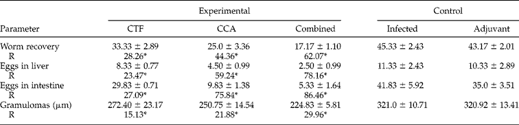
The vaccinated group showed significant reduction in all parasitological parameters when compared with infected and adjuvant-only infected control mice (table 1). Worm reduction was significantly higher in vaccinated groups when compared with infected controls. However, the highest significant reduction was observed with combined antigens followed by CCA when compared with CTF. Reduction in tissue egg counts in both liver and intestine, and the reduction in hepatic granuloma size, followed the same pattern as worm burden reduction. The smallest hepatic granulomata were noted with combined antigens when compared with other vaccinated mice.
Mean values (± SD) of adult worm recovery of Schistosoma mansoni, egg counts in the liver and intestine, and diameter of granulomata (μm) in experimental and control groups of mice, with 18 mice in each group. CTF, cercarial transformation fluid; CCA, crude cercarial antigen; R, % reduction from infected control.

Significance was assessed using ANOVA and a post-hoc test (Scheffé) for pairwise comparisons. Level of significance: *, P ≤ 0.05.
The ultrastructural study revealed that in the infected control mice, the oral sucker of retrieved adult male worms was round to oval with long, well-developed spines (fig. 1A). Their teguments showed tubercles which were arranged regularly in transverse rows with the presence of apically situated spines (fig. 1B). In adult female worms, the tegument was generally smooth but had sparse sensory papillae on the posterior end. In mice receiving CCA, retrieved male worms showed atrophy of the ventral sucker (fig. 1C), together with loss of the tone of their gynaecophoric canal wall (fig. 1D). However, the most significant changes were manifest in worms retrieved from mice receiving combined antigens, where the ventral sucker of the adult male was atrophied with attachment of the leucocytes below it, and spines on the sucker's surface were lost and replaced by dimples (fig. 1E). Tubercles were smooth with deep furrows in between (fig. 1F).
Scanning electron microscopy (SEM) of adult worms of Schistosoma mansoni from mice acting as infected controls, to show (A) sucker (vs) with elongated spines and (B) dorsal surface with tubercles (t) and spines (s); mice receiving CCA, to show (C) atrophy of the ventral sucker (vs) and (D) lost tone of the gynaecophoric canal (gyc); and mice receiving combined antigens, to show (E) atrophy of the ventral sucker (vs) and attachment of leucocytes (l) below it, and (F) smooth tubercles on the dorsolateral surface (t) with deep furrows (f) in between.

Biochemical and immunological analyses
The mean levels of ALT and AST in all groups receiving CTF, CCA and combined antigens showed statistically significant reductions in relation to the infected control mice, but did not reach the levels detected in normal control mice (table 2). Also, mice receiving combined antigens showed the highest significant reduction in the enzyme levels when compared with mice receiving CTF or CCA separately.
Mean values (± SD) of the enzymes alanine transaminase (ALT) and aspartate transaminase (AST) and % CD4+T-cells in experimental and control groups of mice. CTF, cercarial transformation fluid; CCA, crude cercarial antigen.

Significance was assessed using ANOVA and a post-hoc test (Scheffé) for pairwise comparisons. Level of significance: *, P ≤ 0.05.
Regarding CD4+T-lymphocytes, mice receiving CTF showed a statistically significant elevation in splenic CD4+T-lymphocytes when compared with normal mice only. In contrast, mice receiving CCA and mice receiving combined antigens showed a statistically significant elevation of the mean percentage of splenic CD4+T-lymphocytes in comparison to the three control groups. In addition, mice receiving CCA and mice receiving combined antigens showed statistically significant elevation when compared with mice receiving CTF. Mice receiving combined antigens showed the highest significant elevation of splenic CD4+T-lymphocytes when compared with other vaccinated groups.
Discussion
Current schistosomiasis control strategies are mainly based on chemotherapy, yet many researchers believe that the best long-term strategy to control the disease is a combination of improved environmental sanitation, drug treatment and immunization (Gray et al., Reference Gray, McManus, Li, Williams, Bergquist and Ross2010). The good safety index and broad therapeutic profile of PZQ, together with the sharp reduction in its price, have delayed the introduction of other drugs (Soliman et al., Reference Soliman, El Shenawy and El Arabi2008). However, PZQ cures the host of adult worms but does not affect juveniles (Savioli et al., Reference Savioli, Engels, Roungou, Fenwick and Endo2004). Therefore, beside environmental measures, greater efforts are required to identify new antigens and to develop an effective protective vaccine (Capron et al., Reference Capron, Riveau, Bartley and McManus2002).
Crude cercarial antigen proved to be efficient in protection against schistosomiasis (Horowitz et al., Reference Horowitz, Smolarsky and Arnon1982). Thus, it was used in the present work as a reference vaccine candidate. Alum was chosen as an adjuvant as it stimulates type-2 helper (Th2) immune responses and macrophages to produce prostaglandins (PGs), which have an active role in the regulation of immune responses (Kuroda et al., Reference Kuroda, Coban and Ishii2013).
In this study, it was expected that CTF would give a good protective effect due to the wide panel of secretory proteins it contains; however, results of our research showed that CCA was superior upon its sole use as an immunizing agent. The reduced influence of CTF could be explained by the weak effect of the soluble enzymes excreted in this fluid, and by the fact that the membrane-bound form of the proteases present on larvae may be the actual target of protective immune responses, as reported by Ghendler et al. (Reference Ghendler, Parizade, Arnon, McKerrow and Fishelson1996). Moreover, Darani et al. (Reference Darani, Curtis, McNeice, Price, Sayers and Doenhoff1997) did not find clear antibody responses in mice vaccinated against the 27-kDa serine protease enzyme, a major constituent of CTF. In addition, production of antibodies to enzymes could be transient or weak. Thus the role of a humoral immune response is not well proved in case of immunization with CTF. On the other hand, the better results obtained by CCA compared to CTF could be attributed to the components of CCA. This antigen is made from sonicated cercariae that maintain all their structural and functional proteins. It consisted of both soluble antigens, including enzymatically active components, and membrane-associated antigens (Horowitz et al., Reference Horowitz, Smolarsky and Arnon1982).
The current study shows that mice receiving combined antigens have the highest significant reduction in worm load (62.07%), egg count (78.16% and 86.46% in liver and intestine, respectively) and granuloma size (29.96%) followed by those receiving CCA. However, mice receiving CTF showed the lowest reductions in these parameters. In general, many studies have established that T-cell-mediated immunity is fundamental to acquired resistance to schistosomes in mice (McManus & Loukas, Reference McManus and Loukas2008). Based on previous studies, we can suggest that the combination of these two antigens initiated the production of a good cellular effector response, which was mediated by CD4+ T-cells of the Th1 subset, with interferon-gamma (IFN-γ) and tumour necrosis factor as key cytokines. Wilson & Coulson (Reference Wilson and Coulson2006) supported this idea and they suggested that the effector mechanism relies on blocking intravascular migration rather than cytotoxic killing. Also, they stated that with multiple exposures of mice to cercaria vaccines, the immune response progressively develops a Th2 profile. However, in our study, only two doses of vaccines were used, with an interval of 2 weeks, thus the shifting of Th1 to Th2 was unlikely.
Combination strategies in protective vaccine preparation have been a matter of interest for years (Utzinger et al., Reference Utzinger, Chollet, You, Mei, Tanner and Xiao2001). The improved action of the combined vaccines was supported by the work of Ashour et al. (Reference Ashour, Ahmed, Maghraby and Zahran2004), Gabr et al. (Reference Gabr, Mowafy, Ramadan and Kamal2010) and Ewaisha et al. (Reference Ewaisha, Bahey-El-Din, Mossallam, Amer, Aboushleib and Khalil2014). Furthermore combined vaccines were used by Ivory & Chadee (Reference Ivory and Chadee2004), who stated that parasites are complex organisms with multiple life cycles and antigenic variation which enable them to evade the immune system. Also, not all individuals respond to the same antigens in natural infections. Consequentially, multivalent vaccines have a greater number of protective epitopes and could be effective in a greater proportion of the population. Moreover, Etewa et al. (Reference Etewa, Abd El-Aal, Abdel-Rahman and El-Shafey2014) tried three crude antigens, and reported a significant reduction in worm burden and tissue egg count (liver and intestine), when used in combination.
In the current study, splenic CD4+T-lymphocytes were selected as an indicator of the immune status and their levels were measured using flow cytometry. There was a statistically significant elevation of these cells in mice receiving combined antigens, followed by those receiving CCA, whereas mice receiving CTF showed the least percentage elevation. Vignali et al. (Reference Vignali, Crocker, Bickle, Cobbold, Waldmann and Taylor1989) supported our choice when they showed that in vivo depletion of CD4+T-cells resulted in a dramatic reduction in immunity induced by vaccination with irradiated cercariae. However, depletion of CD8+T-cells had no effect on resistance induced by the vaccination.
The increased level of CD4+T-cells denoted improvement of the immune status of mice. This could explain the reduction in granuloma size in the present work. This granuloma down-regulation was attributed to the increase in Th1 cytokines and decrease in Th2 cytokines. The Th1 immune response, which was induced by vaccination, results in production of T-regulatory cells (T-reg). In turn, the latter cells produce interleukin-10 (IL-10), which could help in the prevention of an exacerbated granulomatous reaction (Wynn & Cheever, Reference Wynn and Cheever1995; Jarnicki et al., Reference Jarnicki, Conroy, Brereton, Donnelly, Toomey, Walsh, Sweeney, Leavy, Fletcher, Lavelle, Dunne and Mills2008). Additionally, Wilson et al. (Reference Wilson, Mentink-Kane, Pesce, Ramalingam, Thompson and Wynn2007) suggested that IL-10 plays a key regulatory role in preventing the development of severe pathology. This was also proved by Damian et al. (Reference Damian, Roberts, Powell, Clark, Lewis and Stirewalt1984), who explained change in the granuloma size by immunoregulation and modulation following administration of the vaccine.
In the current work, the significant elevation of the liver enzymes in the infected control mice, when compared to the normal mice, is explained by the production of reactive oxygen species in the liver and the alteration of antioxidant defences, which in turn contribute to hepatocyte disruption (Yang et al., Reference Yang, Yang and Zhang1986). However, reduction in their levels to almost normal following the administration of the combined antigens may be due to amelioration of the oxidant/anti-oxidant balance. This indicates a status of structural integrity of liver cells, which provides further support to the suggestive mechanism of action of the vaccine (Abdel-Hafeez et al., Reference Abdel-Hafeez, Ahmad, Abdulla, Aabdel-Wahab and Mosalem2012).
It is well known that the schistosomal tegument represents a potential target for a drug or a vaccine (Rogers et al., Reference Rogers, Shannon and Reeder1983). It is a unique syncytial layer that is critical for the parasite's survival within the mammalian host. Molecules residing in the tegumental membranes are major targets for the development of drugs and vaccines against the parasite (Abath & Werkhauser, Reference Abath and Werkhauser1996). The tegument is delimited by a membrane of multilaminate appearance comprising an inner classical plasma membrane overlaid by an outer external membrane (Skelly & Alan Wilson, Reference Skelly and Alan Wilson2006). Relevant proteins, such as transporters, enzymes and receptors, are located in the inner plasma membrane where they are protected from recognition by the host immune system; whereas the outer membrane has a predominantly lipid composition with few identified macromolecular constituents, and acts as a barrier against the host immune attack (Sulbaran et al., Reference Sulbaran, Noya, Brito, Ballen and Cesari2013). The tegument represents the interface between host and parasite and its molecules are responsible for essential functions for parasite survival. Therefore, ultrastructural tegumental changes in worms retrieved from vaccinated mice in the present work were noteworthy. The worst deformities were shown in worms retrieved from mice receiving combined antigens, followed by worms retrieved from mice receiving CCA. Adult male worms showed resorption of the ventral sucker with complete loss of its spines, which in turn affected the attachment of males to blood vessels. Moreover, the loss of tubercular spines and appearance of deep furrows on the dorsolateral surface in the same group indicated impairment of tegumental function and, hence, destruction of the defence system of the worm, thereby facilitating the action of the host's immune cells (Xiao et al., Reference Xiao, Binggui, Chollet and Tanner2000). Thus, such changes could explain the significant reduction in the worm burden in vaccinated mice, as supported by Reda et al. (Reference Reda, Ouhtit, Abdeen and El-Shabasy2012). Furthermore, attachment of leucocytes to the dorsolateral surface and below the resorbed ventral sucker was witnessed. Such attachment was similarly described by Yang et al. (Reference Yang, Yang and Zhang1986) and Diab (Reference Diab2012), who used artemether and mefloquine drugs, respectively, against S. mansoni infection. Regarding adult female worms in mice receiving combined antigens, these showed an irregular and collapsed tegument in their posterior ends with loss of their spines and sensory bulbs. Zhang et al. (Reference Zhang, Xiao, Utzinger, Chollet, Keiser and Tanner2009) also observed similar changes. Thus, the potential release of antigenic components of vitelline gland cells might have interacted with the specific anti-Schistosoma antibodies, forming an immune complex that strengthens neutrophilic chemotaxis and subsequent damage of the gland cells. As is well known, the vitelline glands are very important for eggshell synthesis; their damage directly affects egg production and, hence, results in the improvement of pathological changes in vaccinated mice (Tinsley, Reference Tinsley1983).
From the current study, it could be concluded that although CTF gave the poorest results, its combination with CCA augmented its action. Thus, in summary, combined vaccines stimulated specific immunity with a highly significant level of protection, in addition to the anti-pathological effect against S. mansoni. This may provide hope for the development of an appropriate, feasible vaccination approach against such an important human parasite.
Conflict of interest
None.
Ethical standards
The studies on mice were approved by the local government based on the national Egyptian regulations for animal experimentation. Rules of the Ethical Committee, Medical Parasitology Department, Faculty of Medicine, Alexandria University, Alexandria, Egypt were followed as regards housing and sacrifice of mice.