Most vertebrates possess a duplex retina that contains two distinct classes of photoreceptors: the highly sensitive rods that are responsible for scotopic or dim light vision and the less sensitive cones that are responsible for vision at higher light levels, such as daylight. Cones are also responsible for high acuity vision and for color vision. High acuity vision depends on high densities of cones in certain regions of the retina. For example, in humans, high acuity central vision is achieved by a peak cone density at the fovea of around 100,000–200,000/mm2 (Curcio et al., Reference Curcio, Sloan, Kalina and Hendrickson1990), increasing to over a million in some birds of prey. Cones also provide a mechanism for the sensation of color that derives from sampling light at different wavelengths by spectrally distinct cone types. The light sensitive molecule present in all photoreceptor classes comprises an opsin protein with a light sensitive chromophore attached via a Schiff base (SB) linkage to a lysine residue. In vertebrates, the chromophore is derived either from vitamin A1 or vitamin A2 to give, respectively, rhodopsin and porphyropsin pigments. With the vitamin A1-derived chromophore (retinal), rod or Rh1 pigments generally peak around 500 nm, whereas the peak sensitivities (λ max) of the four cone pigments range from 360 nm in the ultraviolet (UV) for the short wavelength-sensitive type 1 (SWS1) class, through 400–470 nm for the short wavelength-sensitive type 2 (SWS2) class, 480–530 nm for the middlewave-sensitive (MWS or rod-like Rh2) class, to a maximum of around 570 nm for the longwave-sensitive (LWS) class (Fig. 1). With the vitamin A2-derived chromophore (3,4-didehydroretinal), these peak values are pushed to longer wavelengths, especially for the LWS pigments which peak at >600 nm. With the important exception of the UV-sensitive (UVS) SWS1 pigments, all other vertebrate visual pigments have an SB that is protonated in the resting state, with a negatively charged residue at site 113 (usually glutamate) acting as a counterion to stabilize the proton of the SB. Spectral cone types are conveniently named by the pigment they contain, for example, more generally S cone or S1 and S2 if both SWS1 and SWS2 pigments are present.
Example absorbance spectra for the four cone classes of visual pigment, SWS1, SWS2, middle wavelength-sensitive (MWS or Rh2) and LWS. An UVS SWS1 pigment is depicted.

Origins and evolution of SWS1 visual pigment
Recent evidence places the evolutionary origin of opsins in the production of light-sensitive molecules in a primitive visual system around 700 mya (Feuda et al., Reference Feuda, Hamilton, McInerney and Pisani2012). Within the vertebrates, the most ancient species with multiple visual opsin classes as described above is the Southern hemisphere pouched lamprey, Geotria australis (Collin et al., Reference Collin, Knight, Davies, Potter, Hunt and Trezise2003b), a member of the gnathostomes or jaw-less vertebrates. This implies that the blueprint for vertebrate color vision was first laid down before the agnathan/gnathostome split in the vertebrate lineage, >540 mya (Davies et al., Reference Davies, Collin and Hunt2012). One of these genes encodes a UVS pigment that is the orthologue of the SWS1 pigment found throughout the vertebrate lineage and defines the most short wavelength-shifted S cone class (Collin et al., Reference Collin, Hart, Shand and Potter2003a). It is inferred from this that the ancestral SWS1 gene encoded a UVS pigment (Hunt et al., Reference Hunt, Carvalho, Cowing, Parry, Wilkie, Davies and Bowmaker2007; Davies et al., Reference Davies, Collin and Hunt2012).
Of the four vertebrate cone opsin classes, mammals have retained only two, the LWS and one of the S opsins; the genes for the other two opsins were lost during mammalian evolution (reviewed in Davies et al., Reference Davies, Collin and Hunt2012). Marsupials and eutherians have retained the SWS1 opsin, monotremes have retained the SWS2 opsin (see Presence of S cones in mammals section; Davies et al., Reference Davies, Carvalho, Cowing, Beazley, Hunt and Arrese2007; Wakefield et al., Reference Wakefield, Anderson, Chang, Wei, Kaul, Graves, Grutzner and Deeb2008). In the Old World primates and a single species of New World primate (Jacobs et al., Reference Jacobs, Neitz, Deegan and Neitz1996a; Dulai et al., Reference Dulai, von Dornum, Mollon and Hunt1999), cone trichromacy was attained de novo by duplication of the LWS opsin gene and subsequent independent mutations in the two copies, leading to the present-day red (L) and green (M) cone opsins of these species. Outside of the primates, the most common mammalian condition is dichromatic color vision based on the presence of L and S cones (review: Jacobs, Reference Jacobs1993). As Jacobs points out, the saliency of color vision differs between species, from the robust dichromacy of those with two cone pigments and a substantial population of cones to the feeble dichromacy of those with two cone pigments but only sparse cone populations.
Presence and distribution of S cones
The presence of S cones in the different vertebrate classes is summarized in Fig. 2 along with their spectral properties (i.e., whether UVS or violet-sensitive) and the retention of the other cone types across species.
A summary cladogram showing the evolutionary retention and loss of the different cone classes within the main branches of the craniate radiation. Four classes of cone opsins first appeared in the agnathan lamprey and were retained by the gnathostome lineage. In all cases, a rod pigment is also present. In the lamprey, Geotria australis, it remains uncertain which of the two Rh pigments present is the orthologue of the cone Rh2 pigment. Most placental mammals have retained both the SWS1 and LWS genes but the loss of the SWS1 gene is seen in many primates, rodents and all dolphins, whales, and seals. Note that in deeper-diving whales, a functional LWS gene is also absent.
Histological identification of S cones
Molecular and sequencing approaches inform us about the presence of the SWS1 opsin gene and whether it is functional or a pseudogene, but these techniques do not tell us in which subset of cones the opsin is expressed, what proportion of all cones that this represents, and how these are distributed across the retina. These population properties are important for the functional role of the S cones in color processing and are most suitably assessed in retinal flatmounts by immunohistochemistry with opsin-specific antibodies that selectively label the respective spectral cone types. Furthermore, the antibodies work in fixed tissue, hence the presence and distribution of cone types can be assessed in species that are not easily available for physiological or behavioral experiments. Studies using opsin antibodies have greatly increased our knowledge about the diversity of cone arrangements across mammals, but less so for other vertebrate classes. Of course, there is a caveat: depending on the epitope(s) that the opsin antibody or antiserum detects, species differences in the amino acid sequence may result in no labeling in certain species even though the opsin is present. Fortunately, a number of reliable antiopsin antibodies are available that robustly label the SWS1 and LWS opsins, respectively, across all mammals studied to date. They are directed against conserved epitopes that are not involved in residue replacements that determine spectral sensitivity or tuning. For other vertebrates, the situation is less comfortable as nonmammalian opsin-specific antibodies are rare and there is less information on S cone densities. Therefore, the account of S cone distributions will largely focus on mammals.
There are other ways to identify the spectral cone types in some nonmammalian vertebrates. One option is in situ hybridization with opsin RNA probes, another is the utilization of morphological differences between the cone types. In many birds and reptiles, cones contain colored oil droplets (see Amphibians and reptiles and Birds sections) that can be used to assess the presence and distribution of the cone types. Such analysis in a range of birds revealed that SWS1 cones constitute roughly 5–10%, and SWS2 cones roughly 10–15%, of the cones across species (Hart, Reference Hart2001a; Kram et al., Reference Kram, Mantey and Corbo2010). In many fish, the cones, and also each spectral cone type, are arranged in near-crystalline mosaics. For example, in zebrafish, the cones form parallel rows where one row contains alternating pairs of M (containing Rh2 pigment) and L (containing LWS pigment) cones that form double cones while the next row contains alternating S1 (containing UV SWS1 pigment) and S2 (containing SWS2 pigment) cones; in this lattice, the S1 and S2 cones each comprise about 15% of the cones (Raymond & Barthel, Reference Raymond and Barthel2004). Across vertebrates, there are certain species where the topographical distributions of the cone types differ, creating retinal regions with different spectral sensitivities; a well-known example is found in the pigeon retina with yellow and red fields (reviews: Hart, Reference Hart2001b; Temple, Reference Temple2011). The European starling and blue tit even show a difference in the distribution of cone types between left and right eye (Hart et al., Reference Hart, Partridge, Cuthill and Bennett2000; Hart, Reference Hart2001b).
Presence of S cones in mammals
The use of opsin antibodies has enabled the detection of even small populations of cones or rods that may have been overlooked in conventional histology. With just one exception so far reported, all mammalian retinae have been shown to possess rods and cones, and there is no modern evidence for older claims that some very nocturnal mammals (e.g., bats) have pure rod retinae and very diurnal mammals (e.g., tree shrews) have pure cone retinae. The exception is found in certain deeper diving whales in which the retinae are devoid of all cone opsins (Meredith et al., Reference Meredith, Gatesy, Emerling, York and Springer2013; see S cone loss in aquatic mammals section). Whereas the retinae of many diurnal nonmammalian vertebrates contain more cones than rods (Walls, Reference Walls1942), the retinae of most mammals—including diurnal species—are rod dominated. This is thought to reflect the nocturnal ancestry of mammals (reviewed in Davies et al., Reference Davies, Collin and Hunt2012). Mammalian cone proportions coarsely correlate with diel activity patterns, from less than one to a few percent cones in very nocturnal species, 2–10% cones in crepuscular and arrhythmic species, 5–30% cones in diurnal species (for summaries, see Ahnelt & Kolb, Reference Ahnelt and Kolb2000; Peichl, Reference Peichl2005). Only a few diurnal species are known to have more cones than rods, for example, ground squirrels have about 85% cones (Kryger et al., Reference Kryger, Galli-Resta, Jacobs and Reese1998) and tree shrews about 95% cones (Müller & Peichl, Reference Müller and Peichl1989). The rod-free fovea of diurnal primates is a regional patch of “all-cone” retina; outside the fovea, cone proportions drop to about 5% (humans: Curcio et al., Reference Curcio, Sloan, Kalina and Hendrickson1990).
With the exception of the egg-laying monotremes, mammals express the SWS1 opsin in their S cones. Monotremes in contrast express the SWS2 opsin, with the SWS1 gene present only as a pseudogene (Davies et al., Reference Davies, Carvalho, Cowing, Beazley, Hunt and Arrese2007; Wakefield et al., Reference Wakefield, Anderson, Chang, Wei, Kaul, Graves, Grutzner and Deeb2008). Most placental and marsupial mammals have both S and L cones, and commonly, each cone exclusively expresses either the LWS or the SWS1 opsin, with the L cones forming a dominant majority, and the S cones forming a 5–15% minority. Examples of such cone complements are illustrated in Fig. 3 for a range of mammals. In the macaque and tree shrew, the S cones are relatively evenly spaced within the cone array. Such a moderately regular S cone mosaic is found in several but not all primates (comparative: Martin & Grünert, Reference Martin and Grünert1999; Martin et al., Reference Martin, Grünert, Chan and Bumsted2000, Reference Martin, Grünert, Chan and Ghosh2001; humans: Curcio & Hendrickson, Reference Curcio and Hendrickson1991). In other mammals, the S cone mosaic is irregular with greatly varying distances between neighboring S cones (Fig. 3; Ahnelt et al., Reference Ahnelt, Fernández, Martinez, Bolea and Kübber-Heiss2000; Schiviz et al., Reference Schiviz, Ruf, Kuebber-Heiss, Schubert and Ahnelt2008). The rather regular S cone spacing in some primates, the tree shrew (Müller & Peichl, Reference Müller and Peichl1989; Petry et al., Reference Petry, Erichsen and Szél1993) and ground squirrels (Ahnelt, Reference Ahnelt1985; Kryger et al., Reference Kryger, Galli-Resta, Jacobs and Reese1998; Galli-Resta et al., Reference Galli-Resta, Novelli, Kryger, Jacobs and Reese1999) is an exception. The functional significance of this regularity is enigmatic, but, at least across primates, it is not related to the diurnal lifestyle (for discussions, see Martin & Grünert, Reference Martin and Grünert1999; Ahnelt & Kolb, Reference Ahnelt and Kolb2000). S cones are also present in most nocturnal and subterranean mammals in addition to L cones; Fig. 3 illustrates this for the nocturnal chinchilla, Chinchilla lanigera, and the subterranean coruro, Spacopus cyanus. Even though their retinae are evolutionarily adapted to low-light vision, both have retained the potential for dichromatic color vision (reviews: Ahnelt & Kolb, Reference Ahnelt and Kolb2000; Peichl, Reference Peichl2005; Nemec et al., Reference Nemec, Cvekova, Burda, Benada, Peichl, Begall, Burda and Schleich2007). Actually, all studied subterranean mammals possess higher cone proportions than nocturnal surface-dwelling species, in the range of 10% cones. Often, the absolute cone densities are not unusually high, but the rod densities in these species are rather low, resulting in a higher cone proportion (reviews: Nemec et al., Reference Nemec, Cvekova, Burda, Benada, Peichl, Begall, Burda and Schleich2007; Glösmann et al., Reference Glösmann, Steiner, Peichl and Ahnelt2008). The share of S cones varies across subterranean species (see Retinal distribution of S cones, S cone loss in terrestrial mammals, and Ospin coexpression sections). It appears that subterranean mammals have not specially adapted to the lightless underground, but retained an “all-purpose” retina for the instances where they chance above ground.
Examples of L and S cone patterning across mammals, visualized in retinal flat-mounts by double immunofluorescence labeling of SWS1 opsin (magenta) and LWS opsin (green), located in the cones’ outer segments. (A) Peripheral retina of the macaque, Macaca fascicularis, with a low cone density and rather regular S cone spacing; (B) cone-dominated retina of the tree shrew, Tupaia belangeri, showing a very regular S cone pattern; (C) peripheral retina of the cat, Felis catus, with irregular S cone spacing; (D) chinchilla, Chinchilla lanigera, with irregular S cone spacing; here, the antibodies also labeled the cone inner segments, giving a 3D impression; (E) the subterranean coruro, Spalacopus cyanus, has a higher cone density than many nocturnal surface dwellers and an irregular S cone spacing; (F) in the elegant fat-tailed mouse opossum, Thylamys elegans (a South American marsupial), a fraction of the L cones occur in pairs (double cones), the single S cones are irregularly spaced. Scale bar 50 μm.
The presence of L and S cones certainly constitutes the basic blueprint of the mammalian retina; for exceptions, see S cone loss in terrestrial mammals and S cone loss in aquatic mammals sections. In marsupials, the pattern is particularly uniform: all American and Australian marsupials studied to date possess L and S cones, although with different retinal distributions (see Retinal distribution of S cones section). Monotremes also have two cone types, but here SWS2 opsin is paired with LWS opsin (see above). Zeiss et al. (Reference Zeiss, Schwab, Murphy and Dubielzig2011) labeled the platypus retina with antisera against the two eutherian opsins and found LWS-labeled as well as SWS1-labeled cones that they interpret as SWS2 (S2) cones; the retinal distribution of these cone types was not determined. It remains to be clarified whether these stainings are specific, and if so, why the polyclonal SWS1 antibody crossreacts with the platypus SWS2 opsin.
Retinal distribution of S cones
In most mammals, the topography of L cones shows a central-peripheral density gradient, and often the S cones constitute a constant low fraction of the cones across the retina. However, studies of the S cone topography in a large range of mammals also found various deviations from concomitant central-peripheral S cone gradients (overviews: Ahnelt & Kolb, Reference Ahnelt and Kolb2000; Lukats et al., Reference Lukats, Szabo, Röhlich, Vigh and Szél2005; Peichl et al., Reference Peichl, Chavez, Ocampo, Mena, Bozinovic and Palacios2005a; Ahnelt et al., Reference Ahnelt, Schubert, Kubber-Heiss, Schiviz and Anger2006; Schiviz et al., Reference Schiviz, Ruf, Kuebber-Heiss, Schubert and Ahnelt2008). Whereas the density of L cones commonly peaks at the area centralis or visual streak (depending on species), many species show a trend for highest S cone densities and hence increased S cone percentages in other retinal regions. In some species, this is the ventral retina, in others the dorsal retina, and/or an S cone-rich rim in the retinal periphery. A strong partition with a dorsal L cone and ventral S cone dominance was first observed in the mouse retina (Fig. 4; Szél et al., Reference Szél, Röhlich, Caffe, Juliusson, Aguirre and van Veen1992), although it later turned out that LWS opsin was not absent from ventral retina, but that most ventral cones coexpressed SWS1 and LWS opsin (for details, see Opsin coexpression section). Strong ventral increases of S cone proportions are also present in a number of other rodents (overviews in Szél et al., Reference Szél, Csorba, Caffe, Szél, Röhlich and van Veen1994a, Reference Szél, Röhlich, Caffé and van Veen1996, Reference Szél, Lukats, Fekete, Szepessy and Röhlich2000), including the subterranean coruro, S. cyanus (Peichl et al., Reference Peichl, Chavez, Ocampo, Mena, Bozinovic and Palacios2005a). The Siberian dwarf hamster, Phodopus campbelli, has a most striking anisotropy, where a sharp horizontal edge separates the dorsal third of the retina with no SWS1 opsin expression, from the ventral two thirds with dominant SWS1 opsin expression in practically all cones (Fig. 4; Huber et al., Reference Huber, Heynen, Imsand, vom Hagen, Muehlfriedel, Tanimoto, Feng, Hammes, Grimm, Peichl, Seeliger and Beck2010). A complementary pattern is found in the subterranean pocket gopher, Thomomys bottae, where LWS opsin is virtually absent from the ventral retina (Williams et al., Reference Williams, Calderone and Jacobs2005). Among other mammalian orders, a ventrally increased S cone proportion is present in members of the Carnivora (cat, lion, tiger, and hyena) (Linberg et al., Reference Linberg, Lewis, Shaaw, Rex and Fisher2001; Calderone et al., Reference Calderone, Reese and Jacobs2003; Ahnelt et al., Reference Ahnelt, Schubert, Kubber-Heiss, Schiviz and Anger2006), and in several Insectivora, including the subterranean European mole, Talpa europaea (Peichl et al., Reference Peichl, Künzle and Vogel2000; Glösmann et al., Reference Glösmann, Steiner, Peichl and Ahnelt2008). In the insectivorous shrews, ventral S cone proportions are up to 50% and in the lesser hedgehog tenrec, Echinops telfairi, up to 70%, without opsin coexpression (Peichl et al., Reference Peichl, Künzle and Vogel2000). In the cone-dominated retina of the tree shrew, Tupaia belangeri, the densities of both L and S cones are highest in the ventro-nasal midperiphery, not in the temporally located area centralis (Müller & Peichl, Reference Müller and Peichl1989; Petry et al., Reference Petry, Erichsen and Szél1993).
Dorso-ventral gradients of SWS1 and LWS opsin expression in the house mouse, M. musculus, and the Siberian dwarf hamster, P. campbelli. Images of the transition zone in midretina, double immunofluorescence-labeled for SWS1 opsin (magenta), and LWS opsin (green). The mouse (left column) shows a gradual change from dorsal retina (up) with a majority of L cones and a minority of S cones to ventral retina (down) with SWS1 opsin expression in most cones and a reduction of LWS opsin expression. The hamster (right column) has a sharp horizontal boundary with no SWS1 opsin expression in dorsal retina, and a reduction of LWS opsin expression in ventral retina. Scale bar 100 μm. Hamster image modified from Huber et al. (Reference Huber, Heynen, Imsand, vom Hagen, Muehlfriedel, Tanimoto, Feng, Hammes, Grimm, Peichl, Seeliger and Beck2010).
Szél et al. (Reference Szél, Csorba, Caffe, Szél, Röhlich and van Veen1994a, Reference Szél, Röhlich, Caffé and van Veen1996, Reference Szél, Lukats, Fekete, Szepessy and Röhlich2000) considered the attractive hypothesis that a ventral increase of S cone proportions may be an adaptation to the natural light distribution. Skylight, projected on to the ventral retina, contains higher proportions of short wavelengths than light reflected from the ground and falling on the dorsal retina. Increased short-wave sensitivity in the ventral retina thus may improve contrast when viewing objects, for example, aerial predators, against skylight. However, they dismissed this hypothesis because some species facing very similar visual environments do not have similarly divided retinae. Even within the genus Mus, some species have a ventral S cone dominance, some have a standard S cone distribution, and some lack S cones completely (Szél et al., Reference Szél, Röhlich, Caffé and van Veen1996). The hypothesis is further challenged by the fact that in the ground squirrel and in four marsupials (tammar wallaby, fat-tailed dunnart, quokka, and quenda), the highest S cone proportions are located in the dorsal peripheral retina (Kryger et al., Reference Kryger, Galli-Resta, Jacobs and Reese1998; Hemmi & Grünert, Reference Hemmi and Grünert1999; Arrese et al., Reference Arrese, Rodger, Beazley and Shand2003, Reference Arrese, Oddy, Runham, Hart, Shand, Hunt and Beazley2005). The role of these S cone-rich dorsal regions for vision has yet to be elucidated. Peter Ahnelt and his colleagues have studied the diversity of S and L cone distributions in a wide range of mammals (Ahnelt et al., Reference Ahnelt, Schubert, Kubber-Heiss, Schiviz and Anger2006; Schiviz et al., Reference Schiviz, Ruf, Kuebber-Heiss, Schubert and Ahnelt2008). In particular, their findings in artiodactyls of differing sizes led them to suggest that increasing eye height (i.e., body size) is associated with a shift in S cone weighting from ventral to dorsal retina, but that habitat and foraging differences may also play a role.
Primate retinae show several peculiarities in S cone patterning. First, there is a range of regularities in the S cone mosaics across species, from the semiregular spacing in humans and macaques to the random spacing in the common marmoset (Martin et al., Reference Martin, Grünert, Chan and Bumsted2000). Second, primate species vary in foveal coverage by S cones (review: Martin et al., Reference Martin, Grünert, Chan and Ghosh2001). In humans and baboons, an area of 100–200-μm diameter in the center of the fovea is free of S cones; this local absence leads to the perceptual phenomenon of small-field tritanopia or foveal blue blindness. In some other primates, S cones are present throughout the fovea, with a local density decrease in the macaque, but no density decrease in two New World primates, the marmoset, Callithrix jacchus, and squirrel monkey, Saimiri sciureus (Martin & Grünert, Reference Martin and Grünert1999; Martin et al., Reference Martin, Grünert, Chan and Ghosh2001). Outside the fovea, S cone topography is rather similar across diurnal primates, with a steep density drop within the first few millimeters from the fovea, and then a more shallow density decrease toward the retinal periphery (Curcio & Hendrickson, Reference Curcio and Hendrickson1991; Martin et al., Reference Martin, Grünert, Chan and Bumsted2000).
In two nocturnal primates, the tarsier Tarsius spectrum (Hendrickson et al., Reference Hendrickson, Djajadi, Nakamura, Possin and Sajuthi2000) and the mouse lemur Microcebus murinus (Dkhissi-Benyahya et al., Reference Dkhissi-Benyahya, Szél, Degrip and Cooper2001), the S cone topography has a “reverse” gradient with highest densities in the retinal periphery and lowest densities in the central retina (9–14% vs. <1% of the cones in tarsier). Central S cones are so sparse that they can hardly contribute to visual processing; presumably, these animals are essentially L cone monochromats (color blind) in the central retina. Some other lemurs have conventional S cone distributions (Peichl et al., Reference Peichl, Rakotondraparany and Kappeler2001b). A similar but weaker reverse S cone gradient exists in a marsupial, the honey possum, Tarsipes rostratus (Arrese et al., Reference Arrese, Rodger, Beazley and Shand2003). Finally, there is a complete absence of S cones in some nocturnal primates (see S cone loss in terrestrial mammals section).
S cone loss in terrestrial mammals
Immunohistochemistry and gene sequencing have detected an increasing number of mammalian species that completely lack SWS1 opsin expression and only possess L cones (review: Jacobs, Reference Jacobs2013). With just one spectral cone type, regular cone-based color vision is precluded. The list of terrestrial L cone monochromats currently includes more than 40 species from four orders (11 rodents, 16 bats, 2 carnivores, and 13 primates; Jacobs, Reference Jacobs2013). Ahnelt et al. (Reference Ahnelt, Moutairou, Glösmann, Kubber-Heiss, Mollon, Pokorny and Knoblauch2003) list some additional species, including the insectivorous European hedgehog, Erinaceus europaeus, and the long tailed pangolin or scaly anteater, Manis tetradactyla. Most likely the list will continue to grow as further species are studied.
A common feature of these terrestrial L cone monochromats is that they are all nocturnal, and that they are relatively isolated in their loss of S cones, often having dichromatic close relatives. For example, in the family Procyonidae the nocturnal raccoons (Procyon lotor and P. cancrivorous) and the nocturnal kinkajou (Potos flavus) lack S cones while the diurnal coati (Nasua nasua) has L and S cones (Jacobs & Deegan, Reference Jacobs and Deegan1992; Peichl & Pohl, Reference Peichl and Pohl2000). Within the muroid genera Mus and Apodemus, S cone absence in M. pahari, M. plathythrix, and A. microps contrasts with the presence of both types in other species (Szél et al., Reference Szél, Csorba, Caffe, Szél, Röhlich and van Veen1994a, Reference Szél, Röhlich, Caffé and van Veen1996). SWS1 opsin is absent in the Syrian hamster, Mesocricetus auratus, but expressed in dwarf hamsters (von Schantz et al., Reference von Schantz, Argamaso-Hernan, Szél and Foster1997; Calderone & Jacobs, Reference Calderone and Jacobs1999; Glösmann et al., Reference Glösmann, Peichl, Neumann and Gattermann2006; Williams & Jacobs, Reference Williams and Jacobs2008; Huber et al., Reference Huber, Heynen, Imsand, vom Hagen, Muehlfriedel, Tanimoto, Feng, Hammes, Grimm, Peichl, Seeliger and Beck2010), and absent in nocturnal flying squirrels, Pteromys volans and Glaucomys sabrinus, but present in their diurnal tree squirrel relatives (Carvalho et al., Reference Carvalho, Cowing, Wilkie, Bowmaker and Hunt2006). Among subterranean rodents, the blind mole rat, Spalax ehrenbergi, completely lacks SWS1 opsin (David-Gray et al., Reference David-Gray, Bellingham, Munoz, Avivi, Nevo and Foster2002). In contrast, the bathyergid mole rats dominantly express SWS1 opsin in most cones, with lower levels of LWS opsin coexpressed in many cones in the Genus Fukomys (Cryptomys), and a curious absence of detectable LWS opsin expression levels in the naked mole-rat Heterocephalus glaber (Peichl et al., Reference Peichl, Nemec and Burda2004). More recently, genome sequencing showed that the naked mole-rat in fact lacks a functional LWS gene and hence is an S cone monochromat (Kim et al., Reference Kim, Fang, Fushan, Huang, Lobanov, Han, Marino, Sun, Turanov, Yang, Yim, Zhao, Kasaikina, Stoletzki, Peng, Polak, Xiong, Kiezun, Zhu, Chen, Kryukov, Zhang, Peshkin, Yang, Bronson, Buffenstein, Wang, Han, Li, Chen, Zhao, Sunyaev, Park, Zhang, Wang and Gladyshev2011). This is a singularity among terrestrial mammals.
The owl monkey, Aotus, a platyrrhine primate is classically described as nocturnal (Levenson et al., Reference Levenson, Fernandez-duque, Evans and Jacobs2007), lacks S cones (Wikler & Rakic, Reference Wikler and Rakic1990), and has an SWS1 pseudogene with deleterious mutations (Jacobs et al., Reference Jacobs, Neitz and Neitz1996b; Levenson et al., Reference Levenson, Fernandez-duque, Evans and Jacobs2007). SWS1 pseudogenes are also present in many species of prosimians, notably in all members of the Lorisiformes (lorises and galagos), and in some species of the Cheirogaleidae family that forms part of the Lemuriformes group (Kawamura & Kubotera, Reference Kawamura and Kubotera2004; Tan et al., 2005). Members of the Tarsiiformes and the remaining species of Lemuriformes, as well as the aye-aye, Daubentonia madagascariensis, the sole extant member of the Daubentoniidae family of the Chiromyiformes, all appear to have an intact SWS1 gene (Tan et al., 2005). The latter species is strictly nocturnal yet possesses an SWS1 gene that would appear to be fully functional, with evidence for purifying or stabilizing selection to reduce genetic variation (Perry et al., Reference Perry, Martin and Verrelli2007); the potential for dichromacy would appear to remain advantageous for the aye-aye despite its nocturnal activity pattern. The retention of an intact SWS1 gene is also seen in the nocturnal colugo, Galeopterus variegatus (also called “flying lemur,” order Dermoptera) (Moritz et al., Reference Moritz, Lim, Netz, Peichl and Dominy2013) with evidence of purifying selection. The Dermoptera belong to the group Euarchonta that also includes Primates and Scandentia (tree shrews).
The conclusion is that the loss of SWS1 opsin has occurred independently several times during mammalian evolution, perhaps as genetic “accidents,” and that it did not jeopardize survival because for nocturnal species mostly using rod vision, color blindness is not a severe handicap. Generally across species, there is no evidence that the loss of SWS1 opsin was adaptive, that is, conveyed an advantage to the respective species (Jacobs, Reference Jacobs2013). After all, most nocturnal mammals have retained both cone types and the potential to see color. In this context, it is noteworthy that no terrestrial mammal, with the above-mentioned exception of the subterranean naked mole-rat, has yet been found to completely lack the LWS opsin. L cones commonly are the more numerous cone type and hence, in addition to their role in color vision, they also provide the dominant input to the luminosity and visual acuity channels in photopic and mesopic viewing conditions. To lose this cone type would severely impede visual performance in any species that chances upon a situation requiring cone-based vision. The functional role of S cones appears to be largely restricted to color vision, hence their loss is less incapacitating (discussions: Mollon, Reference Mollon1989; Jacobs, Reference Jacobs2013). In fact, limited color discrimination is also possible with just one spectral cone type. This has been shown for the L cone monochromatic owl monkey (Jacobs et al., Reference Jacobs, Deegan, Neitz, Crognale and Neitz1993) and for a human cone monochromat (Reitner et al., Reference Reitner, Sharpe and Zrenner1991). The residual color vision is attributed to interactions of the remaining cone type and the rods, as it is restricted to mesopic lighting conditions where both the cones and the rods are in their working range.
S cone loss in aquatic mammals
The situation is quite different in the marine whales (Cetacea) and seals (Carnivora), where an SWS1 opsin loss appears to be the rule rather than the exception. This has been established by immunohistochemistry, electrophysiology, and/or gene sequencing for all species studied to date, comprising odontocete as well as mysticete cetaceans and phocid as well as otariid seals (review: Jacobs, Reference Jacobs2013). The molecular studies carried out in several whale and seal species have shown the presence of SWS1 opsin pseudogenes with deleterious mutations that preclude the expression of the protein.
Pinnipeds belong to the caniform carnivores, and the closest relatives of the cetaceans are the terrestrial artiodactyls (clade Cetartiodactyla). Terrestrial caniforms and artiodactyls have L and S cones, and the presence of an SWS1 opsin pseudogene in seals and whales means that their ancestors also had S cones. It thus appears that the SWS1 opsin loss in all marine members of these two distant orders represents convergent evolution and, most likely, a common selective pressure. The puzzling fact is that in clear open ocean waters, blue light (450–480 nm) penetrates deepest, while longer as well as shorter wavelengths are preferentially scattered and absorbed. Probably color vision is of little use in this monochrome blue underwater world, and two spectral cone types are not really needed. The loss of S cones would seem therefore to be a rather unfitting adaptation, as they would appear to be best suited to collect brightness and contrast information. However, the impact of S cone loss on acuity would be much less than that of L cone loss as they are quantitatively the less frequent cone class. The rod and L cone opsins of seals and whales are tuned to shorter wavelengths than those of their terrestrial relatives, indicating an auxiliary adaptation to the blue-dominated visual environment.
One hypothesis to explain this apparent paradox was that the S cones were lost in an early “coastal” period of cetacean and pinniped evolution. In many coastal waters, the light spectrum is red-shifted due to blue-light absorption by organic and inorganic debris. Under such conditions, a loss of S cones could have been an economical adaptation, simplifying retinal and cortical visual information processing. The scenario surmises that for descendant species that have stayed in coastal waters, the loss probably remains useful or neutral. Descendant species that later moved to open ocean habitats may have profited from a functional S opsin, but they could not reverse the deleterious gene defect. Accounts of the data and hypotheses concerning the SWS1 opsin loss in whales and seals have been given by Peichl et al. (Reference Peichl, Behrmann and Kröger2001a), Griebel and Peichl (Reference Griebel and Peichl2003), and Jacobs (Reference Jacobs2013).
The situation has become more complex by a recent sequence analysis of the complete SWS1 and LWS genes in a broad sample of odontocete and mysticete whales (Meredith et al., Reference Meredith, Gatesy, Emerling, York and Springer2013). First, the study indicates that cetacean SWS1 gene inactivation did not occur as early in evolution as assumed above, and actually occurred independently in mysticete and odontocete whales, after the invasion of open ocean habitats. Second, in many deeper diving mysticete and odontocete species, the LWS gene has also been inactivated to give rod monochromatic vision. Meredith et al. (Reference Meredith, Gatesy, Emerling, York and Springer2013) suggest that deleting the cone pigments with their higher thermal activation rates may have made the rod system even more sensitive. However, losing both cone opsins is a dramatic event for a mammal, likely posing fundamental problems for retinal processing. In mammals, the cone pathway to the retinal ganglion cells via cone bipolar cells is direct, whereas the rod pathway via rod bipolar cells feeds into the cone pathway to reach the ganglion cells (review: Wässle, Reference Wässle2004). Have the whales without cone pigments also lost the cones as cellular entities? If so, have they lost the cone bipolar cells as well because of lacking synaptic input? If so, how do the rod signals reach the ganglion cells? Analysis of the retinal circuitries in these whales would give interesting insights into the evolutionary solution to a very basic wiring problem.
Other aquatic or semiaquatic mammals have retained the SWS1 and LWS opsins. This has been shown for the manatee, sea otter, river otter, polar bear, and pygmy hippopotamus (Griebel & Schmid, Reference Griebel and Schmid1996; Ahnelt & Kolb, Reference Ahnelt and Kolb2000; Peichl et al., Reference Peichl, Behrmann and Kröger2001a, Reference Peichl, Dubielzig, Kubber-Heiss, Schubert and Ahnelt2005b; Levenson et al., Reference Levenson, Ponganis, Crognale, Deegan, Dizon and Jacobs2006; Newman & Robinson, Reference Newman and Robinson2006). In the semiaquatic species, their terrestrial activities probably had some impact on keeping cone dichromacy. The manatee is fully aquatic, but it lives in very shallow waters with spectrally broad lighting.
S cone loss in fishes
As described above, the SWS1 opsin gene can be traced from the jawless vertebrates to modern day representatives of amphibians, reptiles, birds, and mammals. Orthologues of this gene have been retained therefore by these major vertebrate groups and by teleost fishes, but not, it would appear, by members of the Chondrichthyes or cartilaginous fishes that reside at the base of the gnathostome lineage where they share a common ancestor dating from at least 450 mya (Sansom et al., Reference Sansom, Smith and Smith1996). Cartilaginous fishes are divided into two lineages, the holocephalans (chimaeras) and the elasmobranchs (sharks, rays, and skates), and recent work implies that the SWS1 gene has been lost from both lineages. In the former case, only a single species, the elephant shark Callorhinchus milii, has been studied and found to have retained only the Rh2 and LWS cone opsin genes (Davies et al., Reference Davies, Carvalho, Tay, Brenner, Hunt and Venkatesh2009a). Among the elasmobranchs, skates possess a rod-only retina with no evidence for the presence of cones or the expression of cone opsins in the retina (Brin & Ripps, Reference Brin and Ripps1977; Cornwall et al., Reference Cornwall, Ripps, Chappell and Jones1989; O’Brien et al., Reference O’Brien, Ripps and Al-Ubaidi1997). In contrast, rays possess three spectral classes of cone, although none of these has a λ max suggestive of a SWS1 pigment (Hart et al., Reference Hart, Lisney, Marshall and Collin2004; Theiss et al., Reference Theiss, Lisney, Collin and Hart2007), and sharks appear to be L cone monochromats with just a single cone type expressing an LWS pigment (Hart et al., Reference Hart, Theiss, Harahush and Collin2011; Theiss et al., Reference Theiss, Davies, Collin, Hunt and Hart2012). Vision in sharks shows similarities therefore to that in most aquatic mammals in which S cones have been lost.
Although the SWS1 gene has been retained by most teleosts, it is subject to developmental regulation such that it is not continuously expressed in the retina. In the black bream, Acanthopagrus butcheri, the expression of the SWS1 gene is restricted to the larval stage (Shand et al., Reference Shand, Davies, Thomas, Balmer, Cowing, Pointer, Carvalho, Trezise, Collin, Beazley and Hunt2008). It is switched off in the adult retina and this is accompanied by the commencement of LWS gene expression. The overall effect of these changes is to extend the sensitivity of adult fish to longer wavelengths of light, a shift that correlates with a move from relatively clear to reddish-coloured water (Shand et al., Reference Shand, Davies, Thomas, Balmer, Cowing, Pointer, Carvalho, Trezise, Collin, Beazley and Hunt2008). A developmental sequence of opsin gene activation is also found in salmonid fishes, where single cones shift from the expression of a UVS SWS1 pigment to a blue-wavelength sensitive SWS2 pigment during development (Cheng & Flamarique, Reference Cheng and Flamarique2004, Reference Cheng and Flamarique2007), driven by an increase in the level of thyroid hormone (Cheng et al., Reference Cheng, Gan and Flamarique2009; Raine & Hawryshyn, Reference Raine and Hawryshyn2009).
Opsin coexpression
Most mammals follow the rule-book by expressing only one opsin type per cone. This is the canonical way of being spectrally selective and thus useful for color vision. So it came as a surprise when Röhlich et al. (Reference Röhlich, van Veen and Szél1994) first reported the permanent coexpression of SWS1 and LWS opsin in a substantial proportion of the cones in the house mouse, guinea pig, and rabbit retinae. Since then, immunohistochemistry has revealed substantial populations of such “dual pigment” cones in a large number of species, including humans (Ahnelt & Kolb, Reference Ahnelt and Kolb2000; Applebury et al., Reference Applebury, Antoch, Baxter, Chun, Falk, Farhangfar, Kage, Krzystolik, Lyass and Robbins2000; Xiao & Hendrickson, Reference Xiao and Hendrickson2000). It is now well established that these findings are reliable and not just artifacts of antibody crossreactivity. Ágoston Szél, Pal Röhlich, and their colleagues have published thorough surveys of the occurrence and distributions of mammalian dual pigment cones (Szél et al., Reference Szél, Röhlich, Caffé and van Veen1996; Lukats et al., Reference Lukats, Szabo, Röhlich, Vigh and Szél2005).
In many of these species, opsin coexpression is associated with a dorso-ventral partition of cone opsin expression. This has been most thoroughly studied in the mouse where the dorsal retina has an L cone majority and an S cone minority, but the ventral retina is dominated by dual pigment cones with a higher level of SWS1 opsin than LWS opsin expression (Röhlich et al., Reference Röhlich, van Veen and Szél1994; Glösmann & Ahnelt, Reference Glösmann and Ahnelt1998; Applebury et al., Reference Applebury, Antoch, Baxter, Chun, Falk, Farhangfar, Kage, Krzystolik, Lyass and Robbins2000). The zone of dominant SWS1 opsin expression differs between species, for example, in rabbit, it is restricted to the ventral periphery of the retina (Juliusson et al., Reference Juliusson, Bergstrom, Röhlich, Ehinger, van Veen and Szél1994; Röhlich et al., Reference Röhlich, van Veen and Szél1994; Famiglietti & Sharpe, Reference Famiglietti and Sharpe1995). The transition zone between the dorsal and ventral arrangements displays dual pigment cones with individually varying levels of LWS and SWS1 opsin expression (Fig. 5). A strong dorso-ventral gradient in opsin coexpression is also present in the subterranean European mole (Insectivora) (Glösmann et al., Reference Glösmann, Steiner, Peichl and Ahnelt2008).
Coexpression of SWS1 and LWS opsin in the cones of the guinea pig, Cavia porcellus. The guinea pig has a mouse-like dorso-ventral opsin expression gradient. In the midretinal transition zone shown here, most cones coexpress various levels of SWS1 and LWS opsin, as visualized by double immunofluorescence labeling for SWS1 opsin (A; magenta) and LWS opsin (B; green). (C) The merge qualitatively shows the individually differing coexpression levels. Scale bar 50 μm.
In other species, opsin coexpression is found in all cones across the retina, for example, in the pouched mouse Saccostomus campestris and the Siberian dwarf hamster Phodopus sungorus (Lukats et al., Reference Lukats, Dkhissi-Benyahya, Szepessy, Röhlich, Vigh, Bennett, Cooper and Szél2002). Interestingly, the closely related species, P. campbelli, shows substantial opsin coexpression in ventral retina, but no SWS1 opsin at all in the dorsal third of the retina; conversely the subterranean pocket gopher, T. bottae, shows substantial opsin coexpression in dorsal retina, but practically no L opsin in ventral retina (see Retinal distribution of S cones section). In the subterranean mole-rat Cryptomys anselli (now named Fukomys anselli, Bathyergidae, Rodentia), about three quarters of the cones are dual pigment cones, expressing high levels of SWS1 opsin and low levels of LWS opsin (Peichl et al., Reference Peichl, Nemec and Burda2004). Two microchiropteran bat species also show substantial opsin coexpression across the retina (Müller et al., Reference Müller, Glösmann, Peichl, Knop, Hagemann and Ammermüller2009), whereas a study of megachiropteran bats revealed no dual pigment cones (Müller et al., Reference Müller, Goodman and Peichl2007).
In the marsupial tammar wallaby, Macropus eugenii, all cones that were labeled by an SWS1 opsin antiserum were also faintly labeled by an LWS opsin antiserum (Hemmi & Grünert, Reference Hemmi and Grünert1999). The authors were reluctant to interpret this as coexpression rather than a crossreactivity of the LWS opsin antiserum, but it may be a prima facie case of opsin coexpression in marsupial cones. To date, other Australian marsupials have not been checked for dual pigment cones. The mouse opossum Thylamys elegans, a South American marsupial, has no dual pigment cones (Palacios et al., Reference Palacios, Bozinovic, Vielma, Arrese, Hunt and Peichl2010).
Physiological evidence in mouse shows that in the dual pigment cones, both pigments are linked to the phototransduction cascade (Lyubarsky et al., Reference Lyubarsky, Falsini, Pennesi, Valentini and Pugh1999; Ekesten et al., Reference Ekesten, Gouras and Hargitai2002), the components for which are shared by S and L cones, and that the S cone-rich ventral retina indeed is more shortwave-sensitive than the dorsal retina (Calderone & Jacobs, Reference Calderone and Jacobs1995). Opsin coexpression should turn cones into spectral broadband detectors with little use for color vision. On the other hand, spectral tuning seems to be determined by the relative amounts of SWS1 and LWS opsin, so many dual pigment cones could still signal spectral information. In behavioral experiments, mice were able to discriminate colors, although poorly (Jacobs et al., Reference Jacobs, Williams and Fenwick2004). Hence, although a substantial population of dual pigment cones certainly does not help color vision, it does not completely prevent it.
How does opsin coexpression come about? Recent findings suggest that the thyroid hormone system plays an important role. Thyroid hormone (TH) and the cone-specific TH receptor TRβ2, a nuclear transcription factor encoded by the thrb gene have been shown to control SWS1 versus LWS opsin expression during cone development and maturation (see Development of S cones section); in mice with a deletion for thrb, there is a selective loss of L cones with a concomitant increase in S cones (Ng et al., Reference Ng, Hurley, Dierks, Srinivas, Salto, Vennstrom, Reh and Forrest2001), demonstrating that the thrb gene is involved in directing cone development from the default S pathway to form L cones. Glaschke et al. (Reference Glaschke, Weiland, Del Turco, Steiner, Peichl and GlÖsmann2011) have assessed the role of TH in the maintenance of adult cone opsin expression. When adult mice and rats were made pharmacologically hypothyroid, all cones expressed the SWS1 opsin, with a residual LWS opsin coexpression in the former L cones; when the treatment was terminated and the animals became euthyroid again, opsin expression returned to the wildtype pattern (Fig. 6). This unexpected result demonstrates that cone opsin expression in the adult rodent retina is not fixed, but dynamically and reversibly controlled by thyroid hormone.
Effect of thyroid hormone on SWS1 opsin expression in adult rat retina. Double immunofluorescence labeling for SWS1 opsin (top row) and LWS opsin (middle row), the merge visualizes opsin colocalization (bottom row). Left column: Separate populations of S and L cones in a mature (12-week-old) pigmented rat. The one seemingly double-labeled element shows partly overlapping S and L cone outer segments. Middle column: After 12 weeks of pharmacologically induced hypothyroidism (at 24 weeks) practically all cones have switched to SWS1 opsin expression. Many cones maintain a certain level of LWS opsin expression, the coexpression shows up whitish in the merge. Right column: 12 weeks after discontinuing the treatment (at 36 weeks) thyroid hormone levels have returned to normal, and opsin expression also has reverted to normal, with separate populations of S and L cones. Scale bar 50 μm. Modified from Glaschke et al. (Reference Glaschke, Weiland, Del Turco, Steiner, Peichl and GlÖsmann2011).
An interesting aspect of cone development is that across the mouse retina, including the ventral retina with its high density of dual pigment cones, there is a sparse population of “genuine” S cones that do not coexpress LWS opsin, and that are selectively contacted by the S cone bipolar cells (Haverkamp et al., Reference Haverkamp, Wässle, Duebel, Kuner, Augustine, Feng and Euler2005; see Are S cones a separate type section). This strongly suggests that dual pigment cones are L cones that have failed fully to turn off SWS1 opsin expression.
Development of S cones
Photoreceptor development, including that of the cones and their opsin expression, has been intensively studied in a number of vertebrate model species. There are a number of events and factors determining the differentiation of photoreceptor precursors into rods or cones (see Inherited defects in S cones and pigments section; reviews: Livesey & Cepko, Reference Livesey and Cepko2001; Adler & Raymond, Reference Adler and Raymond2008; Lachke et al., Reference Lachke, Zhang and Maas2010; Nasonkin et al., Reference Nasonkin, Cogliati, Swaroop and Dartt2010; Swaroop et al., Reference Swaroop, Kim and Forrest2010). Photoreceptor genesis and functional maturation occur over a relatively long time window, producing partly overlapping waves of cone and rod differentiation. In mammals, cone genesis starts before rod genesis, and in the cones, SWS1 opsin expression starts before LWS opsin expression (review: Nasonkin et al., Reference Nasonkin, Cogliati, Swaroop and Dartt2010; Swaroop et al., Reference Swaroop, Kim and Forrest2010). In their studies of rodent cone development, Agoston Szél and his colleagues observed that in rat and gerbil, all cones initially expressed the SWS1 opsin, and that the majority of them switched to LWS opsin expression within about 2 weeks, transiently coexpressing both opsins during the transition (Szél et al., Reference Szél, van Veen and Röhlich1994b). The authors suggest that SWS1 opsin expression is the default pathway of mammalian cone differentiation, and that specific additional factors affect the transdifferentiation to the L cone phenotype. In vitro experiments under various conditions support the “default S cone pathway” hypothesis (review: Lukats et al., Reference Lukats, Szabo, Röhlich, Vigh and Szél2005). Some of these factors have since been identified, but before they are described, a note of caution is warranted: a developmental cone opsin switch with transient coexpression does not occur in all mammals. In macaque, rabbit and some other species, SWS1 opsin is expressed earlier than LWS opsin, but throughout development, SWS1 expression is restricted to the sparse subpopulation of cones that will remain S cones (macaque: Bumsted et al., Reference Bumsted, Jasoni, Szél and Hendrickson1997; rabbit: Szepessy et al., Reference Szepessy, Lukáts, Fekete, Barsi, Röhlich and Szél2000; overview: Lukats et al., Reference Lukats, Szabo, Röhlich, Vigh and Szél2005). In human retina, most cones exclusively express their adult opsin throughout development, but there is a small fraction of cones that transiently coexpress both opsins (Xiao & Hendrickson, Reference Xiao and Hendrickson2000). Similarly, in the tree shrew, most developing cones exclusively express their adult opsin, but about 5% of the cones show a transient coexpression of both opsins (Lukats et al., Reference Lukats, Szabo, Röhlich, Vigh and Szél2005). These observations can either mean that the opsin switch occurs fast, such that at any one time point only a few such cones are found, or that only a minority of the L cones goes through this transition, with the majority expressing the appropriate opsin from the beginning. Significantly, the density of SWS1 expressing human and tree shrew cones at any developmental stage is not high enough to include all future L cones. It is open what functional role the small fraction of transiently coexpressing cones play or whether this is just a developmental error.
Most work on the mechanisms determining developmental opsin expression patterns has been done in the mouse. The advantages are twofold: the mouse conveniently allows the study of genetically modified animals, and its retina has opposite dorso-ventral gradients of SWS1 and LWS opsin expression as an internal control of the effect of manipulating opsin expression. A disadvantage is that the mouse cone patterning is not representative for the majority of mammals. In the mouse retina, TRβ2 is exclusively localized to cones and has its highest expression level during early cone development (Applebury et al., Reference Applebury, Farhangfar, Glösmann, Hashimoto, Kage, Robbins, Shibusawa, Wondisford and Zhang2007; Ng et al., Reference Ng, Ma, Curran and Forrest2009). A mouse line with a deletion for thrb (TRβ2-knockout) shows a loss of LWS opsin and the expression of SWS1 opsin in all cones, resulting in an all-S cone retina without the normal SWS1 opsin expression gradient (Ng et al., Reference Ng, Hurley, Dierks, Srinivas, Salto, Vennstrom, Reh and Forrest2001). The conclusion is that TRβ2 plays an essential role in suppressing SWS1 opsin expression and supporting an L cone phenotype. Activation of TRβ2 by its ligand TH is important in this mechanism. Studies of various models of congenital hypothyroidism have demonstrated that without TH, all mouse cones develop to dominantly express SWS1 and largely or completely repress LWS expression (see Pessoa et al., Reference Pessoa, Santiago, Santiago, Machado, Rocha, Ventura, Hokoc, Pazos-Moura, Wondisford, Gardino and Ortiga-Carvalho2008; Lu et al., Reference Lu, Ng, Ma, Kefas, Davies, Hernandez, Chan and Forrest2009). A study of the postnatally hypothyroid Pax8−/− (Pax8tm1Pgr) mouse has narrowed down the time window for the TH influence on cone opsin expression to the postnatal period, and has shown that TH substitution in Pax8−/− mice during the first postnatal weeks results in the normal opsin expression pattern (Glaschke et al., Reference Glaschke, Glösmann and Peichl2010). Unexpectedly, TH continues to control opsin expression in mature cones in the adult rodent retina (Glaschke et al., Reference Glaschke, Weiland, Del Turco, Steiner, Peichl and GlÖsmann2011; see Opsin coexpression section).
In addition to thyroid hormone and its receptors, further nuclear receptors, like retinoid X receptor γ (RXRγ), the retinoic acid receptor-related orphan receptors RORα and RORβ, the chicken ovalbumin upstream promoter-transcription factor, the eye-specific homeobox gene Vax2, and other regulators have been shown to influence cone opsin expression (recently reviewed by Forrest & Swaroop, Reference Forrest and Swaroop2012). Inherited defects in S cones and pigments section discusses some developmental S cone regulators that are of clinical relevance. We expect that more intricacies about the developmental path to a given spectral cone type will be uncovered. For example, it still remains unknown what sets the dorso-ventral opsin gradients in wildtype mice, and more generally, how a typical mammalian retina establishes its pattern of L and S cones. If the thyroid hormone system controls that, do L and S cones express different TRβ2 levels, do L and S cones have different deiodinase levels to regulate the availability of biologically active TH at the single cell level? Adler and Raymond (Reference Adler and Raymond2008) have discussed some of the challenges in constructing a unified model of photoreceptor fate specification.
Tuning of SWS1 pigments
One of the major changes that has occurred in the evolution of the vertebrate visual system is the switch from a peak sensitivity of S cones in the UV to longer wavelengths in the violet region of the spectrum (Hunt et al., Reference Hunt, Carvalho, Cowing, Parry, Wilkie, Davies and Bowmaker2007). All visual pigments possess a Lys residue at site 296 (bovine rod opsin numbering) in the seventh transmembrane domain that is covalently linked to the chromophore via a SB. In vertebrate pigments with λ max values >380 nm, the SB is protonated, with the negatively charged residue at site 113 (Glu113) acting as a counterion to stabilize electrostatically the proton of the SB (Nathans, Reference Nathans1990). Consistent with this, the crystal structure of bovine rod opsin shows that Glu113 is located in the vicinity of the SB (Palczewski et al., Reference Palczewski, Kumasaka, Hori, Behnke, Motoshima, Fox, Le Trong, Teller, Okada, Stenkamp, Yamamoto and Miyano2000). Absorption of light causes the isomerisation of the chromophore from 11-cis- to all-trans-retinal in a photobleaching sequence with consequent conformational changes in the opsin protein leading to the activation of the G protein transducin by the activated form, metarhodopsin II (meta II).
The SWS1 class of pigments range from peak values in the UV (around 360 nm) to the violet (>400 nm). Uniquely however, UVS pigments have an unprotonated SB (Babu et al., Reference Babu, Dukkipati, Birge and Knox2001; Shi et al., Reference Shi, Radlwimmer and Yokoyama2001; Fasick et al., Reference Fasick, Applebury and Oprian2002; Mooney et al. Reference Mooney, Szundi, Lewis, Yan and Kliger2012); the protonation state of SWS1 pigments differs therefore and is linked directly to spectral sensitivity. In rod pigments, protonation is stabilized by a hydrogen bond network around the SB. The residues involved in this SB network include those at sites 94, 113, 181, 186, 192, and 268, with a water molecule contributing to the complex counterion of the SB (Janz & Farrens, Reference Janz and Farrens2004). In SWS1 pigments, the residues at four of these sites are identical to those in rod pigments and there are no differences at these sites between UVS and VS SWS1 pigments. Therefore, the mechanism whereby protonation of the SB differs between UVS and VS SWS1 pigments remains uncertain.
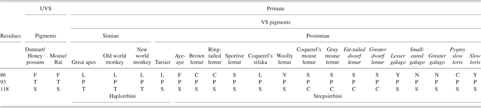
With the one notable exception of avian evolution discussed below, the changes in the spectral tuning of SWS1 pigments have all been toward the loss of UV sensitivity. The molecular basis for the shift from UV at around 360 nm to violet sensitivity was initially elucidated from comparisons between the amino acid sequences of SWS1 pigments in the UVS cones of the goldfish, Carassius auratus, and the common house mouse, Mus musculus, with those in the VS cones of the cow, Bos taurus, and the pig, Sus domesticus. A consistent difference between these pigments is found at site 86; in UVS pigments, Phe is present whereas in VS pigments, Tyr is found (Cowing et al., Reference Cowing, Poopalasundaram, Wilkie, Robinson, Bowmaker and Hunt2002; Fasick et al., Reference Fasick, Applebury and Oprian2002). Site-directed mutagenesis then established that this substitution was sufficient by itself to account for the shift from UVS to VS in the evolution of SWS1 pigments. A feature of UVS pigments is that their peak sensitivities show little variation, with λ max values falling between 355 and 370 nm. It would appear therefore that an unprotonated SB, achieved by the single amino acid substitution at site 86, is responsible for generating the shift in λ max to around 360 nm, with other residue changes having little or no impact. In contrast, VS pigments range from 406 nm as found in the aye-aye (Carvalho et al., Reference Carvalho, Davies, Robinson and Hunt2012) to around 460 nm in the ground squirrel (Jacobs, Reference Jacobs1978). For VS pigments therefore, spectral tuning occurs over a 54 nm range. Yokoyama et al. (Reference Yokoyama, Starmer, Takahashi and Tada2006), in a study of mouse UVS and human VS pigments, identified amino acid substitutions at six sites that generate spectral shifts, but these are largely not conserved across other pigments (see Mammals section). Hence, the substitutions responsible for the spectral shifts among VS pigments largely remain to be identified (Hunt et al., Reference Hunt, Carvalho, Cowing, Parry, Wilkie, Davies and Bowmaker2007).
Fishes
Within the lower vertebrates, such changes in the peak sensitivities of SWS1 pigments are generally not seen; the lamprey SWS1 pigment is UVS with Phe86, and the only example of a switch to VS in teleost fishes is found in the scabbardfish (Lepidopus fitchi) where a deletion of Phe86 results in a λ max at 423 nm (Tada et al., Reference Tada, Altun and Yokoyama2009).
Amphibians and reptiles
The cone photoreceptor complement of the retina of the tiger salamander, Ambystoma tigrinum, comprises double cones and two types of single cones (Sherry et al., Reference Sherry, Bui and Degrip1998), one of which expresses a UVS SWS1 pigment (Ma et al., Reference Ma, Znoiko, Othersen, Ryan, Das, Isayama, Kono, Oprian, Corson, Cornwall, Cameron, Harosi, Makino and Crouch2001a). This pigment again possesses Phe86 (Ma et al., Reference Ma, Kono, Xu, Das, Ryan, Hazard, Oprian and Crouch2001b). In contrast, in frogs, the λ max of the SWS1 pigment is shifted into the violet (Starace & Knox, Reference Starace and Knox1998) and, compared with the putative ancestral UVS pigment, this shift is associated with two substitutions, Phe86Met and Thr93Pro. Interestingly, a Phe86Met substitution into the goldfish UVS pigment failed to generate an LW shift (Cowing et al., Reference Cowing, Poopalasundaram, Wilkie, Robinson, Bowmaker and Hunt2002); the full shift to 423 nm may involve site 93, as proposed for primate VS pigments (see Mammals section), plus additional substitutions at sites 91, 109, 113, 116, and 118 (Takahashi & Yokoyama, Reference Takahashi and Yokoyama2005).
Among the reptiles, the first demonstration of UV sensitivity was made in anoline lizards (Fleishman et al., Reference Fleishman, Loew and Leal1993) where all four cone types are generally present to give the potential for tetrachromacy, with an SWS1 pigment that peaks in the UV (Kawamura & Yokoyama, Reference Kawamura and Yokoyama1998; Loew et al., Reference Loew, Fleishman, Foster and Provencio2002; Fleishman et al., Reference Fleishman, Loew and Whiting2011). UVS cones have also been demonstrated in geckos (Loew, Reference Loew1994), but in these species, the cone classes are reduced to three with the loss of the SWS2 class (Kojima et al., Reference Kojima, Okano, Fukada, Shichida, Yoshizawa and Ebrey1992; Ellingson et al., Reference Ellingson, Fleishman and Loew1995; Taniguchi et al., 1999; Yokoyama & Blow, Reference Yokoyama and Blow2001). Sequences for the SWS1 opsin have been obtained for the tokay gecko, Gekko gecko (Yokoyama & Blow, Reference Yokoyama and Blow2001) and for the anoline lizard, Anolis carolensis (Kawamura & Yokoyama, Reference Kawamura and Yokoyama1996). In all instances, Phe86 is present, consistent with UV sensitivity.
UVS cones are also found among the snakes (Jacobs et al., Reference Jacobs, Fenwick, Crognale and Deegan1992; Sillman et al. 1997, 1999, Reference Sillman, Johnson and Loew2001) and turtles (Loew & Govardovskii, Reference Loew and Govardovskii2001), although the molecular identity of the pigments has only been confirmed in snakes. Molecular analysis of snake UVS pigments is restricted to two species of “basal” henophidian snakes, the sunbeam snake, Xenopeltis unicolor, and the python, Python regius (Davies et al., Reference Davies, Cowing, Bowmaker, Carvalho, Gower and Hunt2009b). Both possess a rod-dominant retina with just two types of single cones with spectral peaks at around 550 and 360 nm. The pigment in the latter S cones was identified as belonging to the SWS1 class with Phe86 retained in both species.
A feature of lizard and turtle retinae (Bowmaker, Reference Bowmaker2008) is the presence in cone photoreceptors of colored oil droplets within the inner segments that contain carotenoid pigments spectrally matched to the absorbance properties of the particular cone class and acting as cut-off filters to reduce noise and increase spectral discrimination (Loew & Govardovskii, Reference Loew and Govardovskii2001; Loew et al., Reference Loew, Fleishman, Foster and Provencio2002). For S cones containing UV SWS1 pigments, these oil droplets are described as transparent as they have essentially zero absorbance; their function may be to act as light waveguides to enhance photon capture.
Birds
Avian cones also contain oil droplets in their inner segments (Bowmaker, Reference Bowmaker1977; Bowmaker & Hunt, Reference Bowmaker and Hunt2006; Hunt et al., Reference Hunt, Carvalho, Cowing and Davies2009a). Birds have generally retained all four-cone opsin classes, with the SWS1 pigment found in a class of single cones. Oil droplets are found in all avian cones but as in reptiles, the oil droplets present in S cones have no significant absorbance above 350 nm (Bowmaker et al., Reference Bowmaker, Heath, Wilkie and Hunt1997; Hart, Reference Hart2001b).
Within the evolution of the Aves, an early event was the loss of UV sensitivity of the SWS1 pigment arising from a Phe86Ser substitution that occurred at the base of the avian lineage (Carvalho et al., Reference Carvalho, Cowing, Wilkie, Bowmaker and Hunt2007). Subsequent to this event, UVS SWS1 pigments reappeared in a number of avian lineages (Wilkie et al., Reference Wilkie, Vissers, Das, Degrip, Bowmaker and Hunt1998; Ödeen & Hästad, Reference Ödeen and Håstad2003); in most cases, the shift back into the UV has been achieved by a different molecular route, the replacement of Ser by Cys at site 90 (Wilkie et al., Reference Wilkie, Robinson, Cronin, Poopalasundaram, Bowmaker and Hunt2000; Yokoyama et al., Reference Yokoyama, Radlwimmer and Blow2000). As for UVS pigments with Phe86, the expectation is that the SB would be unprotonated, but this has not been experimentally verified. Site 86 is also involved since Ser86 is always substituted in UVS pigments, although the actual residue present (Ala, Cys, Ile, Met, or Phe) differs among species (Wilkie et al., Reference Wilkie, Robinson, Cronin, Poopalasundaram, Bowmaker and Hunt2000; Yokoyama et al., Reference Yokoyama, Radlwimmer and Blow2000; Ödeen & Håstad, Reference Ödeen and Håstad2003).
An appreciation of the extent of changes in the spectral sensitivity of avian SWS1 pigments has been extended by molecular studies of the SWS1 opsin gene in 46 bird species distributed across 14 avian orders (Ödeen & Håstad, Reference Ödeen and Håstad2003, Reference Ödeen and Håstad2010; Ödeen et al., Reference Ödeen, Håstad and Alström2010). The approach used largely relies on the supposition that the residues present at sites 86 and 90 in SWS1 pigments are accurate predictors of the λ max of the pigment. Spectral data are, however, largely lacking, so for many species, it has not been possible to verify this. As shown in Fig. 7, Ser90 is present in the SWS1 pigment of most species, consistent with the presence of VS pigments (Carvalho et al., Reference Carvalho, Cowing, Wilkie, Bowmaker and Hunt2007). There are, however, some notable exceptions. Cys90 is present throughout the order Psittaciformes (Carvalho et al., Reference Carvalho, Knott, Berg, Bennett and Hunt2011; Aidala et al., Reference Aidala, Huynen, Brennan, Musser, Fidler, Chong, Machovsky Capuska, Anderson, Talaba, Lambert and Hauber2012), and UVS pigments have been confirmed by spectral analysis in two species, the budgerigar, Melopsittacus undulatus, and the rosella, Platycercus elegans (Wilkie et al., Reference Wilkie, Vissers, Das, Degrip, Bowmaker and Hunt1998; Carvalho et al., Reference Carvalho, Knott, Berg, Bennett and Hunt2011). UVS pigments are also found among species of Passeriformes and Charadriiformes (Ödeen & Håstad, Reference Ödeen and Håstad2003; Ödeen et al., Reference Ödeen, Håstad and Alström2010; Machovsky Capuska et al., Reference Machovsky Capuska, Huynen, Lambert and Raubenheimer2011; Aidala et al., Reference Aidala, Huynen, Brennan, Musser, Fidler, Chong, Machovsky Capuska, Anderson, Talaba, Lambert and Hauber2012), with Cys90 again present. Within the Passeriformes, it would appear that the gain or loss of UVS/VS pigments has occurred on at least eight separate occasions (Ödeen et al., Reference Ödeen, Håstad and Alström2011) and in each case, substitution at site 90 would appear to underlie the spectral shift (Fig. 8A). An example of a particularly localized shift is found among the fairy-wrens (Ödeen et al., Reference Ödeen, Pruett-Jones, Driskell, Armenta and Håstad2012) where UVS pigments are found in several species spread across two clades and requiring two separate Ser90Cys substitutions. In species from the order Charadriiformes (Fig. 8B), which include both sea and shorebirds, UVS pigments are limited to the Gulls, Kittiwake, Noddys, Black Skimmer, and one species of Tern, the white tern Onychoprion fuscatus (Ödeen et al., Reference Ödeen, Håstad and Alström2010; Machovsky Capuska et al., Reference Machovsky Capuska, Huynen, Lambert and Raubenheimer2011); the other 10 species of Tern so far examined all have VS pigments. Overall therefore, Ödeen & Håstad (Reference Ödeen and Håstad2013) have estimated that the shift in avian color vision between VS and UVS has occurred on at least 14 separate occasions.
Evolution of SWS1 pigments in birds. Presence of VS and UVS pigments in the different avian orders. With two exceptions, in UVS pigments, the avian ancestral Ser86 is replaced with a nonpolar residue and Ser90 is replaced by Cys90. The two exceptions are the Trogoniformes (Blue-crowned Trogon), where Phe86 is present with Ser90, and the Struthioniformes, where Phe86 is present with Cys90. Example species with UVS and VS pigments are listed alongside their respective orders. Data from Ödeen and Håstad (Reference Ödeen and Håstad2003, Reference Ödeen and Håstad2010), Ödeen et al. (Reference Ödeen, Håstad and Alström2010, Reference Ödeen, Håstad and Alström2011), Carvalho et al. (Reference Carvalho, Knott, Berg, Bennett and Hunt2011), Ödeen et al. (Reference Ödeen, Håstad and Alström2011), and Aidala et al. (Reference Aidala, Huynen, Brennan, Musser, Fidler, Chong, Machovsky Capuska, Anderson, Talaba, Lambert and Hauber2012). Black lines indicate UVS pigments only, violet lines indicate VS pigments only, and dotted lines indicate both UVS and VS pigments.
Gain/loss of UVS pigments. (A) Within the order Passeriformes. For each lineage, the genus and an example species is shown. Note that UVS pigments have evolved on at least eight separate occasions and in each case, Cys90 is present. The UVS/VS shifts in the pigments of fairy wrens (Ödeen et al., Reference Ödeen, Pruett-Jones, Driskell, Armenta and Håstad2012) are not shown. The tree has been redrawn with additions from Ödeen et al. (Reference Ödeen, Håstad and Alström2011). (B) Within the order Charadriiformes. Data from Ödeen et al. (Reference Ödeen, Håstad and Alström2010) and Machovsky Capuska et al. (Reference Machovsky Capuska, Huynen, Lambert and Raubenheimer2011). Black lines indicate UVS pigments only, violet lines indicate VS pigments only, and dashed lines indicate uncertain ancestral state.
In every UVS pigment mentioned above, Phe86 is also substituted; there is, however, one avian species, the blue-crowned trogon, Trogon curucui, in which Phe86 is present in the SWS1 opsin (Ödeen & Håstad, Reference Ödeen and Håstad2003). In this species, Ser90 is present, so either Phe86 has been retained in this species from the ancestral vertebrate UVS pigment, which would appear unlikely, or this represents a reverse mutation. Site directed mutagenesis of the pigeon and chicken VS pigment confirms that the introduction of Phe86 shifts the λ max into the UV (Carvalho et al., Reference Carvalho, Cowing, Wilkie, Bowmaker and Hunt2007).
Phe86 was also reported in the SWS1 pigment of the common rhea, Rhea americana, by Ödeen & Håstad (Reference Ödeen and Håstad2003), but this time in combination with Cys90, where the SWS1 pigment of the ostrich, Struthio camelus, was identified as VS, based on the presence of Ser90. More recently, data from Aidala et al. (Reference Aidala, Huynen, Brennan, Musser, Fidler, Chong, Machovsky Capuska, Anderson, Talaba, Lambert and Hauber2012) have shown that the ostrich pigment, along with those of other flight-less birds belonging to the order Struthioniformes that includes two species of cassowary, the emu, five species of moa, and six species of tinamou/nothura, not only possess Cys90 but like the common rhea also have Phe86. It would appear therefore that the early paleognathous species combined Phe86 with Cys90, both of which would be expected to generate a UVS pigment. A possible complication with this interpretation is reported by Wright & Bowmaker (Reference Wright and Bowmaker2001) of the presence of VS cones in the ostrich, although this was based on just a single cone. Nevertheless, the presence of VS rather than UVS cones in the common rhea and ostrich is supported by the steep rise below about 370 nm in the absorbance of the ocular media and the high absorbance of the oil droplets in the “blue” (S2) cones containing an SWS2 pigment. In species with UVS S1 cones with a relatively large spectral step to the S2 cones, the C-type droplets generally have a low absorbance, but in species with VS S1 cones, where the spectral separation is smaller (only about 30–40 nm), the droplets have a higher absorbance and probably act as cut-off filters; this is the most likely situation in the rhea and ostrich. It is also the case that short-wavelength light will be focused in a different plane to longer wavelengths, resulting in a blurring of the retinal image (axial chromatic aberration; Mandelman & Sivak, Reference Mandelman and Sivak1983; Rohrer et al., Reference Rohrer, Schaeffel and Zrenner1992); birds with larger eyes, as found in many of the paleognates, might be expected to use VS rather than UVS pigments to lessen the impact on image quality. Is it possible therefore that the SWS1 pigments of these paleognathous species are VS? In those species where sequencing has been extended to include site 93, Met is ubiquitously and uniquely present, which contrasts with Ile, Leu, or Thr at site 93 in all other avian species, whether UVS or VS. As discussed below, Pro93 is present in the VS pigments of all primates and, even when coupled with Phe86, it is sufficient to maintain a VS pigment (Carvalho et al., Reference Carvalho, Davies, Robinson and Hunt2012). The presence of Pro93 would appear to be key therefore to the production of VS pigments in primates, thereby emphasizing the importance of site 93 in the spectral tuning of SWS1 pigments. It remains unknown, however, whether the presence of Met93 in the presence of Phe86 and Cys90 is sufficient to generate a VS pigment; clarification of this will have to await appropriate in vitro studies.
What is the selective basis for these shifts in spectral sensitivity? UV-reflecting plumages are found ubiquitously across all avian lineages, suggesting that UV reflectance is an ancestral characteristic of the avian order (Eaton & Lanyon, Reference Eaton and Lanyon2003; Hausmann et al., Reference Hausmann, Arnold, Marshall and Owens2003; Mullen & Pohland, Reference Mullen and Pohland2008). Consistent with the ubiquitous presence of UVS pigments in parrots (Carvalho et al., Reference Carvalho, Knott, Berg, Bennett and Hunt2011), these birds have the highest percentage of UV reflectance, especially in plumage patches and there is direct evidence that UV reflectance is important for mate selection; female budgerigars prefer males with UV-reflecting plumage over those where UV-reflectance had been removed (Pearn et al., Reference Pearn, Bennett and Cuthill2001). It should be noted, however, that many parrot species are long-lived, with life spans in captivity of >50 years (Brouwer et al., Reference Brouwer, Jones, King and Schifter2000). Since UV radiation is known to damage several components of the eye such as the cornea, lens, retinal pigment epithelium, and photoreceptors (Matsui et al., Reference Matsui, Lin, Ho and Reddy2003; Tratsk & Thanos, Reference Tratsk and Thanos2003; Azzam et al., Reference Azzam, Levanon and Dovrat2004) and is associated with several age-related conditions like cataract and macular degeneration (Tratsk & Thanos, Reference Tratsk and Thanos2003; Azzam et al., Reference Azzam, Levanon and Dovrat2004), the penetration of UV radiation to the neural retina in long-lived parrot species might be expected to cause irreversible damage (Collier et al., Reference Collier, Waldron and Zigman1989; Rogers et al., Reference Rogers, Chan, Sims, Byrd, Hinton and Twining2004). This does not appear to be the case, which raises the question as to how these species tolerate UV exposure. One explanation may be that birds show a lower rate of mitochondrial free-radical production in comparison with mammals (Herrero & Barja, Reference Herrero and Barja1998; Holmes et al., Reference Holmes, Fluckiger and Austad2001). Furthermore, cells of long-lived birds exhibit exceptional resistance to oxidative-induced damage when compared with shorter-lived birds and mammals (Ogburn et al., Reference Ogburn, Carlberg, Ottinger, Holmes, Martin and Austad2001).
The presence of UVS pigments among the passerines may be more complex. An important role for UV reflectance has been demonstrated in mate choice (Hunt et al., Reference Hunt, Cuthill, Swaddle and Bennett1997, Reference Hunt, Cuthill, Bennett and Griffiths1999, Reference Hunt, Cuthill, Bennett, Church and Partridge2001) and foraging (Church et al., Reference Church, Bennett, Cuthill and Partridge1998; Siitari et al., Reference Siitari, Honkavaara and Viitala1999; Martin Schaefer et al., Reference Martin Schaefer, Schaefer and Vorobyev2007) in species with UVS pigments. In other cases, it may be the local habitat that is important with different levels of ambient light and background reflectance. However, the gain of UVS pigments in a subset of fairy wren species (Ödeen et al., Reference Ödeen, Pruett-Jones, Driskell, Armenta and Håstad2012) described above is difficult to interpret since there is no clear change in habitat between species with VS and UVS pigments. It is also surprising that UVS pigments are not found in birds-of-paradise (Ödeen et al., Reference Ödeen, Håstad and Alström2011) with their bright and iridescent male plumages that are clearly important for attracting a mate. The UV reflectance of eggs may be important for protection against parasitism; differences in the parasitic eggs of the brown-headed cowbird, Molothrus ater, a passerine that is likely to possess a UVS pigment, and the eggs of certain species that reject the parasitic eggs, many of which also possess UVS pigments, have been described, indicating that UV sensitivity may be important in protection against parasitism (Underwood & Sealy, Reference Underwood and Sealy2008). However, no significant difference in UV reflectance is present between the eggs of closely related species that either accept or reject the parasitic eggs, so UV-matching does not distinguish between acceptors and rejectors of parasitism.
Among sea and shorebirds, it is only the gulls that ubiquitously possess UVS pigments; unlike other seabirds, gulls forage in terrestrial as well as aquatic environments, and it may be that UV sensitivity is more relevant in a terrestrial setting (Machovsky Capuska et al., Reference Machovsky Capuska, Huynen, Lambert and Raubenheimer2011). UV light reflectance has been demonstrated from the feathers of terns and is thought to play a role in sexual selection (Bridge & Eaton, Reference Bridge and Eaton2005) but the species examined did not include those with UVS pigments.
Mammals
As mentioned above, Tyr86 is found in the VS pigments of the cow and pig (Cowing et al., Reference Cowing, Poopalasundaram, Wilkie, Robinson, Bowmaker and Hunt2002; Fasick et al., Reference Fasick, Applebury and Oprian2002), members of the order Artiodactyla, and this can be extended to the aquatic West Indian manatee, Trichechus manatus (Newman & Robinson, Reference Newman and Robinson2005). However, its close relatives, the elephants (Loxodonta africana and Elephas maximus) have Ser86, a substitution that has been shown to generate the shift into the violet (Yokoyama et al., Reference Yokoyama, Takenaka, Agnew and Shoshani2005). The acquisition of Ser86 in elephants must have occurred within the Proboscidea lineage following a prior Phe86Tyr substitution that occurred in the common ancestor to the Artiodactyla. Tyr86 is also found in the SWS1 pigments of two species of carnivores, the dog, Canis lupus familiaris, and cat, Felis catus (Hunt et al., Reference Hunt, Carvalho, Cowing and Davies2009a).
Light in the Arctic is relatively UV-rich because of scattering of shorter wavelengths and reflection from snow and ice. Given the highly damaging effect of UV light to the retina, it is surprising that the spectral sensitivity of the Arctic reindeer, Rangifer tarandus, extends into the UV as neither the cornea nor the lens block all UV light entering the eye (Hogg et al., Reference Hogg, Neveu, Stokkan, Folkow, Cottrill, Douglas, Hunt and Jeffery2011). This sensitivity is not, however, mediated by a UVS pigment; the reindeer possesses a typical VS pigment with Tyr86 as found in other members of the Artiodactyla, so the UV sensitivity arises solely from the penetration of UV light to the photoreceptor layer of the retina. As in long-lived parrots, it must be presumed that the reindeer has evolved mechanisms to protect the retina against extreme UV exposure.
Within placental mammals or eutherians, only species from the orders Rodentia, Chiroptera (bats) (Wang et al., Reference Wang, Oakley, Mower, Shimmin, Yim, Honeycutt, Tsao and Wen-Hsiung Li2004), and Insectivora (Glösmann et al., Reference Glösmann, Steiner, Peichl and Ahnelt2008) appear to have retained UVS pigments (Fig. 9A). Among the bats, this is inferred from the presence of Phe86 in the SWS1 gene in three species, two members of the megachiroptera, the Philippine dwarf fruit bat, Haplonycteris fischeri, and the flying fox, Pteropus dasymallus formosus, and one species of microchiroptera, the cave myotis, Myotis velifer. In two microchiropterans, Carollia perspicillata and Glossophaga soricina, UV sensitivity was directly shown by electroretinogram recordings, and their SWS1 genes were confirmed to encode Phe86 (Müller et al., Reference Müller, Glösmann, Peichl, Knop, Hagemann and Ammermüller2009). The order Rodentia comprises two suborders, the Sciurognathi and the Hystricognathi. Among members of the former, both mouse and rat have UVS pigments with Phe86 (Shi et al., Reference Shi, Radlwimmer and Yokoyama2001), whereas other species such as the gray squirrel, Sciurus carolinensis, have VS pigments with Tyr86 (Carvalho et al., Reference Carvalho, Cowing, Wilkie, Bowmaker and Hunt2006). Members of the Hystricognathi also show a mixture of UVS and VS pigments, with UVS pigments with Phe86 present in the diurnal degu, Octodon degus, and two subterranean species of tuco-tuco, Ctenomys talarum and Ctenomys magellanicus (Schleich et al., Reference Schleich, Vielma, Glösmann, Palacios and Peichl2010), whereas the guinea pig, Cavia porcellus, has a VS pigment with Val86 (Parry et al., Reference Parry, Poopalasundaram, Bowmaker and Hunt2004). Since Phe86 is retained in both suborders, the Phe86Tyr and Phe86Val substitutions, must have occurred separately within the rodent lineage.
Changes in spectral sensitivity of SWS1 pigments in mammals. (A) Loss of UVS pigments within the different mammalian lineages. The residues present at site 86 of the SWS1 opsin on each lineage are indicated. (B) Phylogenetic relationships of different primate species showing the different residues found at site 86 in SWS1 opsins, together with the corresponding codon sequences. The effects on nucleotide substitution of the alternative hypotheses of ancestral Phe86 or Tyr86 are shown in red and blue, respectively, with black used where both hypotheses are equivalent. Lower case is used to indicate required substitutions in codon 86 under each hypothesis. Crosses on lineages indicate species where SWS1 pseudogenes are present. Black lines indicate UVS pigments and violet lines indicate VS pigments. The tree was modified from the recent molecular phylogeny reported by Perelman et al. (Reference Perelman, Johnson, Roos, Seuanez, Horvath, Moreira, Kessing, Pontius, Roelke, Rumpler, Schneider, Silva, O’Brien and Pecon-Slattery2011).
The retention of UVS pigments and UV-transmitting optics is mostly found in nocturnal mammals, which experience lower less damaging light levels. UVS pigments with Phe86 are also present in species of the rodent family Octodontidae, which contains nocturnal and subterranean species as well as the diurnal degu, Octodon degus (Schleich et al., Reference Schleich, Vielma, Glösmann, Palacios and Peichl2010). The degu has a strongly diurnal retina with about 30% cones among its photoreceptors, and electroretinogram recordings show UV sensitivity (Chavez et al., Reference Chávez, Bozinovic, Peichl and Palacios2003; Jacobs et al., Reference Jacobs, Calderone, Fenwick, Krogh and Williams2003). Degus are very social animals that generously use UV-reflecting urine scent marks, and it has been suggested that their UV sensitivity may be advantageous, allowing them to see as well as smell these scent marks (Chavez et al., Reference Chávez, Bozinovic, Peichl and Palacios2003).
UVS pigments are more common among the marsupials or metatherians. Two major orders, the Diprotodontia and the Polyprotodontia, form the Marsupialia. Members of the Diprotodontia may either have VS or UVS pigments; the Tammar wallaby, Macropus eugenii, and quokka, Setonix brachyurus, have VS pigments with Tyr86 (Deeb et al., Reference Deeb, Wakefield, Tada, Marotte, Yokoyama and Marshall Graves2003; Arrese et al., Reference Arrese, Oddy, Runham, Hart, Shand, Hunt and Beazley2005), whereas the honey possum, T. rostratus, has a UVS pigment (Arrese et al., Reference Arrese, Hart, Thomas, Beazley and Shand2002), with Phe86 retained (Cowing et al., Reference Cowing, Arrese, Davies, Beazley and Hunt2008). Among the Polyprotodontia, UVS pigments are found in the fat-tailed dunnart, Sminthopsis crassicaudata (Cowing et al., Reference Cowing, Arrese, Davies, Beazley and Hunt2008), the South American big-eared opossum, Didelphis aurita (Hunt et al., Reference Hunt, Chan, Carvalho, Hokoc, Ferguson, Arrese and Beazley2009b) and the quenda, Isoodon obesulus (Arrese et al., Reference Arrese, Oddy, Runham, Hart, Shand, Hunt and Beazley2005); in all cases, Phe86 is retained. The Phe86Tyr substitution seen in the Tammar wallaby and quokka must have occurred therefore within the diprotodont marsupial lineage.
Within the primates, all species appear to possess VS pigments (Bowmaker et al., Reference Bowmaker, Astell, Hunt and Mollon1991; Hunt et al., Reference Hunt, Cowing, Patel, Appukuttan, Bowmaker and Mollon1995) and it has been proposed from a comparison with the mouse UVS pigment that tuning from UV to violet required the simultaneous replacement of residues at three sites (Phe86Leu, Thr93Pro, and Ser118Thr) (Yokoyama & Shi, Reference Yokoyama and Shi2000; Shi et al., Reference Shi, Radlwimmer and Yokoyama2001). However, other residue changes at these sites are found in members of the Haplorrhini (New World primates and tarsiers), and in Strepsirrhini (prosimians), with the residues present at sites 86 and 118 being quite variable (Table 1). For example, site 86 is occupied by Leu in Old and New World primates and Tarsiers but differs across the Strepsirrhini with Cys, Ser, Leu, Val, Tyr, and Asn present in at least one species. Surprisingly, Phe86 is found in one species, the aye-aye, a lemur endemic to Madagascar. As described above, Phe86 is generally associated with a UVS pigment (Hunt et al., Reference Hunt, Cowing, Wilkie, Parry, Poopalasundaram and Bowmaker2004) but in vitro expression of the aye-aye pigment gave a peak sensitivity at 409 nm (Carvalho et al., Reference Carvalho, Davies, Robinson and Hunt2012), so Phe86 does not in this case result in a UVS pigment.
Residues present at three sites in transmembrane helices II and III of the VS pigments of primates compared with the UVS pigments of two marsupials, the fat-tailed dunnart (GenBank acc. AY7724723) and honey possum (GenBank acc. AY772472), and two rodents, the mouse (GenBank acc. NM_007538) and rat (NM_031015). Species in italics possess a SWS1 pseudogene
What changes were initially responsible for the generation of the VS pigments seen in all primates? If it is accepted that a common tuning mechanism is likely responsible for the spectral shift into the violet seen in all primate SWS1 pigments, then, as shown in Table 1, the only consistent feature to account for this is Pro93. Substitution at this site may be responsible, therefore, for the long-wavelength shift, regardless of the residue present at site 86. Note that Pro93 is also present in the VS pigment of the clawed frog (Starace & Knox, Reference Starace and Knox1998). This does not, however, rule out a role for site 86 in the initial shift to VS pigments that occurred at the base of the primate lineage. If codon changes at site 86 are placed on to a primate phylogeny (Perelman et al., Reference Perelman, Johnson, Roos, Seuanez, Horvath, Moreira, Kessing, Pontius, Roelke, Rumpler, Schneider, Silva, O’Brien and Pecon-Slattery2011) using the tree shrew as an outgroup (Petry & Harosi, Reference Petry and Harosi1990; Hunt et al., Reference Hunt, Carvalho, Cowing and Davies2009a), then this codon would appear to have passed unchanged into the SWS1 pigment of the aye-aye (Fig. 9B). Since the aye-aye pigment is VS, it is surprising that Phe86 has been retained unaltered, especially since it is replaced in the SWS1 opsin of all other primates. The alternative explanation is that a Phe86Tyr substitution occurred at the base of the primate lineage, and it is this change, as shown in Fig. 9B, that enabled a shift to the violet. Subsequently, a Thr93Pro substitution occurred, a change that also drives the peak sensitivity into the violet, thereby removing the constraint on the residue at site 86. Subsequent changes at this site may be responsible for the fine tuning of the λ max (Carvalho et al., Reference Carvalho, Davies, Robinson and Hunt2012). Under this scenario, the Phe86 in the aye-aye would be a back mutation.
Inherited defects in S cones and pigments
Tritanopia
Mutations in the SWS1 gene in humans (gene symbol OPN1SW) give rise to a rare form of dichromacy called tritanopia; six missense mutations have been reported in the human gene, Gly79Arg, Ser214Pro, Pro264Ser (Weitz et al., Reference Weitz, Miyake, Shinzato, Montag, Zrenner, Went and Nathans1992a,Reference Weitz, Went and Nathansb), Leu56Pro (Gunther et al., Reference Gunther, Neitz and Neitz2006), Arg282Gln (Baraas et al., Reference Baraas, Carroll, Gunther, Chung, Williams, Foster and Neitz2007), and Thr190Ile (Baraas et al., Reference Baraas, Hagen, Dees and Neitz2012), each resulting in a dominant mode of disease inheritance.
Enhanced S-cone syndrome
In an inherited form of retinal degeneration in humans called enhanced S-cone syndrome, an excess number of S cones is present at the expense of other photoreceptor types (Jacobson et al., Reference Jacobson, Marmor, Kemp and Knighton1990; Roman & Jacobson, Reference Roman and Jacobson1991; Hood et al., Reference Hood, Cideciyan, Roman and Jacobson1995) (Fig. 10). The disorder is inherited as a recessive condition and arises from mutations in the NR2E3 gene that encodes a retinal nuclear receptor that acts as a ligand-dependent transcription factor (Kobayashi et al., Reference Kobayashi, Takezawa, Hara, Yu, Umesono, Agata, Taniwaki, Yasuda and Umesono1999). Another gene, NRL that encodes a neural retina-specific leucine zipper protein (Swaroop et al., Reference Swaroop, Xu, Pawar, Jackson, Skolnick and Agarwal1992) is a critical regulator of rod photoreceptor cell fate such that when all function of the NRL gene is removed by targeted gene mutation in the mouse, there is a complete loss of rod function and an enhanced cone function mediated by S cones (Mears et al., Reference Mears, Kondo, Swain, Takada, Bush, Saunders, Sieving and Swaroop2001). Both NRL and NR2E3 are required therefore for the normal development of rod photoreceptors, with NR2E3 also required to suppress cone development (Haider et al., Reference Haider, Demarco, Nystuen, Huang, Smith, McCall, Naggert and Nishina2006). In the developing retina, NRL first activates NR2E3 in newborn photoreceptors, thereby suppressing the cone pathway and leading to the development of rod photoreceptors (Swaroop et al., Reference Swaroop, Kim and Forrest2010). In the absence of NR2E3 expression, normal rod development is blocked by the expression of S cone genes as the default pathway, resulting in a severe reduction in rods and an increase in S cones (Fig. 10). The pattern of expression of NRL and NR2E3 may be responsible therefore for the early appearance of SWS1 opsin in the developing mammalian retina (Szél et al., Reference Szél, Lukats, Fekete, Szepessy and Röhlich2000; Cornish et al., Reference Cornish, Xiao, Yang, Provis and Hendrickson2004; Lukats et al., Reference Lukats, Szabo, Röhlich, Vigh and Szél2005; Arango-Gonzalez et al., Reference Arango-Gonzalez, Szabo, Pinzon-Duarte, Lukats, Guenther and Kohler2010).
Enhanced production of S cones in wildtype mice and mice homozygous for the rd7 mutation in Nr2e3. Immunohistochemistry of WT and rd7 retinal sections (A) and flatmounts (B) at postnatal day 21 stained with anti-S-opsin. Nuclei are stained with bisbenzimide (blue). Note the whorls of S cones in the rd7 retina that ingress into the ONL. RGC, retinal ganglion cell layer; INL, inner nuclear layer; ONL, outer nuclear layer; and OS, outer segments of photoreceptors. Redrawn from Cheng et al. (Reference Cheng, Khan, Roger and Swaroop2011).
Albinism
Albinism, occurring as oculocutaneous albinism or ocular albinism, is a group of genetic defects that impair melanin production due to deficits in tyrosinase function and that seriously affect vision (review: Summers, Reference Summers1996). Studies in various mammals have shown that albinism results in misrouting of retinal ganglion cell axons in the optic chiasm and in an underdevelopment of central retina—including the lack of a fovea in human albinos (review: Jeffery, Reference Jeffery1997). In the photoreceptor layer, the deficit is focused on the rods. A roughly 30% reduction of rod densities was found in albino ferrets and mice, whereas cone densities were similar to those of pigmented animals (Jeffery Reference Jeffery1997; Jeffery et al., Reference Jeffery, Darling and Whitmore1994; Gresh et al., Reference Gresh, Goletz, Crouch and Rohrer2003). These studies did not address cone opsin expression. A more recent study compared L and S cone distributions in pigmented Piebald Virol Glaxo rats and albino Sprague-Dawley rats and found no differences between the strains (Ortín-Martínez et al., Reference Ortín-Martínez, Jiménez-López, Nadal-Nicolás, Salinas-Navarro, Alarcón-Martínez, Sauvé, Villegas-Pérez, Vidal-Sanz and Agudo-Barriuso2010). The total number and topographical distribution of the two cone types, and the existence of only 3% dual pigment cones, was the same. In contrast, Glösmann et al. (Reference Glösmann, Arbogast and Peichl2009) found marked differences in SWS1 and LWS opsin expression between pigmented Brown Norway and albino Wistar rats and between pigmented wildtype and albino deer mice (Peromyscus maniculatus). In Wistar rats, S cone densities at any retinal position were three to four times higher than in Brown Norway rats, and there was a significant proportion of dual pigment cones. In albino deer mice, total cone numbers and the numbers of cones exclusively expressing SWS1 opsin were not significantly different from those in pigmented deer mice. However, in pigmented deer mice, coexpression of cone opsins was rare (0–2% of the cones), whereas in albino animals, SWS1 opsin was coexpressed with LWS opsin in 60–90% of the cones. The difference between the two studies may represent strain differences, or a detection problem at the low magnification used in the automated analysis by Ortín-Martínez et al. (Reference Ortín-Martínez, Jiménez-López, Nadal-Nicolás, Salinas-Navarro, Alarcón-Martínez, Sauvé, Villegas-Pérez, Vidal-Sanz and Agudo-Barriuso2010). Overall, the findings suggest that tyrosinase activity may directly or indirectly affect the regulation of S opsin expression. In human albinos, there may be mild color vision impairments, but the evidence is weak (see Lourenco et al. Reference Lourenco, Fishman and Anderson1983) and their cellular basis is unknown.
Are S cones a separate type?
Traditionally, the spectral cone types have been considered to constitute separate cellular types, differing not only in their visual pigment expression but also in other cellular properties. As described above, reptilian and bird cone inner segments contain colored oil droplets that act as color filters (long-pass filters), and each cone type has an oil droplet color (spectral transmission) tuned to its visual pigment (Hart, Reference Hart2001a,Reference Hartb and literature cited therein). The S cones of sciurids and some primates have inner segments of larger diameter than the M/L cones. In the central retina of primates including humans, the S cone pedicles (synaptic endings) are smaller than the L and M cone pedicles, and they label more strongly with the generic cone marker peanut agglutinin (Ahnelt et al., Reference Ahnelt, Keri and Kolb1990; Lee et al., Reference Lee, Telkes and Grünert2005; Lee & Grünert, Reference Lee and Grünert2007). In ground squirrels, the L cone pedicles are electrically coupled by gap junctions, whereas the S cone pedicles are not (Li & DeVries, Reference Li and DeVries2004); in primates, the gap junctions at S cone pedicles are much sparser than at L and M cone pedicles (O’Brien et al., Reference O’Brien, Chen, MacLeish, O’Brien and Massey2012). Szél et al. (Reference Szél, Röhlich, Caffé and van Veen1996), Ahnelt and Kolb (Reference Ahnelt and Kolb2000), and Calkins (Reference Calkins2001) have summarized some of the cellular differences between mammalian S and M/L cones.
The findings of a developmental switch from SWS1 to LWS opsin expression, with transient coexpression of both opsins in the cones of some mammals, and the permanent coexpression of both opsins in some of these species have questioned the view of two basic cone types (L cones and S cones). Applebury et al. (Reference Applebury, Antoch, Baxter, Chun, Falk, Farhangfar, Kage, Krzystolik, Lyass and Robbins2000) have argued that at least in mouse, there is only one cone type that produces a pattern of subtypes across the retina by regulating the relative expression levels of LWS and SWS1 opsin. This opened a controversial debate (Applebury, Reference Applebury2001; Neitz & Neitz, Reference Neitz and Neitz2001); subsequent studies showing the regulation of opsin expression by TH and its cone-specific receptor TRβ2 seem to favor the “one cone type” hypothesis, as mouse and rat cone opsin expression reversibly switches according to the TH level (see Opsin coexpression and Development of S cones sections). However, the finding of “blue cone” bipolar cells that exclusively contact S cones is strong additional evidence that the S cones have special cellular properties beyond their opsin content. Blue cone bipolar cells were first identified in the macaque monkey and have since been studied extensively in Old World and New World primates (overviews: Martin et al., Reference Martin, Grünert, Chan and Ghosh2001; Puller & Haverkamp, Reference Puller and Haverkamp2011). Blue cone bipolar cells have also been characterized in a number of nonprimate mammals—particularly in ground squirrels—and are considered a basic element of the mammalian retinal blueprint (review: Puller & Haverkamp, Reference Puller and Haverkamp2011). The strongest challenge of the “only one cone type” hypothesis is the S cone-selective bipolar cell of the mouse. Haverkamp et al. (Reference Haverkamp, Wässle, Duebel, Kuner, Augustine, Feng and Euler2005) analyzed a transgenic mouse line where a number of retinal cell types, including the S cone bipolar cells, expressed a fluorescent indicator. The S cone bipolar cells exclusively contacted the small subset of genuine S cones (i.e., cones exclusively expressing the S opsin) also in ventral mouse retina and avoided the many dual pigment cones that dominantly expressed the S opsin. This suggests that even in mouse, the genuine S cones possess additional properties or markers that are a selective attractant for the S cone bipolar cells. The horizontal cells of primates, sciurids, and horse also display specific S cone connectivities, either preferring or avoiding S cones depending on horizontal cell type (reviews: Peichl et al., Reference Peichl, Sandmann, Boycott, Chalupa and Finlay1998; Martin et al., Reference Martin, Grünert, Chan and Ghosh2001; Puller & Haverkamp, Reference Puller and Haverkamp2011). In goldfish, the three horizontal cell types have different connectivities with the different cone types (Stell & Lightfoot, Reference Stell and Lightfoot1975). In summary, their specific circuitry is the most compelling evidence for the S cones constituting a separate cone type.
Bird S cones show a further surprising peculiarity related to magnetic orientation. Birds orientate in the geomagnetic field, and behavioral experiments have shown that their directional magnetic sense only works under appropriate lighting conditions, that the magnetic detector mechanism resides in the eye, and that it involves a light-dependent radical pair process (reviews: Wiltschko & Wiltschko, Reference Wiltschko and Wiltschko2007; Wiltschko et al., Reference Wiltschko, Stapput, Thalau and Wiltschko2010). The flavoprotein cryptochrome was suggested as the magnetic receptor molecule because upon illumination, it forms radical pairs that are sensitive to magnetic field orientation. Double immunolabeling for cryptochrome 1a and SWS1 opsin in the retinae of European robins and chickens showed that cryptochrome 1a is exclusively located in these S1 cones (Niessner et al., Reference Niessner, Denzau, Gross, Peichl, Bischof, Fleissner, Wiltschko and Wiltschko2011). This suggests that bird S1 cones do double duty in color vision and magnetic orientation. The circuitry for processing the magnetic information is largely unknown, and it remains to be studied how the birds are able to separate visual and magnetic information (Bischof et al., Reference Bischof, Niessner, Peichl, Wiltschko and Wiltschko2011).