Introduction
Each year over 39,000 new cases of lung cancer are diagnosed in the United Kingdom, of these about 88% are non-small cell lung cancer (NSCLC).1
Advances in radiotherapy technology are enabling precise targeting and dose-escalation strategies such as stereotactic body radiotherapy treatment (SBRT) which can deliver larger doses in under 2 weeks with reports of fewer acute side effects.Reference Verstegen, Lagerwaard, Haasbeek, Slotman and Senan2, Reference Chi, Liao, Nguyen, Xu, Stea and Komaki3 SBRT is increasingly being used worldwide to treat medically inoperable stage I and II NSCLC and limited pulmonary oligometastases and has been shown to be a more effective treatment than conventional radiotherapy.Reference Fakiris, McGarry and Yiannoutsos4–7 The use of advanced image-guided radiotherapy techniques have been vital in the development of SBRT enabling enhanced target localisation and safety.Reference Hoyer8–Reference van Elmpt, Petit, De Ruysscher, Lambin and Dekker10 Accuracy in position verification is paramount in SBRT as the consequences of any errors is greater due to smaller margins and dose escalation.7
The Royal Marsden NHS Foundation Trust assessed the feasibility of SBRT for solitary lung tumours in 2009 as part of an in-house trial and the technique is now available to a wider range of patients. Imaging practice at the Trust for high dose hypofrationated radiotherapy involved the clinical oncologist, with practitioner status, to perform an online manual cone-beam computed tomography (CBCT) match before each fraction, in line with national and international practice.7, Reference Guckenberger, Krieger and Richter11, Reference Purdie, Bissonnette and Franks12
However, the availability of the clinician can impact on the efficiency of service delivery, creating delays for all patients and disrupting the work flow of treatment units. Delays can compromise efficient patient care, increasing waiting and treatment times which may create discomfort for the patient. Utilising competent therapeutic radiographers could reduce the time taken to deliver each fraction, potentially improving workflow and the efficiency of the service provided to all patients. The expansion of the radiographer’s role to verify SBRT CBCT images could enable more effective use of their knowledge and skills which will benefit both patients and the service.7, Reference James, Beardmore and Dumbleton13
Verification of CBCTs for SBRT requires accurate visualisation of soft tissues; clinical oncologists have extensive experience with viewing and delineating soft tissue images due to their role in outlining computed tomography planning scans and viewing diagnostic images. Therapeutic radiographers are increasing their knowledge and experience with CBCT images on a daily basis with every verification match they perform and have proven advanced CBCT verification competencies in other anatomical sites and techniques such as adaptive bladder treatments.Reference Hutton, Leadbetter, Jain and Baker14, Reference White and Kane15
This study aims to assess the baseline competencies of radiographers for CBCT image verification for patients receiving SBRT for peripheral lung tumours. This will determine the feasibility of radiographer led verification to provide safe, accurate and timely verification in the absence of a clinical oncologist, for patients with solitary lung tumours.
METHOD AND MATERIALS
All radiographers in the department with at least 1 years’ clinical experience were invited to volunteer with the study. They were provided with full information and the opportunity to ask any questions beforehand. The Trusts statistician determined that five radiographers were required for the study and these were selected at random from all volunteers. A consent form agreeing to their participation was completed for each radiographer. Patient and radiographer anonymity were protected by allocating all subjects a study ID, with patients corresponding CBCT images allocated sequential numbers. All data generated through this study is to be held securely in order to maintain anonymity.
Five therapeutic radiographers retrospectively reviewed CBCT image data sets for 20 patients whose images had previously been verified by clinical oncologists and treated with the Active Breathing Control (ABCTM) system as part of the SBRT protocol. The radiographers did not review the clinicians match. CBCT images were reviewed using Elekta Synergy cone beam system (XVI R4·5)16 and details of translational and rotational displacements were recorded. A successful match for a given patient was defined as an agreement within 0·2 cm, in all three planes, between the clinician’s verification and the radiographer’s retrospective match for at least 85% of cases. All verifications that fell outside this definition were defined as a failure. A limit of a translational difference of 0·2 cm was determined as this is the tolerance for daily geometric quality assurance checks on the treatment machines.
All radiographers had completed in-house XVI training and workbook, however SBRT specific training was not provided before the retrospective verifications as baseline competencies were to be assessed.
The time taken by radiographers to manually verify the CBCT images was recorded to determine if their verification time was within the acceptable limits suggested by previous studies of 240 seconds.Reference Higgins, Bezjak and Franks17 The time taken for clinicians to perform online verifications was previously recorded for only 8/20 CBCTs as part of an earlier trial, with the average time taken for as 375 seconds; ranging from 120 to 480 seconds. This timing included the acquisition of the CBCT which took ~75–80 seconds.
Visual analogue scales allowed the radiographers to document confidence in their own ability to interpret and visualise soft tissue CBCT images for SBRT patients.
For all radiographers and images combined, a 93% power would be required to detect a level of success >85% with 100 verifications (exact binomial test, alpha 1-sided=5%). For the technique to be declared a success there should be at least 92 out of 100 correct matches.
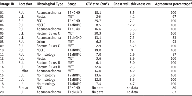
Results (Table 1)
Patient demographics (n=20)
Twelve had NSCLC (four non-biopsy proven) and eight had metastatic lung tumours from rectal and colonic carcinoma (Table 1).
The measured set up errors (i.e., tumour displacement) (Figure 1) were found to be comparable with other studies setup errors.Reference Senan and Lagerwaard9, Reference Higgins, Bezjak and Franks17 The mean (range) of X, Y and Z was; X=0·10 (−1·02 to 0·91) Y=−0·12 (−1·61 to 1·48) and Z=0·22 (−0·37 to 1·48).
Set up errors.

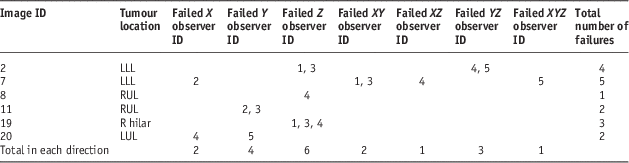
A total of 85/100 verifications were within 0·2 cm translational displacement of the original clinicians match in all three planes (Figure 2a–2c). In the X direction there was 94% agreement in the Y direction 91% agreement and in the Z direction 89% agreement. The lower lobe tumours resulted in the most frequent failures (Table 2).
(a) Difference of observer displacements from clinician in X direction. (b) Difference of observer displacements from clinician in Y direction. (c) Difference of observer displacements from clinician in Z direction.
Patient demographics and agreement percentage between radiographers and clinicians
a Notes: Where 100% equals agreement within 0·2 cm in all observers and all three planes.
Abbreviations: LLL, left lower lobe; LUL, left upper lobe; RLL, right lower lobe; RUL, right upper lobe.
Verifications >0·2 cm by image ID, direction and observer ID
Abbreviations: LLL, left lower lobe; RUL, right upper lobe; LUL, left upper lobe.
The thickness of the chest wall was measured as a possible cause to setup and verification issues. Average chest wall thickness has been found to be ~4·2 cm.Reference Givens, Ayotte and Manifold18, Reference McLean, Richards, Crandall and Marinaro19 There was no data available for GTV size and chest wall thickness for CBCT 19 and 20 and this may limit any comparisons. From the available data all verification failures had a GTV size of 13·1 cm3 or under (Table 1).
Rotational displacements
Previous studies have suggested that rotational displacements <3° is not considered to have a significant impact on verifications for spherical GTV’s.Reference Wang, Zhong and Bai20 Differences between the radiographers rotational match and the clinical oncologists were >2° for CBCT’s 2, 4, 14, 16 and 18. However only CBCT 2 also failed in the translational verification, recording translations >0·2 cm in all three planes when compared with clinical oncologist’s match. CBCT’s 4, 14, 16 and 18 all recorded a 100% translational agreement with the clinical oncologist.
Reproducibility
Interobserver variations were measured using intraclass correlation coefficient produced from a two-way random effects model, with both radiographers and patients being random factors.Reference Shrout and Fleiss21 Secondary endpoint analysis of interobserver variations (reproducibility) between independent therapeutic radiographers were assessed by comparing the means between them. In the X and Z direction there was excellent agreement between radiographers and in the Y direction there was a substantial agreement.
Time taken
Time taken by the radiographers for all methods ranged from 50 to 480 seconds. Automatic matches ranged from 50 to 315 seconds and manually adjusted matches ranged from 70 to 480 seconds. The verification process took an average of 110 seconds for automatic matches and 152 seconds for manual matches and adjustments. An average of 128 seconds was taken for both methods combined.
Confidence in visualisation and match
Where there was 100% agreement with the match, the confidence and visualisation scores were 8 or above (on a 10-point scale). There was no apparent correlation between a longer length of time taken for a verification and poor visualisation and low confidence.
DISCUSSION
Daily online imaging for SBRT treatment is vital to ensure accurate delivery.Reference Bissonnette, Purdie, Higgins, Li and Bezjak22, Reference Grills, Hugo and Kestin23 This study found that therapeutic radiographers were able to retrospectively verify the same CBCT images with registration close to the ‘gold standard’ of the clinicians’ match with no additional training. This suggests that they are able to verify CBCT images for patients treated with SBRT but further training is needed to ensure accuracy and confidence.
As these verifications were completed offline, further assessment and training may be required to ensure radiographers are confident when faced with the pressures of online verification. With the increase in future demand for this type of treatment will come an increase in the number of suitably qualified professionals required to verify SBRT images. It is therefore important to ensure that radiotherapy departments are fully prepared with suitably qualified professionals in accordance with national guidelines National Radiotherapy Implementation Group (NRIG)7.
Of the six CBCTs that failed in one or more direction two were in lower lobes (ID’s 2 and 7) three in upper lobes (ID’s 8, 11, 20) and one in right hilar (ID 19). Furthermore CBCTs 2 and 7 had the highest failure rate with fails in more than one direction and more than one observer; both tumours were located in the left lower lobe. Previous studies have found that the greatest displacement of tumour positionReference Plathow, Ley and Fink24 in the superior/inferior direction, however if the tumour is located in lower lobes it could reasonably be expected to have a higher degree of mobility in all planes.Reference Giraud, De Rycke and Dubray25
In addition patient 2 had small GTV size of 2·6 cm3 and this combined with the location in the lower lobe could contribute to the high failure in agreement rate due to its proximity to the diaphragm.Reference Giraud, De Rycke and Dubray25 However patient 7 had a GTV 13·1 cm3 which was close to the mean GTV size, but a large chest wall thickness of 7·3 cm suggesting that patient 7 could be overweight.Reference McLean, Richards, Crandall and Marinaro19 These two factors may also have caused issues with both patient positioning and the quality of the CBCT images.
Although patients 1, 4, 10 and 17 all had larger chest wall thickness >8·5 cm all four CBCT’s were verified in full agreement with the clinical oncologists match with. These four tumours were all located in upper lobes suggesting that although the patients had a large chest wall thickness the position of the tumour within the thorax has a greater impact on accuracy of verification than chest wall thickness.
A combination of different factors including GTV size, position and chest wall depth could determine tumour visibility and therefore accuracy in verification. If a patient demonstrates a tumour size under 16 cm3 with a chest wall thickness >4·5 cm and the tumour is located in a lower lobe, a clinician will provide patient specific training, before commencement of SBRT.
Rotational displacements were examined to see if they had any impact on the verification accuracy. The results of this study support the suggestion that rotational differences had no perceptible effect on final converted to correction XYZ displacements for mainly spherical lung tumours.
As expected where there was higher visualisation there was generally a higher confidence in the match. In most cases 100% agreement in the match tended to record confidence and visualisation scores of 8 or above. When confidence and visualisation were recorded in the range of 4–7, there was a less clearly defined trend of agreement or failure in the match. This suggests that other factors such as tumour position and size could be contributory factors.
The average time taken for both automatic and manual methods by radiographers to verify the CBCT images fell within the suggested time of 240 seconds,Reference Higgins, Bezjak and Franks17 demonstrating that the time taken was within appropriate limits. Radiographers average time taken was less than that of the clinicians, however, data for clinicians was very limited and they also had the additional pressure of online verification, which could affect any possible comparisons.
Current literature suggests that in order to increase access to SBRT for a greater number of patients, improved use of skill mix and the role expansion of radiographers is required to address limitations and allow greater numbers of patients to benefit.7, Reference James, Beardmore and Dumbleton13 The results of this study seem to suggest that it is feasible for radiographers to undertake CBCT verification for patients receiving SBRT, thus reducing patients treatment time and discomfort and increasing efficiency.
Following this study an advanced competency training workbook has been developed, alongside image analysis sessions with the clinicians. After successful completion of the SBRT workbook and imaging training sessions, clinical implementation of radiographer led online image verification for SBRT has commenced. For SBRT verification two radiographers with advanced training will perform and agree on each match before treatment. A clinician should be present day 1 for close discussion and perform an offline review of the first verification before the second treatment. Re-audit after implementation will be completed to ensure competencies are maintained.
Conclusion
Baseline knowledge of radiographers resulted in registration close to the ‘gold standard’ clinical oncologists. However additional training in cross sectional anatomy and specific training for SBRT CBCT verifications is required to ensure increased accuracy and confidence. The time taken to achieve registration was within acceptable limits; comparison data was incomplete for clinical oncologists and is acknowledged as a limitation.
Acknowledgements
This work was undertaken in The Royal Marsden NHS Foundation Trust who received a proportion of its funding from the NHS Executive; the views expressed in this publication are those of the authors and not necessarily those of the NHS Executive. This work was carried out as part of an MSc in Oncology Practice at Kingston University.
Financial support
This work was supported by the Institute of Cancer Research and Cancer Research UK Section of Radiotherapy (CUK) grant number C46/A2131. We acknowledge NHS funding to the NIHR Biomedical Research Centre.
Conflicts of Interest
None.