Introduction
Toll-like receptors (TLRs) are members of a large family of Pattern Recognition Receptor (PRR) systems (Medzhitov and Janeway, 1997; Akira, Reference AKIRA2004), which detect invading pathogens by virtue of a series of conserved molecular structures known as pathogen associated molecular patterns (PAMPs). PAMPs include various components of pathogens such as lipopolysaccharides (LPS), peptidoglycans, flagellin, bacterial DNA and viral double stranded RNA that are shared by many pathogens but not expressed by hosts. PAMPs such as LPS and peptidoglycan are essential structural components of the bacterial cell wall, and mutations within them are deleterious to microbes. Hence PAMPs are relatively resistant to mutation and are ideal pathogen recognition receptor ligands. Toll-like receptors are highly conserved among both vertebrates and invertebrates.
TLRs perform a vital role as sentinels of the innate immune system. Recognition of PAMPs by TLRs results in an inflammatory immune response characterized by the production of reactive oxygen and nitrogen intermediates (ROI and RNI) and pro-inflammatory cytokines. Further, through the up regulation of co-stimulatory molecules, TLRs also facilitate the development of adaptive immune responses (Medzhitov and Janeway, 2000).
Drosophila Toll (dToll) was the first member of the TLR family to be identified and was initially characterized as a developmental protein governing the formation of dorsal-ventral axis in Drosophila (Stein et al., Reference STEIN, ROTH, VOGELSANG and NUSSLEIN-VOLHARD1991). However, subsequent studies revealed that dToll also play a key role in triggering innate immune responses against fungal infections in adult flies (Lemaitre et al., Reference LEMAITRE, NICOLAS, MICHAUT, REICHHART and HOFFMANN1996). To date, 10 human and 12 murine transmembrane proteins have been shown to belong to the mammalian TLR family (Akira, Reference AKIRA2001; Zarember and Godowski, Reference ZAREMBER and GODOWSKI2002). A series of TLR genes homologous to humans and mice have been identified in other vertebrates including cattle (McGuire et al., Reference MCGUIRE, JONES, WERLING, WILLIAMS, GASS and JANN2006), sheep (Menzies and Ingham, Reference MENZIES and INGHAM2006), dogs (Swerdlow et al., Reference SWERDLOW, KENNEDY, KENNEDY, WASHABAU, HENTHORN, MOORE, CARDING and FELSBURG2006) and pigs (Shinkai et al., Reference SHINKAI, MUNETA, SUZUKI, EGUCHIOGAWA, AWATA and UENISHI2006).
With recent advances in genomics, analyses of chicken expressed sequence tags (ESTs) and genome sequences have revealed TLR genes in the chicken (Leveque et al., Reference LEVEQUE, FORGETTA, MORROLL, SMITH, BUMSTEAD and BARROW2003; Iqbal et al., Reference IQBAL, PHILBIN and SMITH2005a). In this paper, we review chicken TLRs, their structure, role in immunity and disease resistance.
Chicken TLRs
The chicken TLR repertoire consists of ten genes similar to those found in humans and two fewer than in the mouse (Boyd et al., Reference BOYD, GOODCHILD, MORROLL and BUMSTEAD2001; Fukui et al., Reference FUKUI, INOUSE, MATSUMOTO, NOMURA, YAMADA, MATSUDA, TOYOSHIMA and SEYA2001; Leveque et al., Reference LEVEQUE, FORGETTA, MORROLL, SMITH, BUMSTEAD and BARROW2003; Lynn et al., Reference LYNN, LLOYD and O'FARRELLY2003; Smith et al., Reference SMITH, SPEED, LAW, GLASS and BURT2004; Iqbal et al., Reference IQBAL, PHILBIN and SMITH2005a; Philbin et al., Reference PHILBIN, IQBAL, BOYD, GOODCHILD, BEAL, BUMSTEAD, YOUNG and SMITH2005; Roach et al., Reference ROACH, GLUSMAN, ROWEN, KAUR, PURCELL and SMITH2005; Yilmaz et al., Reference YILMAZ, SHEN, ADELSON, XAVIER and ZHU2005; Higgs et al., Reference HIGGS, CORMICAN, CAHALANE, ALLAN, LLOYD and MEADE2006; Keestra et al., Reference KEESTRA, DE ZOETE, VAN AUBEL and VAN PUTTEN2007; Temperley et al., Reference TEMPERLEY, BERLIN, PATON, GRIFFIN and BURT2008). Phylogenetic analyses show these to include six orthologs of mammals and fish, one fish ortholog and three unique to the chicken (Temperley et al., Reference TEMPERLEY, BERLIN, PATON, GRIFFIN and BURT2008). Chicken TLRs 3, 4, 5 and 7 are directly orthologous to those found in other vertebrates. The chicken TLRs show a pattern of gene duplication and gene loss when compared to mammals. In particular, avian specific duplication of both TLR1 and TLR2 was observed. The duplicated genes, TLR2A and 2B found in the chicken are both orthologs of the single TLR2 of mammals. Interestingly, there are remnants of second disrupted TLR2 like gene in tandem with functional TLR2 gene in mice and humans. Hence the duplication of TLR2 might have occurred prior to the divergence of mammals and birds and subsequently lost its functionality in mammalian lineage. Chicken TLR21 is an ortholog of TLR21 in fish and amphibians. It appears that TLRs 1LA, 1LB and 15 are unique to the chicken (Temperley et al., Reference TEMPERLEY, BERLIN, PATON, GRIFFIN and BURT2008). The chicken genome appears to miss a number of TLRs which are present in most mammals. The TLR7, 8 and 9 subfamily is present in fish and mammals but is only represented by TLR7 in chicken (Smith et al., Reference SMITH, SPEED, LAW, GLASS and BURT2004; Iqbal et al., Reference IQBAL, PHILBIN and SMITH2005a; Yilmaz et al., Reference YILMAZ, SHEN, ADELSON, XAVIER and ZHU2005). TLR1 and 2 underwent gene duplication in the chicken, whereas TLR8 and 9 have been lost during the course of evolution. A wide set of TLRs are expressed by immune cells such as macrophages, dendritic cells and B cells as well as by non-immune cells such as epithelial cells which are located at the pathogen entry site (Iqbal et al., Reference IQBAL, PHILBIN and SMITH2005a).
Molecular structure of chicken TLRs
TLRs belong to a family of type-1 transmembrane receptors characterized by an extracellular amino terminus with leucine-rich repeat (LRR) domain and an intracellular carboxy-terminal tail containing a conserved region called the Toll/interleukin-1 receptor homology domain (TIR) (Rock et al., Reference ROCK, HARDIMAN, TIMANS, KASTELEIN and BAZAN1998). LRRs in the extracellular domain vary in number among the members, involve in ligand–binding and required for TLR dimerisation upon ligand binding. The intracellular TIR domain region spans over 200 amino acids and contains three highly conserved regions (Hajjar et al., Reference HAJJAR, EARNST, TSAI, WILSON and S.I.2002). The TIR domain mediates protein-protein interactions between the TLRs and signal transduction components.
Although all TLRs have basic structural features, mild variations are reported. The predicted structure of chicken TLR1LB and TLR7 has been shown to have an additional transmembrane (TM) domain towards N- terminus whereas majority of TLRs have a single TM domain between the extracellular and intracellular domains (Yilmaz et al., Reference YILMAZ, SHEN, ADELSON, XAVIER and ZHU2005; Temperley et al., Reference TEMPERLEY, BERLIN, PATON, GRIFFIN and BURT2008). The predicted structure of TLR1LB is similar to that of TLR1LA except for the two missing LRRs at N-terminus. TLR15 has the unusual feature of many LRRs clustered towards the C-terminus of the receptor and few at the N-terminus.
Expression profile of chicken TLRs
TLRs are expressed by a wide variety of tissues and cell types, where they are specialised to discriminate between varieties of microbial components. Tissue, cellular localisation and distribution of TLRs influence the type of immune response elicited. Most tissues express at least one TLR; many tissues express numerous TLRs ( e.g. spleen and peripheral blood lymphocytes).
Chicken heterophils express TLR1, 2, 3, 4, 5 and 7. TLR1, 3 and 6 are expressed in the blood, kidneys, liver, lungs, oviduct, small intestine (SI), large intestine (LI), testis, spleen, tonsils, bursa and thymus (Kogut et al., Reference KOGUT, IQBAL, HE, PHILBIN, KAISER and SMITH2005). TLR2 is expressed in the spleen, tonsils, bursa, blood, liver, oviduct and intestine (Yilmaz et al., Reference YILMAZ, SHEN, ADELSON, XAVIER and ZHU2005; Iqbal et al., Reference IQBAL, PHILBIN and SMITH2005a). Similar to mammals, chicken monocytes and macrophages are the cell types for TLR4 expression. TLR4 is highly expressed in tissues rich in macrophage like cells such as spleen, tonsil and liver (Iqbal et al., Reference IQBAL, PHILBIN and SMITH2005a). In another study, TLR4 expression was found broadly expressed in the brain, thymus, kidneys, intestine, muscle, lungs, liver, bursa, heart and spleen (Leveque et al., Reference LEVEQUE, FORGETTA, MORROLL, SMITH, BUMSTEAD and BARROW2003).
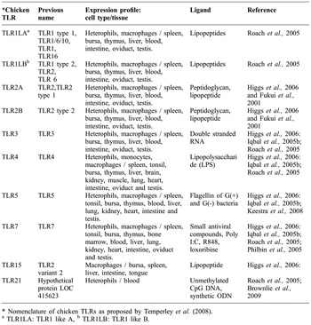
TLR5 is highly expressed in the intestine, tonsils, lungs, liver, kidneys, spleen, heart, testis and immune cells (Leveque et al., Reference LEVEQUE, FORGETTA, MORROLL, SMITH, BUMSTEAD and BARROW2003; Iqbal et al., Reference IQBAL, PHILBIN and SMITH2005a). TLR7 is found highly expressed in the lymphoid associated tissues (spleen, bursa and tonsils), lower in gut associated (small intestine, caecum and colon) and other tissues (thymus, bone marrow, heart, lung, liver, kidney and oviduct) and no TLR7 expression was found in brain, muscle, testis and skin (Philbin et al., Reference PHILBIN, IQBAL, BOYD, GOODCHILD, BEAL, BUMSTEAD, YOUNG and SMITH2005). TLR15 was found abundantly expressed in bone marrow and bursa of Fabricius and moderately in spleen, liver, small intestine, tongue and caecum (Higgs et al., Reference HIGGS, CORMICAN, CAHALANE, ALLAN, LLOYD and MEADE2006). Expression profile of chicken TLRs in various tissue and cell types have been summarized in Table 1.
Chicken TLRs, nomenclature, expression profile and their ligand specificities.

* Nomenclature of chicken TLRs as proposed by Temperley et al. (Reference TEMPERLEY, BERLIN, PATON, GRIFFIN and BURT2008).
a TLR1LA: TLR1 like A, b TLR1LB: TLR1 like B.
TLRs and their ligands
TLRs recognise specific microbial patterns and have ligand specificity. So while individually each TLR respond to limited ligands, collectively the family of TLR can respond to a wide range of proteins associated with bacteria, viruses, fungi and parasites. Until now, ten TLRs have been identified and their ligands have been known for most of them. Chicken TLR2B was shown to act as a receptor for lipoprotein and able to recognize LPS in the presence of MD-2 similar to human TLR2 (Fukui et al., Reference FUKUI, INOUSE, MATSUMOTO, NOMURA, YAMADA, MATSUDA, TOYOSHIMA and SEYA2001). TLR3 recognizes double stranded RNA (ds RNA), associated with viral infections (Alexopoulou et al., Reference ALEXOPOULOU, HOLT, MEDZHITOV and FLAVELL2001). TLR4 is the principal receptor for lipopolysaccaride (LPS), which is a major component of outer membrane of gram-negative bacteria. The LPS receptor complex consists of TLR4, CD14 and MD-2 (Poltorak et al., Reference POLTORAK, HE, SMIRNOVA, LIU, HUFFEL, DU, BIRDWELL, ALEJOS, SILVA, GALANOS, FREUDENBERG, RICCIARDI-CASTAGNOLI, LAYTON and BEUTLER1998). Chicken TLR5 recognizes flagellin, a monomeric subunit of bacterial flagella from both G (+) and G (-) bacteria as in mammals and fishes (Iqbal et al., Reference IQBAL, PHILBIN, WITHANAGE, WIGLEY, BEAL, GOODCHILD, BARROW, MCCONNELL, MASKELL, YOUNG, BUMSTEAD, BOYD and SMITH2005b; Keestra et al., 2008). TLRs 7, 8 and 9 comprise the TLR7 family that is implicated in intracellular recognition of nucleic acids. Chicken TLR7 and mammalian TLR8 have been reported to respond to R848, Poly (I:C), loxoribine and imidazoquinoline compounds with antiviral activity (Jurk et al., Reference JURK, HEIL, VOLLMER, SCHETTER, KREIG, WANGER, LIPFORD and S.2002; Philbin et al., Reference PHILBIN, IQBAL, BOYD, GOODCHILD, BEAL, BUMSTEAD, YOUNG and SMITH2005). Although chicken TLR15 has been sequenced and differential expression in various tissues has been reported, ligand specificity of this TLR is still unknown. As TLR15 is reported to be avian specific and distinctly present in aves in evolutionary map their specific role needs to be explored. In mammals unmethylated CpG DNA and synthetic ODNs are well established ligand for TLR9 which is predicted to be absent in chicken (Yilmaz et al., Reference YILMAZ, SHEN, ADELSON, XAVIER and ZHU2005; Temperley et al., Reference TEMPERLEY, BERLIN, PATON, GRIFFIN and BURT2008). However, similar to mammalian species chicken shows marked immunological responses to CpG oligodeoxynucleotides (ODNs) both in vivo and in vitro. Only recently chicken TLR21 was demonstrated to act as a functional homologue to mammalian TLR9 in the recognition of CpG oligodeoxynucleotides (Brownlie et al., Reference BROWNLIE, ZHU, ALLAN, MUTWIRI, BABIUK, POTTER and GRIEBEL2009). Ligand specificities of various chicken TLRs have been summarized in Table 1.
Different TLRs seem to play crucial roles in the activation of the immune response to PAMPs. TLR1, TLR2, TLR4, TLR5 and TLR6 tend to specialise in the recognition of bacterial components present on the outer surfaces whereas TLR3, TLR7, TLR8 and TLR9 specialise in recognizing nucleic acids especially of viral origin. Strategically these TLRs (3, 7, 8 and 9) are located in intracellular late endosomes–lysosomes compartment enabling them to differentiate host vs. pathogen nucleic acids. In spite of this specificity for the receptor ligand-binding, studies indicate that overall innate immune response is the sum of signals generated by the interaction of multiple TLRs.
Signalling via TLRs
While extra-cellularly, each TLR is distinct in its specificity, intra-cellularly all receptors signal through a common pathway. Each TLR forms either a homodimer or heterodimer for the recognition of a specific or set of specific molecular determinants present on microbes. The binding of a TLR to its appropriate ligand initiates a specific signalling cascade, ultimately resulting in the activation of various transcription factors and the expression of innate immune response genes (Takeda and Akira, Reference AKIRA2004). In silico identification as well as direct sequencing of signalling molecules in chicken confirmed the conserved nature of TLR signalling pathway in this species (Lynn et al., Reference LYNN, LLOYD and O'FARRELLY2003; Karpala et al., Reference KARPALA, LOWENTHAL and BEAN2008; Keestra and van Putten, Reference KEESTRA and VAN PUTTEN2008). Initial TLR activation results in recruitment of one of five adaptor proteins (MyD88, TIRAP/MAL, TICAM-1, TICAM-2 and SARM), while several TLRs are able to utilise multiple adaptors (Watters et al., Reference WATTERS, KENNY and and O'NEILL2007). All TLRs with the exception of TLR3 can signal through the adaptor molecule MyD88 (myeloid differentiation factor 88) (Akira, Reference AKIRA2001). In the MyD88-dependent pathway, once the adaptor is bound to TLR's intracellular domain, it recruits members of the interleukin-1 receptor associated kinase (IRAK) family, which in turn activates the key ubiquitin E3 ligase tumour necrosis factor receptor-associated factor 6 (TRAF6). TRAF6 subsequently activates transforming growth factor-β (TGF-β)-activating kinase (TAK1). TAK-1 activation can ultimately lead to the activation of the transcription factor NF-kB by inducing the phosphorylation of its associated inhibitors or the transcription factor Janus Kinase (JNK) through the kinases MKK 3 and MKK 6. The activated transcription factors subsequently translocate to the nucleus and induce expression of an array of immune genes. In contrast to other family members, TLR3 signals solely through the MyD88-independent pathways. This TLR instead recruits the adaptor molecule TICAM-1, leading to activation of IRF3 and induces an anti-viral response through expression of IFN-β (Karpala et al., Reference KARPALA, LOWENTHAL and BEAN2008).
Although the key features of the TLR signalling pathway are conserved in birds and mammals, some clear differences exist. Chickens lack a functional LPS-specific TRAM-TRIF (TRIF-related adapter molecule/TIR-domain-containing adapter-inducing IFN-β) signalling pathway, which may explain their aberrant response to LPS compared with the mammalian species (Keestra and van Putten, Reference KEESTRA and VAN PUTTEN2008). Comparative analysis of TLR5 function in chickens, humans and mice indicated species specificity of the receptor towards different bacterial flagellin. Activation of TLR5 of these species by flagellin of S. enteric serovar Typhimurium revealed that chicken TLR5 consistently yielded stronger responses than human but not mouse TLR5. This species-specific reactivity was not observed with flagellin of S. enterica serovar Enteritidis (Keestra et al., 2008). These findings show that chicken TLR5 possess different flagellin sensing qualities compared to human and murine TLR5, and that single amino acid substitutions in bacterial flagellin can alter the species-specific TLR5 response, influencing the host range and susceptibility of infection.
Role of TLRs in innate immunity
The basic role of TLRs in innate immunity remains the same in mammals and avian species. Antigen-presenting cells (APC), such as macrophages (Mφ) and dendritic cells (DC) express TLRs on their surface. Upon pathogen infection PAMPs are recognized by TLRs present on APCs and initiate a signalling cascade that stimulates the innate defence through the induction of reactive oxygen and nitrogen intermediates (ROI and RNI). They also initiate adaptive immunity by activating APCs for the production of pro-inflammatory cytokines and up-regulating co-stimulatory molecules (Takeda and Akira, Reference TAKEDA and AKIRA2005). Dentritic cells (DC) play a crucial role in linking the innate and adaptive immune system in TLR stimulatory pathway. Immature DCs are strategically located in peripheral tissues including the potential pathogen entry sites, where they can encounter microbial invaders (Banchereau and Steinman, Reference BANCHEREAU and STEINMAN1998). Immature DCs express a full set of TLRs, which upon stimulation induces DC maturation. Mature DCs express high levels of MHC molecule, co-stimulatory molecules (CD80 and CD86) and migrate to lymphoid organs where they present pathogen derived antigens to naïve T cells. Cell trafficking is mediated by chemokine receptors expressed by DC following TLR stimulation (Rescigno et al., Reference RESCIGNO, GRANUCCI, CITTERIO, FOTI and RICCIARDI-CASTAGNOLI1999, Reference RESCIGNO, GRANUCCI and RICCIARDI-CASTAGNOLI2000; Sallusto et al., Reference SALLUSTO, PALERMO, LENIG, MIETTINEN, MATIKAINEN, JULKUNEN, FORSTER, BURGSTAHLER, LIPP and LANZAVECCHIA1999). TLRs also induce the expression of various pro-inflammatory cytokines by chicken monocytes including IL-1β, IL-6, chemokines IL-8 and macrophage inflammatory protein (MIP)-1β (He et al., Reference HE, MACKINNON, GENOVESE and KOGUT2010) which play a major role in inflammatory as well as innate immune response.
A critical outcome of TLR pathway signalling is the induction of expression of antimicrobial peptides (AMPs) (Birchler et al., Reference BIRCHLER, SEIBL, BUCHNER, LOELIGER, SEGER and HOSSLE2001). AMPs are the direct effecter molecules of the innate immune system that kill pathogens on mucosal surfaces through disruption of bacterial membranes (Wu et al., Reference WU, MAIER, BENZ and HANCOCK1999). They also act as immunomodulators (Yang et al., Reference YANG, CHERTOV, BYKOVSKAIA, CHEN, BUFFO and SHOGAN1999) and chemo-attractants (Bals and Wilson, Reference BALS and WILSON2003). Most of the vertebrates studied contain varying numbers of the two principle families of AMPs, β defensins and cathelicidins (Cormican et al., Reference CORMICAN, MEADE, CAHALANE, NARCIANDI, CHAPWANYA and LLOYD2008). Even between closely related species significant gene gain and loss have been observed in both these class of AMP subtypes, suggesting that each species could carry an armoury of AMPs appropriate to the microbial challenges of its own local environment. In previous studies, chicken ESTs were clustered and searched for orthologs of known TLR pathway components (Lynn et al., Reference LYNN, LLOYD and O'FARRELLY2003) and AMPs (Lynn et al., Reference LYNN, HIGGS, GAINES, TIERNEY, JAMES and LLOYD2004). These studies identified at least partial sequence data for almost half of the TLR pathway components in chickens as well as identified several novel chicken AMPs. In chickens a single cluster of 14 beta-defensin like genes on chromosome 3 as well as a single cathelicidin gene on chromosome 12 have been identified. Recently Cormican et al. (Reference CORMICAN, LLOYD, DOWNING, CONNELL, BRADLEY and O'FARRELLY2009) reported an expanded cluster of 12 novel defensins, which map to the avian beta-defensin locus on chromosome 3. In comprehension chicken TLRs are crucial not only in the early phase of infection when innate immunity is important but also link innate and adaptive immunity throughout the entire course of host defence.
Chicken toll-like receptors and disease resistance
It has been well established that different chicken lines and strains vary in resistance/susceptibility to diseases. Variation in immune response during the course of infection is attributed to the difference in disease susceptibility (Zekarias et al., Reference ZEKARIAS, TER HUURNE, LANDMAN, REBEL, POL and GRUYS2002). TLR expression has also been reported to vary among different lines (Abasht et al., Reference ABASHT, KAISER, VAN DER POEL and LAMONT2009). Significant genetic line effect was found on TLR expression in the spleen of S. enteritidis infected chicken which may be due to gene polymorphisms or varying number of receptors on the immune cells (Dil and Qureshi, Reference DIL and QURESHI2002). In the work carried out in our laboratory it has been observed that TLR5 which recognizes bacterial flagellin was highly expressed in whole blood of Kadaknath chicken in comparison with other pure lines including Dwarf, Naked neck, White Leghorn and Aseel. Whereas TLR3 expression was found higher in White Leghorn than other pure lines studied (Unpublished data).
Chicken TLR4 gene is polymorphic and located in the genomic region associated with the resistance/susceptibility of chicks to Salmonella enterica serovar Typhimurium (Dil and Qureshi, Reference DIL and QURESHI2002; Leveque et al., Reference LEVEQUE, FORGETTA, MORROLL, SMITH, BUMSTEAD and BARROW2003). They also demonstrated TLR4 is required for LPS mediated inducible nitric oxide synthase (iNOS) induction in chicken macrophages. The relative number of TLR4 receptor molecules on the macrophage surface varies between chicken strains, thereby varying the expression of LPS binding receptors among strains (Dil and Qureshi, Reference DIL and QURESHI2002). Single nucleotide polymorphisms in TLR4 gene were identified and their association with Salmonella enteritidis has been established (Malek et al., Reference MALEK, HASENTEIN and LAMONT2004). These results could potentially be used as markers for phenotypic variation in marker-assisted selection to enhance response to Salmonella.
Up regulation of chicken TLR2B, TLR3 and TLR15 mRNA in the caecum of Salmonella enterica serovar Typhimurium infected chicken and in chicken embryonic fibroblasts following stimulation with heat-killed Salmonella enterica serovar Typhimurium have been reported (Higgs et al., Reference HIGGS, CORMICAN, CAHALANE, ALLAN, LLOYD and MEADE2006). Interestingly, down regulation of TLR5 expression was observed after Salmonella infection (Iqbal et al., Reference IQBAL, PHILBIN, WITHANAGE, WIGLEY, BEAL, GOODCHILD, BARROW, MCCONNELL, MASKELL, YOUNG, BUMSTEAD, BOYD and SMITH2005b; Abasht et al., Reference ABASHT, KAISER and LAMONT2008) in chicken gut epithelial cells, most likely attributed to negative feedback needed to limit overstimulation in an area of high bacterial colonization.
The expression of TLR4 and TLR7 was up regulated in spleen of chickens challenged with Clostridium perfringens (Lu et al., Reference LU, SARSON, GONG, ZHOU, ZHU, KANG, YU, SHARIF and HAN2009) but could not appreciate significant changes in MyD88 or TRAF 6 pathway molecules quantified by real time PCR. The authors have hypothesized that chicken TLR may not signal through MyD88 pathway as TLR2 do in mammals, which is contrary to the highly conserved nature of innate immune system. The study might have missed the time window to capture transcription. Therefore, to get a clear picture manipulation of TLR downstream signalling pathway transcription factors such as docking and phosphorylation can be attempted. The expression of TLR3 and TLR7 was enhanced in response to Marek's disease virus infection in the chicken lungs (Abdul-Careem et al., Reference ABDUL-CAREEM, HAQ, SHANMUGANATHAN, READ, SCHAT, HEIDARI and SHARIF S.2009). TLR1 gene expression was decreased and avian beta-defensin (AvBD) gene expression (AvBD3, AvBD10 and AvBD12) was significantly increased in response to Salmonella infection. In contrast, Campylobacter infection increased TLR21 gene expression but significantly reduced expression of seven antimicrobial peptide (AMP) genes (AvBD3, AvBD4, AvBD8, AvBD13, AvBD14, Cathelicidins 2 and 3) (Meade et al., Reference MEADE, NARCIANDI, CAHALANE, REIMAN, ALLAN and O'FARRELLY2009). Significant increase in TLR5 and TLR15 gene expression were detected in response to S. typhimurium but not to C. jejuni. TLR4 and TLR21 gene expression were transiently increased in response to both bacterial species. The differential expression profile of innate immune genes in both infection models shed light on the tailored responses of the host immune system to specific microbes (Shaughnessy et al., Reference SHAUGHNESSY, MEADE, CAHALANE, ALLAN, REIMAN, CALLANAN and O'FARRELLY. C.2009). These reports are based on comparative quantification of respective TLR mRNAs using different housekeeping genes mostly single gene as reference gene. Although these reference genes are stably expressed during development, expression levels can vary between different tissues, cell types and diseases (Thellin et al., Reference THELLIN, ZORZI, LAKAYE, DE BORMAN, COUMANS, HENNEN, GRISAR, IGOUT and HEINEN1999; Schmittgen and Zakrajsek, Reference SCHMITTGEN and ZAKRAJSEK2000; Dheda et al., Reference DHEDA, HUGGET, CHANG, KIM, BUSTIN, JONHNSON, ROOK and ZUMLA2005). To get reliable results validation of various housekeeping genes and their combination, work has to be done under different pathogen challenge conditions.
TLR7 mediated IL-4 secretion was reported in chickens infected by low-pathogenicity avian influenza virus H9N2 (Xing et al., Reference XING, CARDONA, LI, DAO, TRAN and ANDRADA2008). Molecular characterisation of chicken TLR7/8 loci revealed TLR8 gene in chicken is fragmented and disrupted by a CR1 (chicken repeat-1) retrovirus-like element (Philbin et al., Reference PHILBIN, IQBAL, BOYD, GOODCHILD, BEAL, BUMSTEAD, YOUNG and SMITH2005). Further they also showed that this disruption is found in galliform bird species including Red jungle fowl, guinea fowl, Japanese quail, pheasant and turkey, whereas non-galliform bird species like Pekin duck, goose, black swan, penguin and ostrich did not possess the CR1 element tested by nested PCR. Hence, it was concluded that disruption of TLR8 gene only in galliform species might contribute to the difference in viral susceptibility. In contrast a recent study (MacDonald et al., Reference MACDONALD, XIA, SMITH and MAGOR2008) examined TLR8 gene organization of Pekin duck, a non-galliform bird species by sequencing genomic clone spanning the TLR7/8 locus and detected only fragments of TLR8 gene and demonstrated insertion of CR1 element between this gene fragments. The failure of nested PCR used by Philbin et al. (Reference PHILBIN, IQBAL, BOYD, GOODCHILD, BEAL, BUMSTEAD, YOUNG and SMITH2005) to amplify the TLR8 disruption in ducks might be due to differences in the sequence under the primers. Further studies are warranted to explore TLR8 gene organization in other non-galliform bird species. Both TLR7 and 8 are implicated in detection of single stranded viruses in mammals. TLR7 may be involved in detection of influenza virus in birds in the absence of functional TLR8, the critical difference in susceptibility to influenza virus between ducks and chicken remains to be identified.
Application of knowledge on TLRs in the poultry industry
Tremendous progress has been made in egg and meat production in the chicken but traits related to disease resistance remain as the target for breeding programs. Meanwhile, analyses to detect QTLs for disease resistance/susceptibility or immune function have been performed by a few groups. One of the quantitative trait loci (QTLs) for susceptibility to infection with Salmonella enterica is located on the genomic region including the TLR4 gene, and the allelic types of chicken TLR4 have a relationship with susceptibility (Leveque et al., Reference LEVEQUE, FORGETTA, MORROLL, SMITH, BUMSTEAD and BARROW2003). Progress in functional analyses for SNPs in TLR genes will provide clues that may prove the association between TLR alleles and disease susceptibility. It will also contribute to more effective selection aimed at disease resistance.
The potential area of using TLR ligands as adjuvants should not be forgotten. Many compounds that are recognized by TLR molecules possess strong activity as adjuvants. In chicken, researchers are considering using components of microbes such as outer membrane lipoprotein I (OprI) of Pseudomonas aeruginosa and flagellin which are recognized by TLR2/TLR4 and TLR5 respectively, to construct fusion molecules with antigens contained in a vaccine (Loots et al., Reference LOOTS, REVETS and GODDEERIS2008). Moreover in ovo and in vivo administration of CpG oligonucleotides as a potent immunomodulator have also been proved in chicken (Taghavi et al., Reference TAGHAVI, ALLAN, MUTWIRI, VAN KESSEL, WILLSON, BABIUK, POTTER and GOMIS2008; Dar et al., Reference DAR, POTTER, TIKOO, GERDTS, LAI, BABIUK and MUTWIRI2009; Mackinnon et al., Reference MACKINNON, HE, SWAGGERTY, MCREYNOLDS, GENOVESE, DUKE, NERREN and KOGUT2009).
Conclusions
TLRs play a crucial role in determining the type and magnitude of immune response as well as an essential link between innate and adaptive immunity. As TLR genes are proven to be associated with resistance to infectious diseases, these genes can be exploited as molecular marker in genetic selection to develop disease resistant lines. Moreover, TLR ligands could be used as novel molecular adjuvant along with immunogenic antigen. Further studies are warranted to improve the knowledge about TLRs and their role in immunity in the chicken.