




No CrossRef data available.
Article contents
Effect of cyclosporin A on the survival and ultrastructure of Echinococcus granulosus protoscoleces in vitro
Published online by Cambridge University Press: 14 September 2004
Abstract
Surgical treatment of human hydatidsosis involves the use of various scolicidal agents to kill infective Echinococcus granulosus protoscoleces that may disseminate into the peritoneal cavity during surgery and potentially re-infect the patient. Currently, no scolicidal agent is completely effective in killing intracystic protoscoleces in humans. Cyclosporin A (CsA) has previously been found to be lethal for E. granulosus protoscoleces in vitro. In this study, we further assessed the effectiveness of CsA as a scolicidal agent by testing the toxic effect of CsA at higher doses over various time-periods. Experiments were performed on activated and unactivated protoscoleces cultured in nutrient medium or sheep hydatid cyst fluid. All activated protoscoleces were killed following culture in 100 μg/ml of CsA for 3 days and 50 or 20 μg/ml for 5 days. The lethal effect of CsA on unactivated protoscoleces varied but reached 100% over 15 days in culture with 100 or 50 μg/ml of CsA. Pulse treatment of protoscoleces with 50, 20 or 10 μg/ml of CsA for 5 min or 72 h killed all parasites by day 10 and day 5 respectively. Untreated protoscoleces remained greater than 95% viable for the duration of experiments. Changes in protoscolex ultrastructure induced by treatment with 10 μg/ml of CsA over 10 days in in vitro culture was assessed by TEM. Protoscolex alterations observed in treated parasites included an increase in cellular vacuolization, swelling of mitochondria, rounding of cells, damage to the tegument, decrease in glycogen, a breakdown of the extracellular matrix and an increase in lipid globules. The untreated protoscoleces, by comparison, had few changes during the 10-day culture period with the exception of large amounts of extracellular glycogen observed in the protoscoleces at culture days 7 and 10. From these results, CsA is clearly an effective scolicidal agent in vitro that may have potential application as a new therapeutic agent in the treatment of human hydatid disease.
Information
- Type
- Research Article
- Information
- Copyright
- 2004 Cambridge University Press
INTRODUCTION
Cyclosporin A (CsA) is a cyclic peptide isolated from the fungus Tolypocladium inflatum which has been widely used as an immunosuppressant drug for the treatment of organ transplant patients and some auto-immune diseases (Von Graffenreid, 1989; Borel et al. 1996). It is also reported to possess anti-parasitic properties affecting many parasites (Chappell & Wastling, 1992; Page, Kumar & Carlow, 1995) including Echinococcus granulosus (Hurd, McKenzie & Chappell, 1993; Colebrook, Jenkins & Lightowlers, 2002). Humans infected with the larval stage of the tapeworm Echinococcus granulosus develop hydatid disease which can be fatal. Hydatid disease is characterized by the development of fluid-filled cysts in, most commonly, the liver and/or lungs. The disease causes substantial morbidity and some patients require multiple surgical procedures to remove cysts over many years. Mature hydatid cysts may contain numerous protoscoleces which, if released from the cyst into the body cavity or tissues, may develop into new hydatid cysts (secondary hydatidosis). Antihelmintics are commonly used in patients immediately before and after surgery for hydatid disease with a view to minimizing the establishment of secondary echinococcosis. An alternative to excision of hydatid cysts for some hydatid patients is the puncture-aspitation-injection-reaspiration (PAIR) technique (WHO, 1996; Filice et al. 1997) during which a scolicidal agent is injected into the cyst. Various scolicidal agents have been used during the PAIR procedure; however, these are not completely effective in killing protoscoleces and do not eliminate the subsequent growth or regrowth of the cysts. A recent adaptation of the PAIR procedure is percutaneous drug injection without reaspiration (PEDIM) (Deger et al. 2000). However, the limitations with regard to the effectiveness of scolicidal agents applies in the case of this technique also. Development of more potent scolicidal agents would be a valuable adjunct to the treatment of human hydatidosis. For this reason, the finding that CsA has anti-parasitic properties warrants further investigation to assess it's potential application in the treatment of hydatid disease.
The susceptibility of Echinococcus sp. to treatment with CsA differs between species. CsA has little effect on the development of alveolar echinococossus in mice experimentally infected before or after administration of CsA (Liance et al. 1992). In mice experimentally infected with E. granulosus, the administration of CsA prior to infection resulted in the establishment of secondary hydatids with reduced cyst fresh weight and number compared to untreated controls. No effect was observed on established cysts treated with CsA. The results of these studies suggest a limited potential for use of CsA in the treatment of patients with established cysts (Hurd et al. 1993). However, the recent discovery that CsA is toxic to protoscoleces in vitro (Colebrook et al. 2002) suggests that CsA may be a useful scolicidal agent. It was found that protoscoleces were killed following 7 days culture in media containing 10 μg/ml of CsA. The effect was dose dependent because the dose range (0·0001–3 μg/ml) failed to induce parasite death while 5 μg/ml and 8 μg/ml induced 32 and 82% death, respectively, over the same culture-period. However, these studies did not assess parasitocidal activity at higher concentrations of CsA, nor determine the minimum CsA exposure time required to kill the parasite.
Here, we report the results of further investigations of the scolicidal effects of CsA on E. granulosus protoscoleces in vitro, to determine the concentration required to induce 100% lethality, and describe the ultrastructural effects on protoscoleces of exposure to CsA.
MATERIALS AND METHODS
Collection of E. granulosus protoscoleces
Hydatid cysts were collected from liver and lungs of naturally infected sheep slaughtered at an abattoir in southern New South Wales. Protoscoleces were extracted aseptically from hydatid cysts and sedimented. Supernatant was removed leaving approximately 1 ml of packed protoscoleces in 10 ml of cyst fluid. Viability of protoscoleces was assessed microscopically by observing morphology and flame cell activity.
Activation of E. granulosus protoscoleces
Immediately prior to setting up in vitro cultures, protoscoleces (approximately 1 ml packed) were treated with 50 ml of artificial gastric fluid (0·1% pepsin plus 0·5 ml of 32% HCl made up to 100 ml in physiological saline) at 37 °C for 20 min. Protoscoleces were sedimented, the supernatant removed and exchanged with 50 ml of artificial intestinal fluid (Rickard & Bell, 1971) and incubated at 37 °C for 45 min. The percentage of evaginated protoscoleces was determined microscopically.
In vitro culture of protoscoleces in CsA
Activated protoscoleces were washed 3 times in 50× volume of sterile culture medium (Dulbecco's modification of Eagle's medium, DMEM, Flow Labs, pH 7·4, filter-sterilized and supplemented with 100 IU/ml mycostatin and 100 μg/ml gentamicin). A vol. of 2 ml of DMEM plus antibiotics was added to each well of a sterile tissue culture plate (Nunc) under aseptic conditions. CsA (Sigma) (10 μg/μl in 100% ethanol) was added to individual test wells to a final concentration of 100, 50, 20, 10, 5 and 1 μg/ml and the same volume of diluent alone was added to control wells. Cultures containing approximately 1000 evaginated protoscoleces were incubated at 37 °C in 5% CO2 humidified air. Culture medium was changed every 48 h.
Protoscoleces were washed 3 times in 50-fold the volume of filter-sterile sheep hydatid cyst fluid (SHCF) or DMEM supplemented with mycostatin and gentamycin and the parasites allowed to settle at 1 g between washes. The invaginated protoscoleces were transferred to individual wells of a sterile culture dish each containing 2 ml of SHCF or DMEM medium under aseptic conditions, as described for activated protoscoleces. The culture medium was not changed during the experiment.
Time pulse treatment of protoscoleces with CsA
E. granulosus activated protoscoleces were incubated in DMEM plus 50, 20, 10, 5, 1 or 0 μg/ml CsA for 5 min 1, 12 or 72 h. After each period of incubation the protoscoleces were removed from the wells, washed 3 times with fresh medium and transferred back to culture wells containing 2 ml of untreated DMEM medium for the remainder of the experiment.
CsA dose: administered as a single dose or proportionate daily dose.
A total CsA dose of 10 μg/ml was administered to protoscoleces in culture as 2 μg/ml daily for 5 days or as a single dose of 10 μg/ml at day 0. After 5 days protoscoleces were removed from culture and their viability assessed by Toluidine blue staining.
Assessment of protoscolex viability
The condition of the protoscoleces was assessed microscopically every 24 h. Parasite death was determined by collecting approximately 50 protoscoleces from each culture well per day and staining them with 0·1% Toluidine blue in methanol (Vadas et al. 1979; Pons, Adams & Stadecker, 1988). The proportion of protoscoleces excluding the dye was recorded microscopically.
Fixation of protoscoleces for transmission electron microscopy
Samples for electron microscopy were fixed with 2·5% glutaraldehyde in PBS. After fixation, the specimens were washed 4 times in PBS. The samples were then post-fixed in 1% osmium tetroxide in the same buffer for 2 h at room temperature on a vertical rotator. The samples were subsequently washed in distilled water twice and dehydrated through a series of graded acetone solutions before being embedded in procure Araldite (ProSciTech, Australia). The blocks were polymerized at 60 °C for 48 h.
Ultra-thin sections were collected on acetone-cleaned uncoated 200 mesh copper grids. These sections were stained with 5% aqueous solution of uranyl acetate for 10 min, washed with distilled water and stained with Reynold's lead citrate for 10 min. Sections were examined using a Philips 300 electron microscope at 60 kV.
RESULTS
E. granulosus protoscoleces collected for in vitro culture and treated with artificial gastric and intestinal fluids were assessed as 100% viable and evaginated. The protoscoleces were actively motile. Untreated protoscoleces were assessed to be 97% viable and remained invaginated. Non-viable protoscoleces could be identified by staining with Toluidine blue whereas viable protoscoleces excluded the dye.
CsA was found to be toxic for E. granulosus protoscoleces in in vitro culture (Fig. 1). The toxic effect of 100 μg/ml CsA on activated protoscoleces was observed from culture days 1–2 where 45% of parasites were killed in comparison to >95% survival in control cultures, as determined by Toluidine blue dye uptake. By culture day 3 all protoscoleces treated with 100 μg/ml CsA were dead. Susceptibility of protoscoleces to CsA was dose dependent. After 5 days in cultures containing 50, 20, 10 μg/ml CsA the proportion of viable protoscoleces was 0%, 1% and 6% respectively compared with 97% viability in control cultures. Lower concentrations of CsA induced parasite killing over 10 days in which the parasites were observed (Fig. 1).

Fig. 1. Percentage survival of Echinococcus granulosus protoscoleces following 12 days of in vitro culture with various doses of CsA. CsA doses are represented by the symbols [lozf ] 100 μg/ml, [bull ] 50 μg/ml, [utrif ] 20 μg/ml, [dtrif ] 10 μg/ml, [squf ] 5 μg/ml and ○ No CsA.
Survival of E. granulosus protoscoleces after treatment with CsA following a course of either the total dose of CsA at day 0 or 0·2× the total dose administered daily for 5 days revealed little difference in CsA toxicity to the protoscoleces (data not shown). By culture day 5 the percentage survival of protoscoleces was similar in cultures that had received either 50, 20, 10 or 5 μg/ml as a single dose at day 1 or a fifth of that dose administered each day for 5 days. The effect of 1 μg/ml CsA as a single dose at day 1 was more toxic to protoscoleces than using 5×0·2 μg as a proportionate daily dose.
The lethal effect of CsA on protoscoleces in culture was observed following time pulse treatment with various doses of CsA (Fig. 2). All protoscoleces treated for 5 min, 1, 12 or 72 h with 50, 20 or 10 μg/ml CsA were dead after 10 days in culture, as determined by Toluidine blue staining. After 7 days in culture 60, 90, 90 and 98% of protoscoleces were dead following 5 min treatment with 5, 10, 20 or 50 μg/ml CsA respectively. Longer exposure times to CsA induced parasite killing more rapidly. After 72 h of exposure to 50, 20 and 10 μg/ml CsA at least 95% of protoscoleces were killed by day 5, and 100% by day 7. Untreated controls remained at least 95% viable throughout the experiment.

Fig. 2. Survival of Echinococcus granulosus protoscoleces following time pulse treatment with various doses of CsA. Data from culture day 5 show survival of protoscoleces when treated for 5 min, 1, 12 and 72 h in culture with CsA. In each time group the doses of CsA are represented by individual bars. The untreated controls are represented by black bars and doses of CsA at 50 (), 20 (□), 10 () and 1 μg/ml () are plotted on the right of the controls respectively.
Invaginated protoscoleces cultured in DMEM or SHCF survived longer in culture when treated with comparable doses of CsA than activated protoscoleces. The toxic effect of 100 and 50 μg/ml CsA on protoscoleces was observed by culture day 15 where 100% of parasites were killed in comparison to >95% survival in control cultures. Some variation in toxicity between replicate cultures was found where 100% of protoscoleces treated with 100 μg/ml CsA in SHCF were dead by culture day 5 but not until day 14 in a replicate test. In both cases there was a progressive decline in the percentage of protoscoleces surviving relative to the control parasites. After 15 days in cultures containing 1 or 10 μg/ml CsA the proportion of viable protoscoleces in replicate was 19 and 90% and 0% and 54 respectively. Invaginated protoscoleces cultured in DMEM plus CsA had similar survival rates as those cultured in SHCF. All protoscoleces were dead following 15 days of culture in 100 or 50 μg/ml CsA and lower doses of CsA at 1 and 10 μg/ml killed 84% and 68% and 30% and 33% (in replicate) of protoscoleces respectively. Control cultures containing either media or media plus ethanol (as the vehicle for CsA delivery) remained greater than 90% viable for 15 days of culture (Fig. 3).

Fig. 3. Percentage survival of invaginated Echinococcus granulosus protoscoleces following (A) 15 days in culture with SHCF medium or (B) 20 days in DMEM medium. Cultures contained either medium plus the CsA vehicle alone ([utrif ]), medium ([bull ]) or various doses of CsA represented in replicate by □ or [squf ]. Each of the graphs within group A or B represents data from protoscoleces treated with different CsA doses. Data from cultures containing 1 μg/ml of CsA are represented in the graph at the top left of group A or B, 10 μg/ml in the top right, 50 μg/ml in the bottom left and 100 μg/ml in the bottom right.
The effect of administration of 10 μg/ml CsA on E. granulosus protoscolex ultrastructure was assessed by TEM in protoscoleces cultured in vitro. A minimum of 1000 activated protoscoleces cultured in DMEM alone or DMEM plus CsA in vitro were collected and processed for TEM on days 1, 2, 3, 5, 7 and 10 to determine the early morphological changes induced by the drug in comparison with untreated controls. Untreated protoscoleces cultured for 5 days were morphologically indistinguishable from controls collected at day 0. After 7 and 10 days in cultures, the untreated protoscoleces appeared to display increased levels of parenchymal glycogen content. The glycogen, present as β-glycogen granules, was extensive in the cytoplasm of parenchymal cells of the somal (posterior) region of the protoscoleces.
Treated protoscoleces showed appreciable differences compared with control specimens fixed at identical times. The structure of the tegument and its associated glycocalyx appeared to be little changed after CsA administration, although increasing lucency of the distal cytoplasm occurred after 7 days, when most protoscolex cells were substantially necrotic. The first observed effect of CsA administration was the appearance of lucent regions in the basal zone of the distal cytoplasm (Fig. 4). These lucent regions were apparent 1 day after treatment, appeared to be membrane bound and may represent a loss of integrity of the basal invaginations of the distal cytoplasm. These focal regions swelled and coalesced over days 2 and 3 of culture until there appeared an almost complete separation of the distal cytoplasm from its underlying basal matrix.

Fig. 4. TEM ultrastructure of Echinococcus granulosus protoscoleces in in vitro culture. (A–D) Untreated protoscoleces sampled from cultures at days 1, 3, 7 and 10. (A,B) Protoscoleces from culture days 1 and 3 respectively, note there are no distinct morphological differences in ultrastructure from specimens at day 0. (C,D) Protoscoleces from days 7 and 10 respectively, glycogen (G) has disseminated in the extracellular matrix. (F–I) Ultrastructure of protoscoleces treated with CsA in vitro. (F,G) Specimens treated for 1 and 2 days respectively. The basal laminar is separating from the distal cytoplasm of the tegument forming electron-lucent regions with no apparent matrix and vacuoles (V) are forming in the tegumentary cytons. (H) Specimens from culture day 5, vacuoles forming in teguments cells are more extensive, some cells appear to be rounded and lipidic structures are present. (I) Specimens from culture day 10, showing degradation of the tegument (T), detached microtriches (Mt) and large electron-lucent areas in the soma region. (E) Mitochondria (M) and lipid globules (L) from untreated protoscolex and (J) Higher magnification of enlarged mitochondria (M) from CsA-treated protoscolex.
Extensive vacuolization of the cytoplasm of insunken tegumentary cytons was observed after 1 day of CsA treatment. The perinuclear vacuoles had a slightly granular matrix and probably represent expanded regions of endoplasmic reticulum. The nuclei, as well as other cytoplasmic constituents of the cytons, remained morphologically unchanged. Mitochondria of parenchymal cells were often enlarged in protoscoleces treated for 3 days with CsA (Fig. 4). The mitochondria had a generally granular matrix and a peripheral array of christae. It was found that 90% of the parasites were dead and necrotic after 5 days of treatment with CsA, and the specimens that remained intact showed progressive degradation of the tegument and distal cytoplasm. In all specimens, glycogen was much less abundant than in control specimens, there was an increased abundance of lipidic granules throughout the cells and extracellular matrices and the mitochondria were further enlarged. All protoscoleces were dead by culture day 10 and, as demonstrated by TEM, were so necrosed that few structures were discernible.
DISCUSSION
This study provides further evidence that CsA is an effective scolocidal agent against E. granulsosus protoscoleces in vitro. Previous studies have shown that treatment of protoscoleces with 10 μg/ml CsA kills 100% of the protoscoleces within 7 days in in vitro culture (Colebrook et al. 2002). A range of lower doses induced parasite damage but were not completely effective in killing the parasite. In this study 100 μg/ml of CsA killed 100% of protoscoleces within 72 h of treatment. A single dose of 50, 20 or 10 μg/ml given for 5 min resulted in a complete scolicidal effect over 7 days. The time and dose of CsA required to attain maximum scolicidal effect is important when considering the application of this finding to the surgical treatment of hydatid patients.
The results of this study also showed that the scolicidal effect varied between activated or unactivated protoscoleces in vitro. Unactivated and invaginated protoscoleces cultured in DMEM and SHCF had a longer survival time when exposed to CsA in vitro than those activated and cultured in DMEM. The active protoscoleces are highly motile and may take up and metabolize CsA at a faster rate than the relatively dormant invaginated protoscoleces. There was some variation in the survival rates of the replicate cultures of invaginated protoscoleces exposed to the same drug concentration. The variation may be due to sampling errors or to the fact that some unactivated protoscoleces remained in brood capsules and were less susceptible to CsA toxicity. Irrespective of the variation in CsA toxicity between replicates, all unactivated protoscoleces died when exposed to 100 or 50 μg/ml CsA in SHCF. There was no difference in parasite mortality whether the CsA dose was given as a single dose or as a fractional daily dose.
TEM investigation demonstrated that administration of CsA (10 μg/ml) in vitro resulted in a progressive pathological effect compared with untreated protoscoleces maintained in culture over the same period. It is of note that untreated protoscoleces displayed increasing levels of β-glycogen in parenchymal cells over the culture period. In cestodes, glucose is taken up by active transport, involving glucose transporters expressed in the tegument (Rodriguez-Contreras et al. 1998). Cestodes are capable of taking up glucose against a concentration gradient (McManus & Smyth, 1982) and the enhanced amounts of glycogen (the stored form of glucose) in untreated cultured protoscoleces on day 10 compared with worms fixed on day 0, possibly reflects the abundance of glucose in vitro compared with that available in cyst fluids. It is of interest that there was less glycogen in protoscoleces treated with CsA than in untreated controls, possibly reflecting impairment of glucose transport across the tegument. Glycogen depletion has been observed in the ultrastructure of CsA-treated Hymenolepis microstoma in vitro (McLauchlan, Roberts & Chappell, 2000) and CsA has been shown to impair the uptake of glucose through the tegument of H. microstoma in vivo (Wastling & Chappell, 1994).
Major ultrastructural effects of CsA include the separation of the distal cytoplasm of the tegument from its underlying basal matrix and the appearance of vacuoles in the tegumentary cytons. These vacuoles appear to arise from expanded endoplasmic reticulum in the cytons, and it seems likely, therefore, that one effect of CsA on protoscoleces is to disrupt translational activity and protein synthetic activity of the cells that support the tegument. Whether this is a direct effect or down-stream consequence remains to be determined.
The mitochondria in CsA-treated protoscoleces became enlarged. Swelling of the mitochondria has been described following CsA treatment of H. microstoma (McLauchlan et al. 2000). Changes to the appearance of mitochondria may result from changes in the permeability of tegument damage and subsequent fluid build up. A direct effect by the drug may also be the cause of swelling mitochondria as CsA reportedly inhibits respiration in rat mitochondria (Fournier, Ducet & Crevat, 1987) and augments mitochondrial damage in cardiac muscle tissue of animals receiving tissue organ transplants (Jurado et al. 1998). An inhibition of normal mitochondrial function and glycogen depletion would impair the ability of the parasite to function through the inhibition of glycolysis and could account for the lethal effects of CsA on protoscoleces.
The finding that CsA is a potent scolicidal agent may have application in the treatment of cystic Echinococcosis. Injection of CsA as a scolicidal agent into an hydatid cyst during PAIR could potentially kill intracystic protoscoleces and eliminate the risk involved with peritoneal dissemination of protoscoleces and relapses. Application of CsA may be more appropriate in the PEDIM procedure (Deger et al. 2000). Our results show that 5 min of exposure to CsA at various doses can effectively kill activated and unactivated E. granulosus protoscoleces in vitro. The relevance of these data needs to be further investigated in animal models to assess the efficacy of CsA as a scolicidal agent when delivered into cysts by percutaneous injection. Ivermectin has been investigated as a potential scolicidal agent for use in treatment of hydatid disease. The drug is effective in killing E. granulosus protoscoleces in in vitro culture (Casado et al. 1989) and does have deleterious effects on hydatid cysts in both sheep (Hokelek et al. 2002) and jirds (Ochieng-Mitula & Burt, 1996). Similarly, albendazole sulphoxide has been successfully used percutaneously in the treatment of hydatid cysts in sheep (Deger et al. 2000). The toxicity of perenterally administered ivermectin in humans has yet to be assessed; however, these studies highlight the potential for development of novel scolicidal therapies for treatment of hydatid disease. CsA has been widely used in the treatment of human autoimmune disease and as an immunosuppressive agent in organ transplant patients (Borel et al. 1996). A large amount of information is available on the toxic effects and doses of the drug to be safely administered to humans. The drug is known to be anti-parasitic at subimmunosuppressive levels (Munro & McLaren, 1990; Page et al. 1995) and is suggested to mediate it's effects through inhibition of normal parasite cyclophilin function. However, no direct association between the anti-parasitic effects of CsA and parasite cyclophilin has yet been established (Khattab et al. 1998; McLauchlan et al. 2000). Further work with CsA or these other potential scolicidal drugs is required before their application to treating humans could be recommended.
This work was funded with research grants from the National Health and Research Council.
References
REFERENCES
BOREL, J. F.,
BAUMANN, G.,
CHAPMAN, I.,
DONATSCH, P.,
FAHR, A.,
MUELLER, E. A. &
VIGOURET, J. M. (1996).
In vivo pharmacological effects of ciclosporin and some analogues.
Advances in Pharmacology 35,
115–246.CrossRefGoogle Scholar
CASADO, N.,
RODRIGUEZ-CAABEIRO, F.,
JIMENEZ, A.,
CRIADO, A. &
DE ARMAS, C. (1989).
In vitro effects of levamisole and ivermectin against Echinococcus granulosus protoscoleces.
International Journal for Parasitology 19,
945–947.CrossRefGoogle Scholar
CHAPPELL, L. H. &
WASTLING, J. M. (1992).
Cyclosporin A: antiparasite drug, modulator of the host–parasite relationship and immunosuppressant.
Parasitology 105 (Suppl.),
S25–S40.CrossRefGoogle Scholar
COLEBROOK, A.,
JENKINS, D. &
LIGHTOWLERS, M. (2002).
Anti-parasitic effect of cyclosporin A on Echinococcus granulosus and characterization of the associated cyclophilin protein.
Parasitology 125,
485–493.CrossRefGoogle Scholar
DEGER, E.,
HOKELEK, M.,
DEGER, B. A.,
TUTAR, E.,
ASIL, M. &
PAKDEMIRLI, E. (2000).
A new therapeutic approach for the treatment of cystic echinococcosis: percutaneous albendazole sulphoxide injection without reaspiration.
American Journal of Gastroenterology 95,
248–254.CrossRefGoogle Scholar
FILICE, C.,
BRUNETTI, E.,
D'ANDREA, F. &
FILICE, G. (1997).
Minimal invasive treatment for hydatid abdominal cysts: PAIR (Puncture, Aspiration, Injection, Reaspiration) – state of the art.
World Health Organization, Geneva WHO/CTD/SIP/97.3,
1–41.Google Scholar
FOURNIER, N.,
DUCET, G. &
CREVAT, A. (1987).
Action of cyclosporine on mitochondrial calcium fluxes.
Journal of Bioenergy and Biomembranes 19,
297–303.CrossRefGoogle Scholar
HOKELEK, M.,
DEGER, B. A.,
DEGER, E.,
TUTAR, E. &
SUNBUL, M. (2002).
Ivermectin used in percutaneous drug injection method for the treatment of liver hydatid disease in sheep.
Gastroenterology 122,
957–962.CrossRefGoogle Scholar
HURD, H.,
MACKENZIE, K. S. &
CHAPPELL, L. H. (1993).
Anthelmintic effects of cyclosporin A on protoscoleces and secondary hydatid cysts of Echinococcus granulosus in the mouse.
International Journal for Parasitology 23,
315–320.CrossRefGoogle Scholar
JURADO, F.,
BELLON, J. M.,
PAREJA, J. A.,
GOLITSIN, A.,
MILLAN, L.,
PASCUAL, G. &
BUJAN, J. (1998).
Effects of ischaemia-reperfusion and cyclosporin-A on cardiac muscle ultrastructure.
Histology and Histopathology 13,
761–774.Google Scholar
KHATTAB, A.,
PICA-MATTOCCIA, L.,
KLINKERT, M. Q.,
WENGER, R. &
CIOLI, D. (1998).
Cyclosporins: lack of correlation between antischistosomal properties and inhibition of cyclophilin isomerase activity.
Experimental Parasitology 90,
103–109.CrossRefGoogle Scholar
LIANCE, M.,
BRESSON-HADNI, S.,
VUITTON, D. A.,
LENYS, D.,
CARBILLET, J. P. &
HOUIN, R. (1992).
Effects of cyclosporin A on the course of murine alveolar Echinococcosis and on specific cellular and humoral immune responses against Echinococcus multilocularis.
International Journal for Parasitology 22,
23–28.CrossRefGoogle Scholar
McLAUCHLAN, P. E.,
ROBERTS, H. C. &
CHAPPELL, L. H. (2000).
Mode of action of cyclosporin A against Hymenolepis microstoma (Cestoda): relationship between cyclophilin binding and drug-induced damage.
Parasitology 121,
661–670.CrossRefGoogle Scholar
McMANUS, D. P. &
SMYTH, J. D. (1982).
Intermediary carbohydrate metabolism in protoscoleces of Echinococcus granulosus (horse and sheep strains) and E. multilocularis.
Parasitology 84,
351–366.CrossRefGoogle Scholar
MUNRO, G. H. &
McLAREN, D. J. (1990).
Toxicity of cyclosporin A (CsA) against developmental stages of Schistosoma mansoni in mice.
Parasitology 100,
29–34.CrossRefGoogle Scholar
OCHIENG-MITULA, P. J. &
BURT, M. D. (1996).
The effects of ivermectin on the hydatid cyst of Echinococcus granulosus after direct injection at laparotomy.
Journal of Parasitology 82,
155–157.CrossRefGoogle Scholar
PAGE, A. P.,
KUMAR, S. &
CARLOW, C. K. S. (1995).
Parasite cyclophilins and antiparasite activity of cyclosporin A.
Parasitology Today 11,
385–389.CrossRefGoogle Scholar
PONS, H. A.,
ADAMS, S. &
STADECKER, M. J. (1988).
Schistosoma mansoni: the basis for the antischistosomal effect of cyclosporine A.
Experimental Parasitology 67,
190–198.CrossRefGoogle Scholar
RICKARD, M. D. &
BELL, K. J. (1971).
Successful vaccination of lambs against infection with Taenia ovis using antigens produced during in vitro cultivation of the larval stages.
Research in Veterinary Science 12,
401–402.Google Scholar
RODRIGUEZ-CONTRERAS, D.,
SKELLY, P. J.,
LANDA, A.,
SHOEMAKER, C. B. &
LACLETTE, J. P. (1998).
Molecular and functional characterization and tissue localization of 2 glucose transporter homologues (TGTP1 and TGTP2) from the tapeworm,
Taenia solium. Parasitology 117,
579–588.CrossRefGoogle Scholar
VADAS, M. A.,
DAVID, J. R.,
BUTTERWORTH, A.,
PISANI, N. T. &
SIONGOK, T. A. (1979).
A new method for the purification of human eosinophils and neutrophils, and a comparison of the ability of these cells to damage schistosomula of Schistosoma mansoni.
Journal of Immunology 122,
1228–1236.Google Scholar
VON GRAFFENRIED, B. (1989).
Sandimmun (ciclosporin) in autoimmune diseases. Overview on early clinical experience (status September 1987).
American Journal of Nephrology 9 (Suppl. 1),
51–56.CrossRefGoogle Scholar
WASTLING, J. M. &
CHAPPELL, L. H. (1994).
Cyclosporin A: drug treatment in vivo affects the kinetics of [14C] glucose transport in Hymenolepis microstoma in vitro.
Parasitology 108,
223–228.CrossRefGoogle Scholar
WORLD HEALTH ORGANIZATION (1996).
Guidelines for the treatment of cystic and alveolar echinococcosis in humans.
Bulletin of the World Health Organization 74,
231–242.
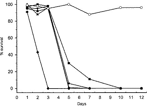
Fig. 1. Percentage survival of Echinococcus granulosus protoscoleces following 12 days of in vitro culture with various doses of CsA. CsA doses are represented by the symbols [lozf ] 100 μg/ml, [bull ] 50 μg/ml, [utrif ] 20 μg/ml, [dtrif ] 10 μg/ml, [squf ] 5 μg/ml and ○ No CsA.
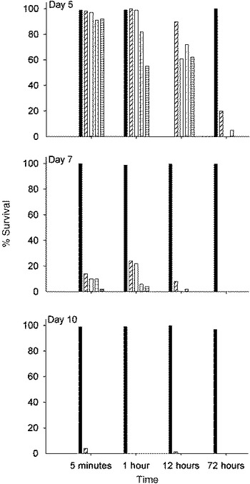
Fig. 2. Survival of Echinococcus granulosus protoscoleces following time pulse treatment with various doses of CsA. Data from culture day 5 show survival of protoscoleces when treated for 5 min, 1, 12 and 72 h in culture with CsA. In each time group the doses of CsA are represented by individual bars. The untreated controls are represented by black bars and doses of CsA at 50 (), 20 (□), 10 () and 1 μg/ml () are plotted on the right of the controls respectively.
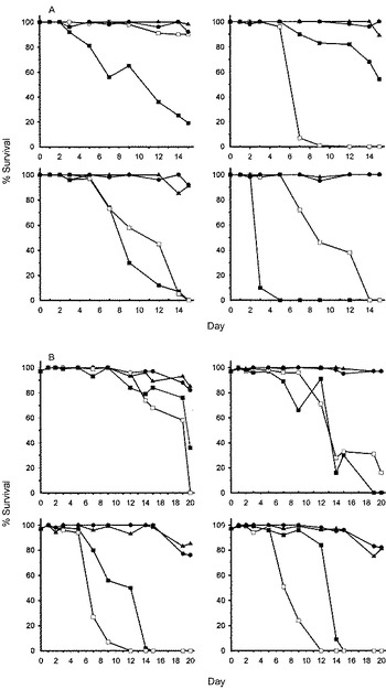
Fig. 3. Percentage survival of invaginated Echinococcus granulosus protoscoleces following (A) 15 days in culture with SHCF medium or (B) 20 days in DMEM medium. Cultures contained either medium plus the CsA vehicle alone ([utrif ]), medium ([bull ]) or various doses of CsA represented in replicate by □ or [squf ]. Each of the graphs within group A or B represents data from protoscoleces treated with different CsA doses. Data from cultures containing 1 μg/ml of CsA are represented in the graph at the top left of group A or B, 10 μg/ml in the top right, 50 μg/ml in the bottom left and 100 μg/ml in the bottom right.
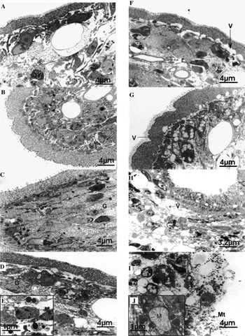
Fig. 4. TEM ultrastructure of Echinococcus granulosus protoscoleces in in vitro culture. (A–D) Untreated protoscoleces sampled from cultures at days 1, 3, 7 and 10. (A,B) Protoscoleces from culture days 1 and 3 respectively, note there are no distinct morphological differences in ultrastructure from specimens at day 0. (C,D) Protoscoleces from days 7 and 10 respectively, glycogen (G) has disseminated in the extracellular matrix. (F–I) Ultrastructure of protoscoleces treated with CsA in vitro. (F,G) Specimens treated for 1 and 2 days respectively. The basal laminar is separating from the distal cytoplasm of the tegument forming electron-lucent regions with no apparent matrix and vacuoles (V) are forming in the tegumentary cytons. (H) Specimens from culture day 5, vacuoles forming in teguments cells are more extensive, some cells appear to be rounded and lipidic structures are present. (I) Specimens from culture day 10, showing degradation of the tegument (T), detached microtriches (Mt) and large electron-lucent areas in the soma region. (E) Mitochondria (M) and lipid globules (L) from untreated protoscolex and (J) Higher magnification of enlarged mitochondria (M) from CsA-treated protoscolex.
 You have
Access
- 22
- Cited by