INTRODUCTION
Infection of sheep with the sheep scab mite, Psoroptes ovis, results in a profound intradermal inflammatory response with the generation of IgE and IgG antibodies (Van den Broek et al. 2000, 2003). P. ovis allergens have therefore been implicated in the development of the pathology and/or the development of immunity to infection and may also offer new candidates for novel therapies, including vaccine development (Nisbet and Huntley, 2006). Expressed Sequence Tag (EST) analysis of P. ovis has shown the presence of transcripts encoding several proteins with close homology to house dust mite (HDM) allergens (Kenyon et al. 2003). In addition, proteomic and Western blotting analyses, using sera from hyperimmune sheep, or specific antibodies to the HDM allergens Der p 10 and Der p 11, have provided firm evidence for the presence, and immunodominance, of these allergens in extracts of the Psoroptes mite (Huntley et al. 2004).
The group 10 HDM allergen has been identified as tropomyosin, an allergenic, actin-binding protein involved in the regulation of actin/myosin interactions which is highly conserved amongst arthropods and possesses high immunological cross-reactivity to shellfish and other invertebrate tropomyosins (Reese et al. 1997).
The group 11 HDM allergen, paramyosin, is a structural protein of thick filaments in invertebrate muscles and, like tropomyosin, is a major allergen associated with HDM allergy (Thomas and Smith, 1998). Western blot studies of dust mite extracts with sera from HDM allergic humans have shown the presence of an IgE binding protein of 90–100 kDa, which is most likely to be paramyosin and, recently, Maldi-ToF analysis combined with anti-Der p 11 immunoblotting have confirmed the presence of this allergen in the sheep scab mite (Huntley et al. 2004).
The presence and antigenicity of tropomyosin and paramyosin during sheep scab mite infestation suggests that they may also be good candidates for the development of diagnostic tools for mite infestation. The applicability of such a diagnostic tool has recently been demonstrated by Jacober et al. (2006) where accurate and early diagnosis of P. ovis infestation was used to selectively treat individual sheep, leading to the potential benefits of reduced drug resistance, exposure and environmental contamination. Current immunodiagnostics for P. ovis, however, rely on the use of native antigen extracts (e.g. Lonneux et al. 1996) which may introduce problems of repeatability and sustainability of production. The potential use of defined, recombinant proteins would alleviate these problems.
The aims of the current paper were to characterize the tropomyosin and paramyosin-encoding genes of P. ovis, to determine their usefulness in diagnosis of psoroptic mange and to determine the tissue distribution of the proteins encoded by these genes.
MATERIALS AND METHODS
Mites
Mites (adults, nymphs and larvae) were collected from donor infestations in sheep (cross-bred Suffolk sheep aged 1–2 years), and the live mites washed by vortexing for 5 min in ice-cold phosphate buffered saline (PBS, pH 7·2), followed by 1% sodium dodecyl sulphate (SDS) at room temperature and then 10 washes in ice-cold PBS. Mites were then snap-frozen in liquid nitrogen and stored at −80 °C prior to RNA extraction. For immunolocalization studies, live mites were fixed in Carnoy's fixative (60% ethanol, 30% chloroform, 10% acetic acid) and stored at 4 °C until ready for processing.
Amplification of cDNAs encoding tropomyosin and paramyosin
Interrogation of the P. ovis EST database (http://nema.cap.ed.ac.uk/Chelicerates/Chel_anno.php) (Kenyon et al. 2003) revealed the presence of a 390 bp contig with significant sequence identity to Der p 10 from Dermatophagoides pteronyssinus. Based on this sequence, an oligonucleotide primer was designed to incorporate the putative initiation codon of the EST (PoTropF: ATGGAGGCCATCAAGAAGAAAATGCAG), with a vector-specific anti-sense primer to amplify the whole open reading frame (ORF) and 3′ untranslated region using a preparation of phage from which the original EST sequence had been determined as a template (Po_ad_0406, kindly supplied by Claire Whitton, Nematode Genomics Laboratory, Edinburgh University). PCR conditions were as follows: primer concentrations (final concentration in 50 μl reaction) 0·2 μM; reaction buffer, polymerase and dNTPs all as described in manufacturer's instructions for BD Advantage™ 2 (BD Biosciences). Cycling parameters: 95 °C for 1 min followed by 30 cycles of 95 °C for 15 sec, 68 °C for 1 min and a final extension of 68 °C for 10 min. The resulting amplicon was ligated into pGEM®-T Easy vector and transformed into Escherichia coli JM109 competent cells according to the manufacturer's protocol (Promega). Plasmids isolated from cDNAs cloned in this way were subjected to automated sequencing.
For the amplification of paramyosin cDNA, degenerate primers were designed based on conserved regions of the published sequences of acarine paramyosins. The sequences of these primers were: PoParaDgF; G(A/T)CT(C/T)GAGGACAA(A/G)ATTCG and PoParaDgR; AGACTTGTCGAGT(A/G)CCGC. Total RNA was extracted from mixed-stage P. ovis under liquid nitrogen by homogenization in TriPure™ reagent (Roche) according to the manufacturer's protocol. Reverse transcription and cDNA amplification were performed using 1 μg of the RNA, each degenerate primer (final concentration 0·4 μM) and the SuperScript™ One-Step RT-PCR System (Invitrogen) in accordance with the manufacturer's instructions. The single, approx. 2·5 kb product, was cloned and sequenced as described above. Based on the resulting sequence, an oligonucleotide primer (PoPAR3p GCTAATGCATTGGCCGGTGGCA) was designed to amplify the 3′ portion of the gene when used in conjunction with a vector-specific primer and, as template, a heat-denatured, phage lysate preparation of a cDNA library made in λTriplEx2 by long distance PCR following the manufacturer's instructions for SMART™ cDNA Library Construction (BD Biosciences) and packaged in Gigapack Gold III packaging extract (Stratagene). The resulting amplicon was cloned and sequenced as described above. Based on an alignment of this sequence with an existing EST encoding the putative 5′ portion of P. ovis paramyosin (Po_ad_03D08, Kenyon et al. 2003), two oligonucleotide primers were designed to amplify the complete ORF of paramyosin (Paraflfor ATGTCTGCACGTACAGCAAAATATATGTACC and Paraflrev ATTAATAATTTGTTTGTTCCTCTGTCGTCGTG). Reverse transcription and cDNA amplification were performed using 1 μg of total RNA, each primer (final concentration 0·4 μM) and the SuperScript™ One-Step RT-PCR System (Invitrogen) in accordance with the manufacturer's instructions.
Full-length cDNA sequences of P. ovis tropomyosin and paramyosin were translated to amino acid sequences and the relationships between these and homologous proteins were examined by neighbour-joining analysis using Clustal X (Jeanmougin et al. 1998) and TreeView (Page, 1996), using default settings.
Expression of recombinant tropomyosin and paramyosin
cDNAs encoding tropomyosin, paramyosin and a truncated version of paramyosin (first 1032 nucleotide bases of the 5′ end) were subcloned into expression vector pET-22b(+) (Novagen) after amplification by PCR using the plasmids in pGEM®-T Easy as template. Primer sequences were:
TroppETF 5′-AATTCGGATCCGATGGAGGCCATCAAGAAGA-3′
TroppETR 5′-GAGTGCGGCCGCATATCCAGTAAGTTCGGCAA-3′
ParapETF 5′-TAATTCGGATCCGATGTCTGCACGTACAGCA-3′
ParapETR 5′-TCGAGTGCGGCCGCATAATTTGTTTGTTCCTCTGTCG-3′
TruncParapETR 5′-TCGAGTGCGGCCGCCAATGAGCTG CATTTGTTCA-3′
(BamH1 and Not1 restriction sites underlined, initiation codons in italics). PCR conditions were as follows: primer concentrations (final concentration in 50 μl reaction) 0·2 μM; reaction buffer, polymerase and dNTPs all as described in the manufacturer's instructions for BD Advantage™ 2 (BD Biosciences). Cycling parameters: 95 °C for 1 min followed by 30 cycles of 95 °C for 15 sec, 50 °C for 1 min, 70 °C for 3 min and a final extension of 68 °C for 10 min. The PCR products were subcloned into pET-22b(+) (Novagen) between the vector sequence encoding an amino-terminal pelB leader and a carboxyl-terminal (His)6 tag, cultivated in E. coli JM109 (Promega) competent cells and plasmids were extracted using Wizard® Plus SV Miniprep kits (Promega). The plasmids were then used to transform E. coli BL21-CodonPlus® (DE3)-RIL competent cells (Stratagene). Recombinant protein expression was induced in the presence of 1 mM isopropyl-β-D-thiogalactopyranoside (IPTG) at 37 °C. Soluble, recombinant tropomyosin (rPoTRO), paramyosin (rPoPAR) and truncated paramyosin (trPoPAR) were purified from cell lysates by nickel column affinity chromatography employing a step-wise gradient of imidazole and HisTrap™ HP columns (GE Healthcare) according to the manufacturer's instructions.
Identification of immunoreactivity by Western blotting
Purified rPoTRO, rPoPAR and trPoPAR were electrophoresed on NuPAGE® Bis-Tris 4–12% gels under reducing conditions employing NuPAGE® MES SDS running buffer and proteins were transferred to a nitrocellulose membrane according to the manufacturer's instructions (Invitrogen). After transfer, the membranes were washed briefly in Tris- buffered saline (TBS; 25 mM Tris, 137 mM NaCl, 2·7 mM KCl) containing Tween 20 (0·1%) (TBST) then incubated in TBST containing 5% Marvel non-fat milk powder (Premier International Foods Ltd) for 4 h at room temperature (RT) on an orbital shaker (100 cycles/min) to block non-specific protein adsorption. To detect rPoTRO, rPoPAR and trPoPAR, the blotted proteins were incubated with either polyclonal serum raised in mice against HDM tropomyosin (diluted 1[ratio ]1000 in TBST 5% Marvel) or a mouse monoclonal antibody produced by DNA immunization using Blo t 11, a paramyosin from Blomia tropicalis (Ramos et al. 2004), diluted 1[ratio ]250 in TBST 5% Marvel. Blotted proteins were also incubated in pooled serum (1[ratio ]250 dilution) obtained from sheep with a 6-week duration P. ovis infestation or from sheep with no history of P. ovis infestation. Blots were incubated for 1 h at RT with constant rocking. Control blots were performed by omitting the test sera (mouse or sheep) during this primary incubation step. The blots were washed (10 min/wash) 3 times in TBST, before being incubated in serum raised in rabbit against mouse immunoglobulins (DakoCytomation) or donkey anti-sheep IgG (Sigma) as appropriate. Both sera were horse-radish peroxidase (HRP) conjugates and were diluted 1[ratio ]1000 in TBST, 5% Marvel. For the detection of IgE in sheep sera, the secondary antibody was a mouse monoclonal antibody raised against ovine IgE (1[ratio ]1000 in TBST, 5% Marvel) in which blots were incubated for 1 h, prior to washing in TBST and incubation in rabbit anti-mouse HRP conjugate as a tertiary antibody. Following incubation at RT for 1 h, the blots were washed a further 3 times in TBST and peroxidase activity was revealed by chemiluminescent detection using EC Plus™ Western Blotting Detection Reagents (GE Healthcare) according to the manufacturer's instructions.
Immunolocalization
Fixative was removed and mites were sedimented in 1% molten agarose, then left to solidify before embedding in paraffin wax. Sections (5 μm) were cut and dried at 40 °C, dewaxed in xylene and rehydrated. Sections were treated to remove endogenous peroxidase (EnVision Kit™, DAKO) and blocked in 25% normal goat serum in TBS for 30 min before immunolabelling using mouse polyclonal anti-HDM tropomyosin (1[ratio ]1000 diluted in TBS) or mouse monoclonal anti-Blo t 11 (1[ratio ]100) and incubated at 4 °C overnight. The EnVision™+ System-HRP(DAB) (DAKO) was used, according to the manufacturer's instructions, for detection of the mouse antibodies. Negative controls were prepared by omitting the primary antibodies. Sections were washed in water, counterstained in haematoxylin for a few sec, washed in water, dehydrated, cleared and mounted in a resin-based mountant.
RESULTS
Amplification of cDNAs encoding tropomyosin and paramyosin
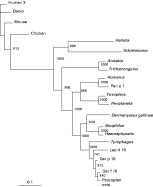
The PCR product obtained by amplification with PoTropF and a vector-specific primer contained an ORF of 852 bp (nucleotide sequence data available in the EMBL, GenBank™ and DDJB databases under Accession number AM114276) which encoded a predicted protein with 98%, 97% and 95% identity to Der f 10, Der p 10 and Lep d 10 respectively (Fig. 1). The predicted protein contained 284 amino acids residues, characteristic of a muscle isoform rather than the smaller non-muscle isoforms of the molecule, and the consensus C-terminal signature sequence for tropomyosins [LK(EAD)AExRA(ET)] was located at L232 to E240 in the predicted sequence. The heptad repeat of amino acids (a-b-c-d-e-f-g, where a and d are generally apolar residues, e.g. L232 and A235 at positions a and d in the signature sequence) that characterize coiled-coil molecules (Cohen and Parry, 1990) is found in 40 repetitive units across the length of the predicted protein. In common with the tropomyosins of vertebrate striated muscle, more than 30% of the residues at the ‘d’ position of the heptad are filled by alanine in the predicted P. ovis tropomyosin homologue and there are 7 clusters of d-position alanines in addition to a regular distribution of negatively charged residues in these predicted ‘α-zones’. These α-zones are involved in the interaction of each tropomyosin molecule with the 7 actin monomers it spans in muscle tissue (Brown et al. 2001; Hitchcock-DeGregori et al. 2002) and, overall, the conservation of features in the P. ovis homologue is strongly suggestive of conserved function and structure between this molecule and vertebrate tropomyosins.

Fig. 1. Relationships between selected tropomyosin proteins. The neighbour-joining tree was bootstrapped 1000 times using Clustal X (Jeanmougin et al. 1998) and the resulting tree viewed with TreeView (Page, 1996). Each node is annotated with a figure indicating the degree of bootstrap support for each branch. Abbreviations and Accession numbers are as follows: Psoroptes ovis (CAJ38272), Dermanyssus gallinae (AM167555), Der p 10 (Dermatophagoides pteronyssinus AAB69424), Der f 10 (Dermatophagoides farinae Q23939), Lep d 10 (Lepidoglyphus destructor CAB71342), Tyrophagus (Tyrophagus putrescentiae AAT40866), Haemaphysalis (Haemaphysalis longicornis AAN05633), Boophilus (Boophilus microplus AAD17324), Homarus (American lobster Homarus americanus O44119), Pen a 1 [Farfantepenaeus aztecus (brown shrimp) AAZ76743], Toxoptera [Toxoptera citricida (brown citrus aphid) AAU84930], Periplaneta [Periplaneta americana (American cockroach) AAD19606], Anisakis (Anisakis simplex CAB93501), Trichostrongylus (Trichostrongylus colubriformis A44980), Haliotis [Haliotis rufescens (California red abalone) CAA53028], Schistosoma (Schistosoma haematobium Q26503), human3 (Homo sapiens tropomyosin 3 CAH71269), Danio [Danio rerio (zebrafish) NP_571180], mouse (Mus musculus CAA46043), chicken (Gallus gallus AAA48580).
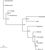
The complete paramyosin ORF generated by RT-PCR with Paraflfor and Paraflrev (Accession number AM114275) was 2625 bp in length and encoded an 875aa predicted protein of 102·6 kDa with 97%, 95% and 89% identity to the paramyosins of D. pteronyssinus (Der p 11), Sarcoptes scabiei and Blomia tropicalis (Blo t 11) respectively (Fig. 2). As with S. scabiei paramyosin, a coiled-coil secondary structure is predicted for P. ovis paramyosin (predicted using BCM Search Launcher: Protein Secondary Structure Prediction at http://searchlauncher.bcm.tmc.edu/seq-search/struc-predict.html) and, with the exception of the 27 amino terminal amino acids and the 30 carboxy terminal residues, the heptad periodicity and characteristic 28-residue meta-repeat of alternating charge are retained in P. ovis paramyosin.

Fig. 2. Relationships between selected paramyosin proteins. The neighbour-joining tree was bootstrapped 1000 times using Clustal X (Jeanmougin et al. 1998) and the resulting tree viewed with TreeView (Page, 1996). Each node is annotated with a figure indicating the degree of bootstrap support for each branch. Abbreviations and Accession numbers are as follows: Psoroptes ovis (CAJ38271); Der p 11 (Dermatophagoides pteronyssinus AAO73464); Boophilus (Boophilus microplus AF479582); Sarcoptes (Sarcoptes scabiei AF317670); Blomia (Blomia tropicalis AF525465); Drosophila (Drosophila melanogaster CAA41557); Ancylostoma (Ancylostoma caninum ABC86903); Dictyocaulus (Dictyocaulus viviparus AAT36324); Brugia (Brugia malayi Q01202); Onchocerca (Onchocerca volvulus A48575); Apis (Apis mellifera XP_393281).
Expression of rPoTRO, rPoPAR and trPoPAR and identification of immunoreactivity by Western blotting
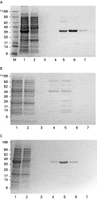
rPoTRO, rPoPAR and trPoPAR were expressed in soluble form with carboxyl-terminal (His)6 tags which were used to purify the recombinant proteins from soluble bacterial material (Fig. 3). Full-length recombinant paramyosin appears to be poorly expressed and/or readily degraded in this expression system (Fig. 3B). However, the truncated version of the molecule, representing the amino-terminal 344 amino acid residues, was expressed in large amounts and appeared stable (Fig. 3C). MALDI-ToF-ToF analysis confirmed the identity of rPoTRO and trPoPAR, but insufficient rPoPAR could be generated to allow definitive identification by this technique.

Fig. 3. Expression and purification of recombinant Psoroptes ovis tropomyosin (rPoTRO, Panel A), paramyosin (rPoPAR, Panel B) and truncated paramyosin (trPoPAR, Panel C). Recombinant proteins were expressed as hexa-His-tagged fusion proteins in a prokaryotic expression system and purified by nickel column affinity chromatography. Samples of starting material and eluates from the purification procedure were separated by electrophoresis. Each panel depicts the stepwise purification from whole soluble bacterial extract (lane 1), flow-through of unbound material from the affinity column (lane 2), and elution with increasing concentrations of imidazole from 50 mM (lane 3), through 200 mM (lane 4), 350 mM (lane 5) and 500 mM (lanes 6 and 7). ‘M’ represents molecular mass markers. *Molecular mass in kDa.
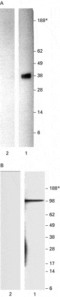
rPoTRO was readily detected in Western blots by the IgG component of polyclonal serum raised in mice against HDM tropomyosin (Fig. 4A, lane 1). IgG in serum from P. ovis-infested sheep reacted weakly with a protein of this molecular mass but serum from P. ovis-naïve sheep also reacted weakly with this band (not shown) suggesting cross-reactivity between P. ovis tropomyosin and tropomyosins of other mite species to which the sheep were likely to have been exposed (forage or harvest mites). IgE in serum from P. ovis-infested sheep failed to react with rPoTRO (not shown).

Fig. 4. Western blots of recombinant Psoroptes ovis tropomyosin (rPoTRO) and paramyosin (rPoPAR) with sera used in immunolocalization experiments. Panel A: rPoTRO was detected by incubation with polyclonal serum raised in mice against house dust mite tropomyosin (Lane 1). Secondary antibody was rabbit anti-mouse IgG (DakoCytomation), horse-radish peroxidase (HRP) conjugate. Negative control (no primary antiserum) is shown in lane 2 (secondary antibody; rabbit anti-mouse IgG). Peroxidase activity was revealed by chemiluminescent detection using ECL Plus™ Western Blotting Detection Reagents (GE Healthcare) according to the manufacturer's instructions. Panel B: rPoPAR was detected by incubation with monoclonal antibody P2E7-G1 raised against Blomia tropicalis paramyosin (Ramos et al. 2004) diluted (Lane 1). Secondary antibodies, detection and negative controls, as for panel A.
A mouse monoclonal antibody generated against B. tropicalis paramyosin reacted strongly with a band of approx. 100 kDa on Western blots of rPoPAR, reflecting the calculated molecular mass of the recombinant protein (Fig. 4B). In spite of the potential degradation of rPoPAR during bacterial expression (Fig. 3B), the monoclonal antibody only detected the full-length molecule rather than degradation products. The monoclonal antibody used (P2E7-G1, Ramos et al. 2004) recognizes epitope f5 (amino acid residues 425 to 647) of the B. tropicalis paramyosin molecule. A combination of purification of rPoPAR by the use of a C-terminal His-tag and the use of a monoclonal antibody that recognizes a specific epitope explains the lack of reactivity with potential degradation products on these Western blots. In addition, trPoPAR could not be detected by this monoclonal antibody as the recombinant protein only represents amino acid residues 1-344. Neither rPoPAR or trPoPAR were detected by IgG or IgE in serum from P. ovis-infested sheep (not shown).
Immunolocalization
Staining with antibodies directed against tropomyosin and paramyosin was observed throughout the sections in mite tissues, reflecting the ubiquitous nature of these proteins and their biological roles.
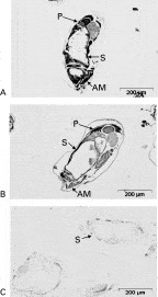
Sections of P. ovis incubated with antisera to Der p 10 and monoclonal antibodies to Blo t 11 each showed similar patterns of immunostaining. For example, in the anterior region, immunoreactivities for the two allergens, tropomyosin and paramyosin, was evident in the anterior muscle bundles associated with the legs (Fig. 5A,B). The immunoreactivity observed throughout the mid- and posterior regions of the mite is related to the numerous, small muscle bundles associated with the digestive and reproductive systems. No specific immunolabelling was observed in the control material (Fig. 5C).

Fig. 5. Immunolocalization of tropomyosin and paramyosin in sections of Psoroptes ovis. Panel A, staining with anti- Der p 10 as primary antibody; Panel B, staining with monoclonal anti-Blo t 11 as primary antibody; C, Control (no primary antibody). The EnVision™+ System-HRP(DAB) (DAKO) was used, according to the manufacturer's instructions for detection of the mouse antibodies. Annotation: ‘AM’anterior muscle bundles associated with legs; ‘S’ stomach muscle blocks; ‘P’ posterior muscle blocks associated with reproductive and digestive tracts.
DISCUSSION
The tropomyosin and paramyosin-encoding genes of P. ovis were characterized and the tissue distribution of the proteins encoded by these genes was determined by immunolocalization. The predicted proteins encoded by these genes possessed very high homologies to the group 10 and group 11 allergens of house dust mites and also to the tropomyosin and paramyosin proteins of other parasitic species of the Acari.
Tropomyosin is a strongly allergenic, actin-binding protein which has previously been shown, by Western blotting using anti-Der p 10 sera, to be present in P. ovis extracts and to provoke the production of an IgE response in hyperimmune sheep host during infestation (Huntley et al. 2004). When used in vaccination trials in other species of parasite, tropomyosin has shown mixed levels of success. For example, immunization of jirds with a 41 kDa tropomyosin-like protein purified from the filarial nematode Acanthocheilonema viteae reduced adult worm burden by >60% and circulating microfilariae by up to 93% (Hartmann et al. 1997). However, DNA immunization of mice with plasmid expressing the Onchocerca volvulus antigen, Ov-TMY-1 (tropomyosin) induced strong Th1/Th2 IgG isotype profiles but failed to protect against infection on challenge (Harrison and Bianco, 2000).
Paramyosin has been identified from both P. ovis and Sarcoptes scabiei as an important immunogen, though its role in the development of natural protective immunity is unclear (Huntley et al. 2004; Mattsson et al. 2001). In the case of S. scabiei, sera from rabbits that had been immunized with high molecular weight (90–200 kDa) proteins from S. scabiei were used to identify paramyosin-encoding genes from a cDNA library by immunoscreening. Re-screening of the library with sera from scabies-infested dogs identified further clones encoding paramyosin and recombinant paramyosin expressed on a larger scale from these clones was recognized by sera from infested animals, suggesting that it is an immunogenic protein exposed to the host's immune system during the course of natural infestation (Mattsson et al. 2001). This also appears to be true of P. ovis paramyosin (Huntley et al. 2004).
A cDNA encoding paramyosin has also been described from the tropical cattle tick Boophilus microplus, an ectoparasite against which no natural immunity develops, and the tissue-specific expression of the native molecule was described by Western blot analysis (Ferreira et al. 2002). Interestingly, paramyosin was found in tick organs with little associated musculature, e.g. fat body and gut as well as in the salivary glands. However, in spite of the presence of the molecule in salivary glands, it was absent from saliva harvested from ticks and therefore probably not exposed to the host during tick infestation. The presence of the tick paramyosin in the gut, it's potential exposure to ingested host blood and the demonstrated ability to interact with components of the host's immune system (e.g. collagen) led the authors to conclude that it may represent an important new ‘concealed’ antigen for vaccination studies (Ferreira et al. 2002). Paramyosins from some species of helminth endoparasites have also shown considerable promise as vaccine candidates (for recent comprehensive review see Gobert and McManus, 2005). Vaccination of mice with both native and recombinant versions of Schistosoma mansoni paramyosin (Sm97) resulted in significant reductions in worm burden (Pearce et al. 1988) and vaccination with native S. japonicum paramyosin induced 62–86% resistance against cercarial challenge (Ramirez et al. 1996). Clearly there are important differences between helminth endoparasites and acarine ectoparasites in the presentation of paramyosin during natural infestation that may affect the ability of an immune response to interact with the parasite during infestation – schistosome paramyosin is present in the tegument and therefore exposed to host immune responses much more readily than mite or tick paramyosins which are associated with internal organs. However, both ticks and mites ingest host immunoglobulins and host-derived immune cells (Pettit et al. 2000), making vaccination with muscle-related proteins such as paramyosin and tropomyosin an intriguing possibility. Future vaccination studies with recombinant forms of the molecules described herein will determine the applicability of these targets as vaccine candidates.
The applicability of rPoTRO, rPoPAR and trPoPAR to immunodiagnosis of sheep scab mite infestation seems limited as there appears to be cross-reactivity between rPoTRO and the tropomyosins of other invertebrate species, in agreement with previous studies on other tropomyosins (Reese et al. 1997) and the paramyosins are not detected by sera from 6-week duration primary infestations. It is likely that infestations of a longer duration would lead to sera which were capable of detecting paramyosin (e.g. see Huntley et al. 2004); however, clinical disease may be obvious by then. Future research in this area will focus on molecules which are presented to and detected by the immune response of the sheep during these early stages of infestation.
A.J.N., J.F.H. and A.McK. gratefully acknowledge funding by the Department for Environment Food and Rural Affairs, UK Research Contract OD0544 and the Scottish Executive Environment and Rural Affairs Department. We are grateful to Claire Whitton, Nematode Genomics Laboratory, Edinburgh University for the provision of plasmids from the P. ovis EST sequencing project.