

Introduction
The term myeloproliferative disease (MPD) was first used in 1951 to describe: polycythaemia vera (PV), essential thrombocythaemia (ET), primary myelofibrosis (PMF) and chronic myeloid leukaemia (CML).Reference Dameshek1 It is perhaps surprising that the most recent WHO classification, published 57 years later, still retains these 4 main disease categories albeit with the addition of some rarer conditions such as systemic mastocytosis, chronic neutrophilic leukaemia, chronic eosinophilic leukaemia and chronic myelomonocytic leukaemia.Reference Swerdlow, Campo and Harris2 This article will focus on the four commonest conditions: PV, ET, CML and PMF. Given that haematocrit and platelet count elevations are both relatively common clinical scenarios, rarely caused by primary haematological problems, we will discuss the diagnosis of MPD in the context of a general approach to patients presenting with these blood count abnormalities.
Size of the problem
MPDs are rare disorders with a combined incidence of approximately 6 per 100,000. Incidence increases with increasing age: a recent US study found that the combined incidence of PV, ET and PMF ranged from 0.27 per 100,000 in those under 40 years, to 13.25 in those over 80 years old.Reference Rollison, Howlader, Smith, Strom, Merritt, Ries, Edwards and List3
The impact of increasing age
In broad terms, the MPDs are characterized by:
(i) bone marrow hypercellularity. The cell line predominantly affected correlates with the subtype of MPD: in CML an excess of granulocyte forms is seen, in PV an excess of red cells, in ET an excess of megakaryocytes and thus platelets, and in PMF an excess of marrow fibrosis;
(ii) thrombotic risk;
(iii) haemorrhagic risk;
(iv) risk of progression to marrow fibrosis;
(v) risk of transformation to acute leukaemia.
Whilst age itself does not fundamentally affect the biology of MPD, it is clear that the natural history of the disease for an individual patient is affected by a range of variables including age. Taking thrombosis in PV as an example, the presence of cardiovascular risk factors, a previous history of thrombosis and age >60 years are all identified risk factors for recurrent thrombosis. Older patients unsurprisingly experience poorer overall survival than those diagnosed at a younger age.Reference Wolanskyj, Schwager, McClure, Larson and Tefferi4, Reference Cervantes, Passamonti and Barosi5
Advances in molecular understanding of MPDs
In recent years, a string of discoveries in the molecular biology of the MPDs have led to important changes in the diagnostic approach and, in some cases, to changes in therapy.
The presence of the Philadelphia (Ph) chromosome in CML was first described in 1960.Reference Nowell and Hungerford6 This translocation between chromosomes 9 and 22 results in the generation of an oncogenic protein, BCR/ABL. Reference Sawyers7 The BCR/ABL protein is a constitutively active tyrosine kinase, which increases activity in various cellular signalling and control pathways such as the JAK-STAT pathway. Excessive activation of such pathways results in the hallmarks of malignancy in this disease:
(i) increased cellular proliferation;
(ii) failure of cell death (apoptosis);
(iii) lack of cell–cell adhesion.
In 2005, a Janus kinase 2 (JAK 2) mutation, JAK2V617F, was discovered in >95% of patients with PV and in approximately 50–60% of patients with ET and PMF.Reference Vannucchi, Guglielmelli and Tefferi8 Since then, an additional mutation in JAK 2 exon 12 has also been reported.Reference Scott, Beer, Bench, Erber and Green9 This mutation is found in a proportion of the 5% of PV cases that are negative for JAK2V617F mutation. The presence of these JAK 2 mutations results in loss of control within cellular signalling pathways.
In 2006, a mutation at codon 515 of the MPL gene was described. MPL (named after the myeloproliferative leukaemia virus homologous oncogene) is a thrombopoietin receptor found on megakaryocytes and primitive myeloid cells. MPL 515 mutations are demonstrable in approximately 8% of patients with PMF and 8% of JAK2V617F-negative patients with ET.Reference Pardanani, Levine, Lasho, Pikman, Mesa, Wadleigh, Steensma, Elliott, Wolanskyj, Hogan, McClure, Litzow, Gilliland and Tefferi10 These mutations confer hypersensitivity to the growth factor thrombopoietin and, like the JAK2V617F mutation, result in constitutive activation of the JAK-STAT pathway.
These data suggest that dysregulation of cellular signalling pathways is a common feature amongst the MPDs. Current work focuses on how best to use this information. The discovery of these mutations has changed the way that we diagnose MPDs, with a move away from bone marrow biopsy and towards mutation testing in peripheral blood samples. The mutations also serve as potential molecular targets for treatment; use of tyrosine kinase inhibitors in CML has revolutionized treatment of this condition. Other mutations, such as those in JAK 2, are an ongoing focus for drug development. These issues will be addressed as we discuss each of the MPDs in more detail.
Polycythaemia vera (PV)
Approach to the patient with a high haematocrit
The term polycythaemia can be applied to any raised haematocrit (Hct) and is a common clinical finding, but rarely is PV the cause. The diagnosis of PV, also termed primary polycythaemia, is only made when an intrinsic clonal bone marrow defect is present. Frequently, the polycythaemia will instead be secondary to another systemic condition, which drives excessive red cell production in an otherwise normal marrow.
In some cases the raised haematocrit may simply reflect a reduced circulating plasma volume (termed spurious or apparent polycythaemia) rather than a genuine increase in total red cell mass (true or absolute polycythaemia). A spurious or apparent polycythaemia is most often associated with dehydration, use of diuretics, hypertension, diabetes mellitus, smoking, excess alcohol consumption and obesity. True polycythaemia can be diagnosed with confidence when the haematocrit is >0.6 in a male or >0.56 in a female, though it can be present with a less markedly raised Hct.
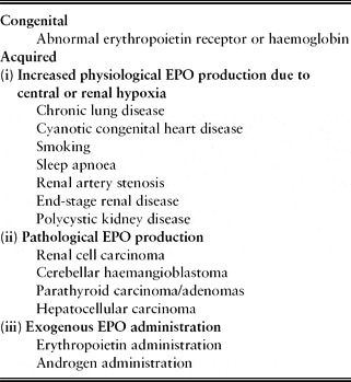
There are many congenital and acquired causes of a secondary true polycythaemia; some of these are listed in Table 1. Red cell production in the normal bone marrow is regulated by the production of erythropoietin (EPO) by the kidney. EPO synthesis is stimulated by renal hypoxia. Any process causing central or local renal hypoxia can therefore lead to increased EPO production and a secondary polycythaemia. EPO may also be produced in excess in the presence of particular tumours including renal cell carcinoma, hepatocellular carcinoma, cerebellar haemangioblastoma, and phaeochromocytoma. Exogenous, often illicit, EPO administration can also lead to a secondary polycythaemia.
Causes of secondary polycythaemia

The discovery of JAK 2 mutations have changed the traditional approach to the investigation of polycythaemia. Initial assessment of the patient includes a careful history and examination, considering potential causes and risk factors for a secondary polycythaemia. In many cases, a secondary cause will be immediately apparent, e.g. chronic hypoxic lung disease, smoking, or end stage renal disease. However, if no obvious secondary causes are present, it is appropriate to test for the JAK2V617F mutation. This is found in >95% of patients with PV. Many centres routinely test for the JAK 2 exon 12 mutation and MPL gene mutations simultaneously. The presence of a JAK2V617F mutation in a patient with a raised Hct is sufficient to establish a diagnosis of PV.
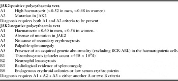
In patients with true polycythaemia who are negative for JAK 2 mutations, further investigations are required. Chest X-ray, oxygen saturations, renal ultrasound, and serum erythropoietin level should all be assessed to exclude a secondary cause. A bone marrow biopsy should also be considered and may be required to establish a diagnosis in the small proportion (<5%) of patients with PV in whom JAK 2 mutations are absent (Table 2).Reference McMullin, Reilly, Campbell, Bareford, Green, Harrison, Conneally and Ryan11, Reference McMullin12
Diagnostic criteria for polycythaemia vera

Incidence of PV
The approximate incidence of PV is 2–3 per 100,000.Reference Swerdlow, Campo and Harris2 Incidence increases with age and there is a slight male predominance. The mean age at diagnosis was 60 years in a large prospective study.Reference Landolfi, Marchioli, Kutti, Gisslinger, Tognoni, Patrono and Barbui13
Clinical features specific to PV
Patients may be asymptomatic; a high haemoglobin and haematocrit may simply be an incidental finding when a blood count is done for other reasons. Some patients suffer troublesome pruritis or may present with gout due to a hypermetabolic state. Headache, dizziness and fatigue are also common along with constitutional symptoms including weight loss and night sweats. Erythromelalgia may be present; transient occlusions of the microvasculature in the extremities cause burning pain and erythema. Patients may experience major thrombotic events such as stroke or myocardial infarction. Thromboses in unusual sites, such as mesenteric or portal veins, should prompt consideration of PV as an underlying trigger. Occasionally, patients may present with haemorrhage of the skin or gastrointestinal tract. Examination may reveal plethoric facies and hepatomegaly or splenomegaly.
Therapy and complications
The aims of therapy in PV are to:
(1) reduce the risk of thrombotic complications due to hyperviscosity;
(2) minimize the risk of transformation to acute leukaemia or myelofibrosis;
(3) reduce other associated symptoms of PV, e.g. pruritis, sweats and weight loss.
There have been six major randomized clinical trials of treatment in PV since the 1970s, and these have been well summarized.Reference McMullin, Bareford and Campbell14 These trials have compared venesection with or without various cytoreductive agents and the addition of low and high dose anti-platelet agents.
The correlation between increasing haematocrit and increasing thrombotic risk is striking and well documented. A target haematocrit of 0.45 is considered optimum to reduce hyperviscosity and thus the associated vascular complications.Reference Pearson and Wetherley-Main15 This target can sometimes be achieved by the use of venesection alone, whilst other patients also require cytoreductive agents. The indications for such cytoreductive therapy include: symptomatic or progressive splenomegaly; the presence of constitutional symptoms such as sweats or weight loss; a failure of venesection as a result of intolerance, technical difficulties or lack of efficacy.
Hydroxycarbamide is the first-line cytoreductive agent in PV patients over the age of 40 years. It is given orally and is usually well tolerated. Potential side-effects include skin ulceration, pigmentation and myelosuppression. There have been concerns regarding an increased risk of acute leukaemic transformation in patients treated with hydroxycarbamide, but there is currently no conclusive evidence regarding its leukaemogenicity in MPD patients.Reference McMullin, Bareford and Campbell14
Given the risk of major thrombotic episodes, assiduous management of other thrombotic risk factors, including hypertension, diabetes and lipids is essential. The ECLAP study (European Collaboration on Low Dose Aspirin in Polycythaemia Vera) has demonstrated the benefits and safety of low dose aspirin in PV patients in whom there is no contraindication. Significant reductions in non-fatal stroke, myocardial infarction and major venous thromboembolism were seen in aspirin-treated patients of all ages. Over a quarter of the patients were aged >70 years.Reference Landolfi, Marchioli, Kutti, Gisslinger, Tognoni, Patrono and Barbui13
Prognosis
Historically, the prognosis of PV has been poor. Morbidity and mortality are typically consequent upon serious thrombotic episodes or disease progression to myelofibrosis or acute leukaemia.
The introduction of venesection, cytoreductive agents and anti-platelet agents have all improved outcome. An Italian population-based study, however, demonstrated that overall mortality is 1.6-fold higher in patients with PV compared with the general population.Reference Passamonti, Rumi, Pungolino, Malabarba, Bertazzoni, Valentini, Orlandi, Arcaini, Brusamolino, Pascutto, Cazzola, Morra and Lazzarino16
Advancing age is associated with an increased risk of PV-related complications. The ECLAP study demonstrated that age over 65 years, and history of previous thrombosis, were the most significant risk factors for further thrombotic events (10.9 events/100 persons/year in those aged over 65 with a previous history of thrombosis). This study demonstrated an overall incidence of cardiovascular death in those over 65 years as 2.7/100 persons/year. Age over 70 years was associated with a 5-fold increase in the risk of developing acute leukaemia, compared with PV patients under the age of 60.Reference Landolfi, Marchioli, Kutti, Gisslinger, Tognoni, Patrono and Barbui13 Longer disease duration was associated with the highest risk of marrow progression to myelofibrosis.Reference Marchioli, Finazzi, Landolfi, Kutti, Gisslinger, Patrono, Marilus, Villegas, Tognoni and Barbui17
Essential thrombocythaemia (ET)
Approach to the patient with a high platelet count
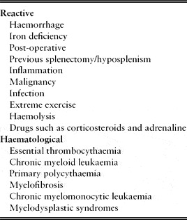
High platelet counts always require explanation. They are common in hospitalized patients and are usually reactive. There are many causes of a reactive thrombocytosis (Table 3). In one study, over 80% of unselected patients with persistent thrombocytosis had no evidence of a clonal bone marrow disorder.Reference Beer, Erber, Campbell and Green18 In some patients with a reactive thrombocytosis, the cause may be immediately obvious, for example a patient recovering from major surgery. In some cases, however, thrombocytosis may be the only manifestation of an occult problem such as a malignancy or bleeding. A persistently raised platelet count should, therefore, always be investigated and explained.Reference Harrison, Bareford, Butt, Campbell, Conneally, Drummond, Erber, Everington, Green, Hall, Hunt, Ludlam, Murrin, Nelson-Piercy, Radia, Reilly, Van der Walt, Wilkins and McMullin19
Causes of thrombocytosis

ET is essentially a diagnosis of exclusion; reactive causes should be carefully considered in the first instance. Other haematological diagnoses, including other MPDs or myelodysplastic syndromes, should also be excluded before a diagnosis of ET is made (see Table 2). PV can be associated with thrombocytosis, and occasionally this may be seen in the absence of a raised Hct if the patient has co-existent iron deficiency. Cytogenetic studies should exclude the presence of the Ph chromosome, which would suggest a diagnosis of an alternative haematological malignancy.
As with PV, the discovery of the JAK2V617F mutation, which is present in approximately 50% of cases of ET, and the MPL gene mutation, present in 10% of ET patients, has altered the diagnostic approach to ET.
The revised diagnostic criteria for ET, proposed by the British Committee for Standards in Haematology (BCSH),Reference Harrison, Bareford, Butt, Campbell, Conneally, Drummond, Erber, Everington, Green, Hall, Hunt, Ludlam, Murrin, Nelson-Piercy, Radia, Reilly, Van der Walt, Wilkins and McMullin19 take these mutations into account and the diagnosis of ET thus requires the presence of A1–A3, or A1 and A3–A5:
A1 Sustained elevated platelet count > 450 × 109/l;
A2 Presence of an acquired pathogenetic mutation in either the JAK2 or MPL genes;
A3 No other haematological malignancy. CML, PRV, PMF and MDS should specifically be excluded;
A4 No reactive cause for thrombocytosis; normal iron stores;
A5 Bone marrow aspirate and trephine showing typical features of ET with no features to suggest an alternative haematological malignancy.
Incidence of ET
ET is uncommon, with an incidence of 1–2 cases per 100,000 population. The disease can occur at any age but is more common in late middle-aged and elderly patients. The average age of onset is 50–60 years.
Clinical features specific to ET
There are no specific clinical features that define ET, and it is often asymptomatic. Patients with ET may present with thrombosis, typically arterial. Paradoxically, patients may also present with bleeding and bruising due to platelet dysfunction, despite the thrombocytosis. Erythromelalgia can be a presenting feature and cold peripheries, acrocyanosis and even areas of gangrene can occur. Some patients experience vague neurological symptoms such as headaches and dizziness, whilst others describe left upper quadrant pain due to splenomegaly and splenic infarction.
Therapy and complications
The major threat to patients with ET is arterial or venous thrombosis. It is therefore crucial that other risk factors including hypertension, diabetes mellitus, obesity and smoking are aggressively managed. Low dose aspirin should be offered to all patients.
In addition, patients with ET should be stratified according to their risk of thrombosis, to establish those most likely to benefit from the use of cytoreductive agents. By definition, older people fall into the high-risk group and should be considered for cytoreductive treatment, as the identified high-risk features comprise: age over 60 years, platelets > 1500 × 109/l, or a previous ET-related thrombotic or haemorrhagic event.
As with PV, hydroxycarbamide is the first-line cytoreductive agent used in ET. The dose is titrated to reduce the platelet count to within the normal range. Anagrelide is an effective second-line therapy, but it is associated with more side-effects, including cardiovascular problems and an increased risk of transformation to myelofibrosis. Busulphan may be used but can produce significant myelosuppression which can be difficult to manage. Busulphan has also been associated with a greater risk of leukaemic transformation.
Prognosis
In around 10–20% of patients, ET evolves into secondary myelofibrosis and a small number of patients (<5%) progress to acute myeloid leukaemia (AML). In the remaining patients, well-controlled ET has very little impact on life expectancy.
A long-term study of 322 patients with a median age of 54 years showed a median survival of 18.9 years. Survival in the first decade after diagnosis is similar to that of the control population, but worsens thereafter, and increasing age is associated with poorer outcome. Age >60 years at diagnosis, leukocytosis, and concomitant diabetes were predictors of poorer survival. In addition, age, leukocytosis and a history of thrombosis were independent predictors of major thrombotic events. The risk of transformation to leukaemia or myelofibrosis is low in the first 10 years (1.4 and 9.1%, respectively) but increased substantially in the second (8.1 and 28.3%, respectively) and third (24.0 and 58.5%, respectively) decades after diagnosis.Reference Wolanskyj, Schwager, McClure, Larson and Tefferi4
Primary myelofibrosis (PMF)
PMF is characterized by clonal proliferation of myeloid cells, resulting in bone marrow fibrosis and splenomegaly. It has previously been termed chronic idiopathic myelofibrosis or agnogenic myeloid metaplasia. The fundamental abnormality is marrow fibrosis that inhibits normal haematopoietic marrow function. The spleen and later the liver attempt to compensate by expanding extra-medullary haematopoiesis. This causes significant organomegaly. Whilst extra-medullary haematopoiesis may improve cytopenias in the short term, the expanding spleen acts as an expanding reservoir for blood cells and further reduces peripheral blood counts.
Incidence
PMF is the rarest of the chronic myeloproliferative diseases with an incidence of 0.4 per 100,000.Reference Mesa, Silverstein, Jacobsen, Wollan and Tefferi20 Median age at diagnosis is 67 years and there is a slight male predominance.
Clinical features
Patients most often present with symptoms of anaemia and severe fatigue. Weight loss, sweats and fever due to a hypermetabolic state are commonly seen. Some patients notice an abdominal mass due to splenomegaly, which can be massive. Patients often describe pain in the left upper quadrant either due to capsular stretch or splenic infarction. Early satiety, due to stomach compression by an enlarged spleen, is common.
Although the liver and spleen are the most common sites of extra-medullary haematopoiesis, this phenomenon can occur in any organ. It may result in pericardial, pleural or abdominal effusions or cause neurological symptoms and signs. Rarely, the skin is involved, resulting in dermal infiltration (Figure 1).Reference Osborne, Wilkins and Lennard21 Skeletal changes can also occur. Some, such as osteosclerosis, are asymptomatic but can be confused with metastatic carcinoma radiologically.
Extra-medullary haematopoiesis affecting the skin

Venous thromboembolism occurs at the same frequency in PMF as in ET.Reference Barbui, Carobbio, Cervantes, Vannucchi, Guglielmelli, Antonioli, Alvarez-Larrán, Rambaldi, Finazzi and Barosi22 Portal vein thrombosis is described and may be a presenting feature.
Approach to diagnosis
PMF should be considered in the differential diagnosis for any patient with anaemia and palpable splenomegaly. Anaemia is common, with a haemoglobin (Hb) level of less than 10 g/dl in >50% of patients.Reference Visani, Finelli and Castelli23 The platelet and white cell count can vary significantly and can be either elevated or suppressed. Thrombocytopenia becomes more common with disease progression.
Blood film examination is essential. Typically the film is leukoerythroblastic (immature white cells and nucleated red cells are present). A similar blood film picture can also be seen in metastatic malignancy and severe infections. In PMF, characteristically shaped cells referred to as tear drop poikilocytes, are also seen. These reflect the presence of bone marrow fibrosis. In the presence of these features, a bone marrow aspirate and trephine biopsy are indicated. Marrow aspiration is often difficult due to extensive fibrosis. The trephine typically confirms the diagnosis with significant marrow fibrosis demonstrated on the reticulin stain. The level of marrow cellularity correlates with the phase of disease. In early phase disease, when blood counts are preserved, marrow cellularity is typically good but later, as fibrosis progresses and peripheral counts fall, cellularity is poor.
Therapy and complications
PMF is generally regarded as an incurable condition and is managed with supportive therapy and cytoreductive treatment. Supportive therapy includes blood transfusion and prompt treatment of infection. Allogeneic haematopoietic stem cell transplantation (HSCT) is the only treatment with curative potential but is associated with high transplant-related morbidity and mortality. The median age of patients at transplant is 40 years, markedly below the median age at diagnosis.
Hydroxycarbamide can be effective in reducing splenomegaly and controlling blood counts. It also improves constitutional symptoms but is most effective in patients who are JAK2V617F-positive. Sirhan et al.Reference Sirhan, Lasho, Hanson, Mesa, Pardanai and Tefferi24 observed that 48% of patients with the JAK2V617F mutation respond to hydroxycarbamide in contrast to only 8% of those who were JAK 2-negative.
Steroids, thalidomide and androgens have all been shown to improve anaemia in some patients.Reference Besa, Nowell, Gella and Gardner25 All patients receiving androgens should have their liver function tests monitored and men should be screened for prostate cancer. Interferon-alpha can improve counts and spleen size but most patients are unable to tolerate the influenza-like and mood-altering side-effects associated with this drug.
Radiotherapy has been used to treat problematic splenomegaly and in some instances splenectomy can bring about symptomatic benefit. Twenty-five to 50% of patients experience an initial reduction in transfusion requirements after splenectomy. Splenectomy is, however, associated with a peri-operative mortality risk of 9% in PMF, rising to 26% at 3 months post-procedure.Reference Mesa, Nagorney, Schwager, Allred and Tefferi26 Hepatomegaly often worsens after splenectomy, resulting in increased transfusion requirements in the longer term.
Prognosis
Survival rates vary between studies, with a large population-based epidemiological study demonstrating overall survival of 52% at 3 years.Reference Mesa, Silverstein, Jacobsen, Wollan and Tefferi20 In the case series used to develop the International Prognostic Scoring System (IPSS), the median survival was reported at 69 months.Reference Cervantes, Dupriez, Pereira, Passamonti, Reilly, Morra, Vannucchi, Mesa, Demory, Barosi, Rumi and Tefferi27 The differences between these outcome data probably reflect differences in the study populations. Population-based cohorts typically include poorer risk patients such as older people and those not considered eligible for clinical trial entry.
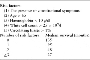
The IPSS defines five adverse prognostic factors at diagnosis. The scoring system is defined in Table 4. As shown, advancing age has a significant negative impact on prognosis. Transformation to acute myeloid leukaemia occurs in a few patients and is associated with a poor prognosis. This complication is associated with high treatment-related mortality and median survival of only 2.5 months.Reference Mesa, Li, Ketterling, Schroeder, Knudson and Tefferi28
International Prognostic Scoring System for PMF

Chronic myeloid leukaemia (CML)
CML is a clonal disorder of the pluripotent stem cell. The presence of the Ph chromosome, which causes CML, results in increased myeloproliferation and differentiation with a loss of cellular response to normal apoptotic signals.
Incidence
The incidence of CML is approximately 1–2 per 100,000. It can occur at any age, but is most commonly diagnosed in the fifth or sixth decade of life. There is a slight male predominance.Reference Swerdlow, Campo and Harris2
Clinical features
Some patients are asymptomatic at presentation and the diagnosis is made incidentally after an unexpected high white blood cell count is found. Symptomatic patients present with weight loss, lethargy and night sweats. Splenomegaly is often present and abdominal pain, due to capsular splenic stretch, may be another presenting feature. Occasionally symptoms of gout or reduced visual acuity and headaches may occur due to whole blood hyperviscosity.
Approach to diagnosis
A careful history may elicit constitutional symptoms or the symptoms of hyperviscosity, described above. Physical examination frequently reveals splenomegaly. Lymphadenopathy is not a feature of CML. Retinal changes with haemorrhage and exudates are present in some patients who present with very high white cell counts.
Inspection of the peripheral blood film typically reveals numerous neutrophils and myelocytes (immature neutrophil precursors). Basophilia is invariably present, eosinophilia and thrombocytosis can also be seen. It is important to note that an isolated thrombocytosis may be the only presenting feature of CML. Similarly, an unexplained basophilia or eosinophilia should prompt a request for peripheral blood film inspection to see if other features of CML are present.
Once the diagnosis is suspected, cytogenetic testing of bone marrow or peripheral blood can confirm or refute the presence of the Philadelphia chromosome, which is almost invariably present in CML. The BCR-ABL gene is the product of the classic Philadelphia chromosome t(9;22) translocation, and can be detected using PCR (polymerase chain reaction) techniques.29 A bone marrow biopsy has typically always been performed at diagnosis, but expert opinion increasingly suggests this is no longer required as genetic studies on peripheral blood can confirm the presence of the BCR-ABL gene.
Therapy and complications
Treatment of CML has been revolutionized in the last 10 years by the use of tyrosine kinase inhibitors (TKIs), such as imatinib, which is now the standard first-line treatment. In our view, this represents the most successful translation to date of the understanding of oncogenesis to therapeutics. Bone marrow transplantation used to be considered a standard treatment, but has become virtually obsolete with the emergence of the TKIs. The possibility of ‘cure’ with these oral agents alone is now being tentatively considered.
TKIs directly target the abnormal BCR-ABL gene fusion protein and its dysregulated tyrosine kinase activity. Prior to the use of TKIs the median survival in CML was 4–5 years, but the IRIS study (International Randomized Study of Interferon and STI571) demonstrated the improved cytogenetic and haematological response in patients treated with imatinib rather than standard chemotherapeutic agents.Reference O'Brien, Guilhot, Larson, Gathmann, Baccarani, Cervantes, Cornelissen, Fischer, Hochhaus, Hughes, Lechner, Nielsen, Rousselot, Reiffers, Saglio, Shepherd, Simonsson, Gratwohl, Goldman, Kantarjian, Taylor, Verhoef, Bolton, Capdeville and Druker30 Five-year follow-up of this study has shown 89% 5-year survival, and this figure continues to improve.Reference Druker, Guilhot, O'Brien, Gathmann, Kantarjian, Gattermann, Deininger, Silver, Goldman, Stone, Cervantes, Hochhaus, Powell, Gabrilove, Rousselot, Reiffers, Cornelissen, Hughes, Agis, Fischer, Verhoef, Shepherd, Saglio, Gratwohl, Nielsen, Radich, Simonsson, Taylor, Baccarani, So, Letvak and Larson31 Over 20% of patients included in the IRIS study were over the age of 60, though patients over the age of 70 years were excluded.
Imatinib is also well tolerated. Serious toxicities are rare, but oedema (peripheral and peri-orbital) is seen in up to 60% of patients, and gastrointestinal side-effects (including nausea and diarrhoea) in approximately half of patients. Cytopenias are common.
Prognosis
Untreated, CML will ultimately proceed from a ‘chronic phase’ to ‘accelerated phase’ and then ‘blast phase’ which is equivalent to acute leukaemia.
Most patients present in chronic phase. A rising white cell count, progressive splenomegaly, and the appearance of increasing numbers of blast cells may herald disease progression. Accelerated and blast phase CML carry a poorer prognosis. The Sokal prognostic scoreReference Sokal, Cox and Baccarani32 defines spleen size, platelet and peripheral blood blast count as the relevant prognostic features. Older age is also associated with a poorer prognosis. In the TKI era, the Sokal score remains relevant, as it appears to influence the probability of achieving complete cytogenetic and major molecular response to treatment.Reference Goldman33
Future approaches to treatment
Recently, improved understanding of the pathogenesis of MPDs has raised expectations that targeted therapy may improve the outcome, morbidity and mortality associated with these diseases. The importance of abnormalities in the JAK-STAT pathway, and particularly the prevalence of the JAK2V617F mutation in MPDs, has led to great interest in the development of JAK 2 inhibitors.
Several JAK inhibitors are currently in clinical trial and have demonstrated some disease-modifying effects. In PMF, for instance, treatment with JAK inhibitors has reduced the systemic effects of the disease and some reduction in the degree of splenomegaly has been shown. Tolerability has, however, been limited by myelosuppression. The duration of responses has been relatively short and no impact on overall survival has yet been demonstrated. In some cases, cessation of treatment has been associated with a rapid return of constitutional and disease-related symptoms. Given the long natural history of PV and ET, any useful therapy will need to be efficacious in the short term but will also require a good long-term safety profile.Reference Chen and Prchal34
As primary and secondary myelofibrosis are incurable, progressive illnesses associated with an inextricable decline in marrow function, allogeneic HSCT can be considered as a treatment option. Reduced intensity HSCT can be considered as a potentially curative therapy for these diseases but, due to treatment-related toxicity, will only rarely be considered for patients over the age of 65–70 years. The procedure is associated with a significant mortality, morbidity and relapse risk giving 5-year disease-free survival rates of around 30%.Reference Stewart, Pearce, Kirkland, Bloor, Thomson, Apperley, McQuaker, Marks, Craddock, McCann, Russell, Cook and Kottaridis35
Conclusion
Elevated red cell, white cell and platelet counts are common clinical findings usually with a clear precipitant. In the minority of patients, however, the cause is MPD.
MPDs become more common with increasing age. They are characterized by bone marrow hypercellularity and a risk of progression to acute leukaemia or marrow fibrosis. Patients with MPD are at increased risk of both thrombosis and haemorrhage.
For most patients treatment is supportive, aimed at controlling but not curing the disease and minimizing the risk of complications. Venesection, cytoreductive drugs and anti-platelet therapy are the mainstays. The prognosis for patients with CML has dramatically improved with the use of TKIs as oncogene-targeted therapy. Recent advances in our understanding of the genetic pathogenesis of the remaining MPDs, has allowed JAK2 mutation testing to become part of the diagnostic process for MPD and raises expectations that targeted drugs could be developed to improve the natural history of the remaining MPDs.
Conflicts of interest
The authors declared no conflicts of interest.