Significant outcomes
∙ Compared to healthy volunteers, patients with anxious depression had significantly less activation in several neural networks during a working memory task condition with the highest level of demand.
∙ Whole group differences in research may be driven, in part, by depression subtypes (e.g. anxious depression).
∙ MEG may be a useful modality for neurobiological exploration of mental illness.
Limitations
∙ The analysis was post-hoc in nature.
∙ Anxious depression can be defined many other ways.
∙ The results of this small study are preliminary and require replication.
Introduction
Major depressive disorder (MDD) is a common, heterogeneous disease with many subtypes, including anxious depression (Reference Rush1). When defined dimensionally – using Diagnostic and Statistical Manual of Mental Disorders (DSM) criteria of a diagnosis of MDD (2) plus concurrent high levels of anxiety (i.e. baseline Anxiety/Somatisation Factor Score of ≥7 from the Hamilton Depression Rating Scale; HAM-D) (Reference Cleary and Guy3) – ~50% of patients with MDD have anxious depression (Reference Fava, Rush and Alpert4). Studies have suggested that anxious depression may represent a more severe illness than nonanxious depression, and has a distinct clinical profile that typically includes a longer duration of current episode, higher endorsement of melancholic symptoms, and more medical co-morbidities; poorer treatment outcomes have also been observed (Reference Fava, Rush and Alpert4–Reference Papakostas, Fan and Tedeschini7). Even with an initial positive response to medications or therapy, patients with anxious depression often do not have a sustained response, nor do they generally achieve sustained remission (Reference Fava, Rush and Alpert4,Reference Farabaugh, Alpert and Wisniewski8–Reference Wu, Chen and Yuan13). In addition, anxious depression patients may be at risk for a greater medication side effect burden compared with depressed patients without anxiety (Reference Fava, Rush and Alpert4,Reference Farabaugh, Alpert and Wisniewski8,Reference Wu, Chen and Yuan13–Reference Thase, Demyttenaere, Earley, Gustafsson, Udd and Eriksson15), further emphasising the clinical importance of this depression subtype.
Despite the clinical heterogeneity of depressive illnesses, depressed patients are often studied as a homogeneous group in the research literature. However, based on clinical presentation, treatment response, and long-term outcome, anxious depression represents a clinically distinct subtype of depression (Reference Ionescu, Niciu, Mathews, Richards and Zarate16). Perhaps more importantly, anxiety is a pervasive symptom in a large proportion of MDD patients, and thus this clinical feature may disproportionally influence the differences observed between depressed patients and healthy volunteers in research. There is a notable paucity of studies examining whether specific subtypes of depression (i.e. anxious depression) may underlie the neurobiological characteristics attributed more broadly to all patients with depression. This is a critical question, specifically because distinguishing various subtypes of depression from one another may lead to improved diagnosis and treatment of depressive illnesses, and ultimately refine the ongoing search for the pathophysiological substrates of depression.
Neurobiological explorations are crucial to the more careful characterisation of depressive subtypes. In particular, magnetoencephalography (MEG) is a functional neuroimaging technique that provides improved localisation of neural activity compared with electroencephalography, and better temporal resolution than functional magnetic resonance imaging (fMRI), and is useful for studying the neurobiology of mental illness. Beta-band activity is related to cognitive processes during performance of the N-back working memory task (Reference Deiber, Missonnier and Bertrand17–Reference Pesonen, Hamalainen and Krause19), and can be measured by MEG. Under basal conditions, a baseline level of beta power is present, reflecting populations of neurons firing in synchrony. Under conditions requiring cognitive functions, beta power decreases as neuronal populations fire asynchronously in response to task demands. In other words, beta-band frequency power has been shown to decrease (suggesting increased desynchronisation, and therefore increased activation of neural networks) as memory load increases (Reference Pesonen, Hamalainen and Krause19,Reference Brookes, Wood and Stevenson20). Indeed, the N-back task is a useful tool for studying working memory in depression, particularly because depressed patients have previously been found to have a wide range of executive dysfunctions (Reference Austin, Mitchell and Goodwin21), including working memory (Reference Harvey, Le Bastard and Pochon22–Reference Xu, Lin and Rao24).
Clinically, cognitive impairments, including memory complaints (Reference Antikainen, Hanninen and Honkalampi25), are common in depressed patients. As such, previous research has used the N-back as a cognitively-demanding task for studying potential biomarkers of treatment response to antidepressants. For example, treatment-resistant patients with MDD who displayed the least pretreatment engagement of pregenual anterior cingulate cortex (as measured by MEG) with increasing working memory load on the N-back task showed the greatest symptomatic improvement within 4 h following a single-infusion of ketamine, an experimental rapidly-acting antidepressant(Reference Salvadore, Cornwell and Sambataro26). Thus, the N-back task may function as a useful tool for studying the neurobiology of depression and its treatments.
Aims of the study
The goal of this post-hoc study was to determine whether individuals with treatment-resistant anxious depression had neurobiological differences (as measured by MEG) in brain response compared with both healthy volunteers and individuals with treatment-resistant nonanxious depression, and whether this subgroup mediated differences between depressed subjects as a whole and healthy controls. Beta-band oscillations were specifically targeted, as they had been previously shown to desynchronise (suggesting increased activation) with escalating memory load specifically associated with this N-back working memory task.
Methods
Participants
Participants were informed about the purpose of the study and risks involved, and gave written consent as approved by the National Institutes of Health Combined CNS Institutional Review Board. Data were analysed from 28 age- and sex- matched healthy volunteers and 30 unmedicated patients with treatment-resistant MDD, currently experiencing a major depressive episode lasting at least 4 weeks and without psychotic features. All data were obtained as part of larger experimental treatment projects and analysed post-hoc. Patients were between the ages of 18 and 65; 18 patients had anxious depression and 12 patients had nonanxious depression. Current MDD diagnosis was confirmed by the Structured Clinical Interview for DSM-IV Axis I Disorders (SCID) (Reference First, Gibbon and Williams27) and an unstructured interview by a psychiatrist. Although anxiety disorders were permitted, MDD was the primary psychiatric diagnosis. Severity of depression was assessed using the Montgomery–Åsberg Depression Rating Scale (MADRS) and the HAM-D; before participation, all patients were required to have a MADRS score of ≥20. In addition, depressed patients had previously failed at least two adequate antidepressant trials and were considered treatment-resistant.
All participants were determined to be in good physical health following physical examination, medical history, laboratory assessment, electrocardiogram, urinalysis, and toxicology screen. Participants were free of comorbid substance use disorders (excluding nicotine or caffeine) for at least 3 months. Exclusion criteria for all patients included any serious unstable medical or neurological disorder or condition, concomitant treatment with psychotropic medications or electroconvulsive therapy in the 2 weeks before testing (5 weeks for fluoxetine), serious suicidal ideation, or psychosis. As part of the research protocol in which they were enrolled, all patients were tapered off of psychotropic medications to ensure medication-free status at baseline, and patients were medication-free for at least 14 days before testing. Females could not be pregnant or breastfeeding. Healthy volunteer status was determined by the same procedures, and healthy subjects were recruited through advertisements. Exclusion criteria for healthy volunteers further included any Axis I diagnosis as determined by SCID-NP (non-patients) or first-degree relative diagnosed with a major psychiatric disorder.
Anxious depression definition
Anxious depression was defined as MDD (currently experiencing a major depressive episode) plus an Anxiety/Somatisation Factor Score of ≥7 on the 17-item HAM-D at baseline screening. Thus, patients were dichotomised as having either anxious depression (Anxiety/Somatisation Factor Score of ≥7) or nonanxious depression (Anxiety/Somatisation Factor Score of <7). This score, derived from a factor analysis of the HAM-D (Reference Cleary and Guy3), comprises six items: anxiety (psychic), anxiety (somatic), somatic symptoms (gastrointestinal), somatic symptoms (general), hypochondriasis, and insight. For both clinical work and research, the HAM-D Anxiety/Somatisation Factor Score has been used by several groups as a way of delineating anxious from nonanxious depressed patients (Reference Fava, Rush and Alpert4,Reference Wu, Chen and Yuan13,Reference Chan, Rush and Nierenberg14), and has been deemed an acceptable measure for identifying anxious symptoms in depression (Reference McClintock, Husain and Bernstein28), making it a clinically relevant objective score that can be used by clinicians for applying research findings into clinical practice.
N-back task
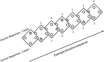
We used a modified N-back task, which is a parametric working memory task that requires monitoring of sequentially presented stimuli, as well as maintaining information until recall (Reference Fletcher and Henson29). During MEG scanning, participants completed the N-back task at three difficulty levels (0-back, 1-back, and 2-back). Task details are provided in Fig. 1. In this modified N-back task, participants were asked to recall a number (1, 2, 3, or 4) presented N number of trials ago, regardless of the number currently being presented on the screen. Each stimulus was presented for 0.5 s duration with an inter-trial interval of 1.8 s. Following a practice run, participants performed a run of 18 blocks, lasting 22 s each, repeating in order between 0-back, 1-back, and 2-back conditions; 11 stimuli were presented per run. Because of the inherent nature of the task, no responses were recorded for the first two stimuli of the 2-back or the first stimulus of the 1-back; therefore, only the last nine stimuli were analysed for all conditions so that there were an equal number of trials for all task conditions. A total of 54 trains were recorded and analysed for each of the three conditions. Accuracy and reaction times were recorded.
Participants completed the N-back task at three difficulty levels (0-back, 1-back, and 2-back) while undergoing magnetoencephalography. In this modified N-back task, participants were asked to recall a number (1, 2, 3, or 4) presented N number of trials ago, regardless of the number currently being presented on the screen. Increasing N levels represent tasks of increasing difficulty.

Data acquisition, source power analysis, and processing
MEG recordings were acquired in a magnetically-shielded room on a 275 sensor whole-head CTF system (CTF Systems, Coquitlam, Canada) and digitised at 1200 Hz with 0–300 Hz bandwidth. Background noise was removed with active noise cancellation using synthetic third-gradient balancing. Throughout the scan, head position was monitored using fiducials placed on the nasion, and the left and right preauricular points. Trial data was discarded if subjects moved >5 mm. Co-registration and source localisation was achieved with T1-weighted anatomical magnetic resonance imaging (MRI) scans obtained at a separate session on a 3T General Electric (GE) scanner (Milwaukee, WI, USA); MRI visible fiducials were placed in the same locations as for MEG scanning in most cases (verified with photographs) to facilitate registration. Structural MRIs were acquired as part of several imaging studies, and were not all acquired on the same scanner. Parameters were (TR=4.96 ms, TE=2.05 ms, inversion time=725 ms, FA=12, resolution 0.94×0.94×1.5 mm) or similar.
Scans were preprocessed using Analysis of Functional NeuroImages software. The 3dSkullStrip routine was used to remove non-brain matter and calculate the surface representing the inner skull. The three fiducial points were marked on the images, and the inner skull surface was reoriented to the fiducial plane and used for source analysis. The images were transformed to Talairach space, and the transformation matrices were retained to apply to the MEG contrast images. The MEG recordings were visually inspected and responses overlapping with stimuli or occurring during artifacts were removed. Source localisation via synthetic aperture magnetometry (Reference S. E. Robinson30) was used to reconstruct oscillatory power from the beta-band (14–30 Hz) in source space on a 5 mm isotropic grid. Following the calculation of signal covariance across all sensors, weights were calculated (using a Nolte realistic head model for the forward solution)(Reference Nolte31) specific to each load condition (0-back, 1-back, 2-back) for estimation of the oscillatory power at each voxel in source space.
In order to determine weights at each point in source space, the total source power from all other points was minimised. A data window of −0.75 to 0.5 s around each response was used to calculate beamformer weights, and this window was increased to −0.75 to +0.75 s around the responses if the shorter window produced artifactual results. Responses were collected via a button box; pressing one of the response buttons caused a trigger to be placed in the MEG file. A thresholding algorithm was used to detect each button press, and all marks were visually assessed to ensure accurate placement. Because we wanted to see changes in power most closely connected to the response itself, power was calculated around a window of −0.25 s before the response (when the subject is presumably deciding upon their response), and 0.25 s after the response. All measurements of oscillatory power were taken between two cognitive load conditions, and Mann–Whitney U maps were created of power differences across the following conditions: 2-back versus 1-back, 2-back versus 0-back, and 1-back versus 0-back.
For each of the contrasts identified above, a t-test was conducted between all MDD patients (anxious and nonanxious) compared with healthy volunteers. Only those contrasts that produced significant results were assessed among the three groups [significance was defined based on whole brain correction using false discovery rate (FDR), p<0.05]. For contrasts in which healthy volunteers and all MDD subjects differed, a mask was created and used to limit the search volume of subsequent comparisons among sub-groups, for which three contrasts were conducted (anxious depressed vs. healthy volunteers, nonanxious depressed vs. healthy volunteers, anxious depressed vs. nonanxious depressed). Significance for sub-group comparisons were defined using an FDR to control for multiple comparisons (p<0.05, two-tailed) based on small volume correction (SVC) defined by the extent of the mask. This post-hoc procedure was intended to determine the effects of the anxious depressed subgroup on the overall results between all MDD subjects and healthy volunteers. While a preferable approach would be to compare the three subgroups directly, we were underpowered to detect differences in this manner. Furthermore, it should be stated that by using this statistical approach, we can only state which significant differences between healthy and depressed patients are mediated by those patients with significant anxiety. We can make no statement regarding subgroup differences in other brain regions.
Performance accuracy and reaction time data were examined for each level of task difficulty (0-back, 1-back, and 2-back) and each group (healthy volunteers, patients with anxious depression, and patients with nonanxious depression). The main effects and their interaction were evaluated using repeated measures analysis of variance, where task difficulty was a within-subjects factor. In the presence of a significant interaction or main effect, and because two outcomes were evaluated, Bonferroni corrected simple contrasts were used to further characterise the effects. Significance was considered at p<0.025, two-tailed. All behavioural data p values are reported unadjusted.
Results
Demographic results
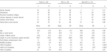
Demographic information for patients is given in Table 1. No significant differences were observed between healthy volunteers and MDD patients (both with and without co-morbid anxiety) with regard to age (38.7 years vs. 43.3 years, respectively; df=31, p=0.38) or gender (13 females vs. 10 females, respectively; p=0.31).
Demographics

AD, anxious depression; HAM-A, Hamilton Anxiety Rating Scale; HAM-D, Hamilton Depression Rating Scale; HAM-D A/S, Hamilton Depression Rating Scale Anxiety/Somatisation Factor Score; MADRS, Montgomery–Åsberg Depression Rating Scale.
* Statistically significant for p<0.05.
Anxious depressives had significantly higher scores on total HAM-D17 (24.4±5.3 vs. 19.7±2.8; F=6.408, p=0.003) and HAM-D Anxiety/Somatisation Factor Score (8.3±1.7 vs. 5.4±0.67; F=3.028, p=0.000), as well as higher total MADRS scores (35.4±5.5 vs. 30.7±3.8; F=3.513, p=0.01) at baseline than those with nonanxious depression. However, no significant differences were noted between patients with and without anxious depression with regard to demographic characteristics, total Hamilton Anxiety Rating Scale score (23.4±5.2 vs. 19.7±5, respectively; F=0.012, p=0.07), or lifetime diagnosis of anxiety disorder (nine vs. eight, respectively; p=0.37).
Behavioural results
As expected, (Reference Callicott, Mattay and Bertolino32) increasing working memory load (i.e. increasing task difficulty) was associated with decreased accuracy across subjects (F=36.24, df=2,110, p<0.001); accuracy was higher in the 0-back condition compared with the other conditions (1-back: t=5.24, p<0.001; 2-back: t=7.10, p<0.001), and significantly higher accuracy in the 1-back condition than the 2-back condition (t=4.59, p<0.001). No significant group effect (F=1.53, df=2,55, p=0.22) or interaction between group and task difficulty (F=1.60, df=4,110, p=0.18) was observed in conjunction with performance accuracy.
For reaction time, there was a significant main effect of group (F=4.34, df=2,55, p=0.018), but the group X task difficulty interaction did not remain significant following correction for multiple comparisons (F=2.82, df=4,110, p=0.028). The healthy volunteers (mean=0.483, SE=0.024) were significantly faster than patients with nonanxious depression (mean=0.606, SE=0.037; t=2.77, p=0.023), but not those with anxious depression (mean=0.557, SE=0.030; t=1.90, p=0.19).
MEG results
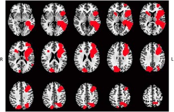
No significant difference in beta-band power was detected between the MDD patients and healthy volunteers for the 1-back versus 0-back contrast, or the 2-back versus 0-back contrast. Significant differences in beta-band power were observed between MDD patients and healthy volunteers in the 2-back versus 1-back contrast; a mask identifying the brain regions that differed between the combined MDD group (combined anxious and nonanxious patients) versus healthy volunteers is shown in Fig. 2 (FDR, p<0.05, two-tailed) and included nonspecific regions of the precuneus, cuneus, and insula extending into the inferior and middle frontal cortex.
This mask identified the brain regions that significantly differed between patients with major depressive disorder (both anxious and nonanxious patients) versus healthy volunteers (false discovery rate, p<0.05) during the 2-back versus 1-back contrast.

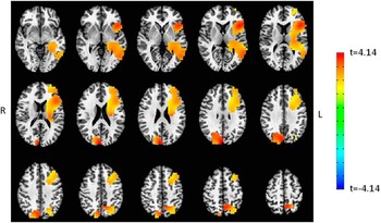
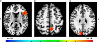
The group comparisons conducted within the 2-back versus 1-back condition found lower beta power (indicating increased desynchronisation, and therefore more activation) in healthy volunteers compared with patients with anxious depression (SVC; FDR, p≤0.05) throughout regions from the mask, including the left precuneus, bilateral cuneus, and left insula extending into the inferior and middle frontal cortex (Fig. 3). Given that these regions are large and nonspecific, p (FDR corrected) was further reduced to 0.025 to yield discrete regions, located specifically (based on centre of mass) in the left insula extending into the inferior frontal cortex, left precuneus, and right cuneus (Talairach coordinates (Reference Talairach33) were: left insula/inferior frontal cortex (37 voxels; x=−45.9, y=+19.6, z=+11.5); right cuneus (17 voxels; x=+9.4, y=−82.0, z=+28.9); left precuneus (33 voxels; x=−5.6, y=−55.6, z=+51.7) (Fig. 4).
Specific major depressive disorder group comparisons (patients with anxious depression vs. healthy volunteers and patients with nonanxious depression vs. healthy volunteers) within the 2-back vs. 1-back condition. Increased desynchronisation (more activation/less beta power) was observed in healthy volunteers compared with patients with anxious depression (small volume correction; false discovery rate, p≤0.05) throughout regions included in the mask, including the left precuneus, bilateral cuneus, and left insula extending into the inferior and middle frontal cortex.

Further reducing p to 0.025* was done in order to yield discrete regions in significant regions, located specifically (based on centre of mass) in the left insula extending into the inferior frontal cortex (a), left precuneus (b), and right cuneus (c). *False discovery rate applied.

No significant difference in beta-band power was detected when comparing healthy volunteers and patients with nonanxious depression, or when comparing patients with anxious and nonanxious depression.
Discussion
This MEG study is the first to compare activation patterns during a working memory N-back task in patients with anxious depression versus those with nonanxious depression and healthy volunteers. The results indicate widespread decreased beta-band power (indicating increased desynchronisation/more activation) between healthy and depressed subjects, in general, during a demanding working memory task. Post-hoc subgroup analysis found that, specifically, healthy volunteers showed increased activation compared with the anxious subgroup in the left precuneus, right cuneus, and left insula extending into the inferior and middle frontal cortex during the more demanding condition of the N-back working memory task, relative to a less demanding condition. All of these regions previously have been implicated in working memory or visual processing/visuospatial attention(Reference Brookes, Wood and Stevenson20,Reference Hahn, Ross and Stein34–Reference Pernet, Franceries, Basan, Cassol, Demonet and Celsis36). Note that no significant differences were found in beta-band activity between anxious depressives and nonanxious depressives, nor between nonanxious depressives and healthy volunteers. This suggests that results from anxious depressives preferentially drove the differences seen between the combined MDD group and healthy volunteers.
Healthy volunteers showed an increased activation of the left precuneus region in association with increasing task demands that was not observed in anxious depressives; this may suggest that anxious depressives use maximum levels of neural response in this area to working memory demands at lower difficulty levels. The precuneus was previously implicated in working memory and found to activate consistently during performance of the N-back task (Reference Owen, McMillan, Laird and Bullmore35). Similarly, healthy volunteers showed an increased activation of the right cuneus region during increasingly difficult N-back testing (between the 1-back vs. 2-back conditions) compared with anxious depressives. Interestingly, the cuneus has been implicated in visual processing and visuospatial attention (Reference Hahn, Ross and Stein34), and the right cuneus has specifically been implicated in tasks involving structural-visual memory representations (Reference Pernet, Franceries, Basan, Cassol, Demonet and Celsis36). Researchers previously suggested that ceiling levels of neural response are reached during working memory tasks when the brain is not able to generate additional activation as task difficulty is further increased (Reference Schneider-Garces, Gordon and Brumback-Peltz37). Indeed, our findings suggest that anxious depressives may reach ceiling level of neural response in these regions at the 1-back task, which has a smaller working memory load than the more demanding 2-back task. Of note, while one may expect to see larger differences between the 0-back and 1-back or the 0-back and 2-back, the 0-back task condition has no memory requirement and thus is a qualitatively different task and basically functions as a sensory motor control condition. The 1-back versus 2-back comparison is the only contrast between memory loads.
Other significant effects were found in the left insular region, extending into the inferior and middle frontal cortex, where healthy volunteers again showed more activation than the anxious depressives. Previous MEG studies found that beta-band power decreases (i.e. more activation) in the middle frontal cortex as memory load increases in the N-back task in healthy volunteers (Reference Brookes, Wood and Stevenson20). This finding may be particularly important for the study of depression, as MDD patients often report memory difficulties (2).
However, despite finding neuronal activation deficits in the left precuneus, right cuneus, and left insula extending into the inferior and middle frontal cortex in anxious depressives, working memory performance deficits were not observed in anxious depressives compared with healthy volunteers. In fact, patients with nonanxious depression were significantly slower than healthy volunteers and slower at a trend level compared with anxious depressives during the task with the highest difficulty (2-back task). One possible explanation for this is that anxious depressives may use different cognitive strategies at higher task levels. As predicted by the valence-arousal hypothesis(Reference Heller38,Reference Heller, Etienne and Miller39), anxiety alone causes a hyperarousal state and depression alone causes a hypoarousal state. Perhaps the hypoarousal of pure depression is offset by the anxiety factor in anxious depression. Thus, the anxiety component of anxious depression would cause a hyperarousal to counterbalance the predicted hypoarousal of pure depression. In keeping with this hypothesis, anxious depressives performed faster than nonanxious depressives during the 2-back condition, though only at the trend level. In contrast, those patients with nonanxious depression would exclusively have a hypoarousal component to their illness, as reflected in a slower mean reaction time associated with the task, particularly at the more difficult 2-back level. The fact that anxious depressives and healthy volunteers had detectable activation differences on MEG scanning, but no such differences were reflected in the behavioural/performance data, suggests a compensatory mechanism in the combined anxiety-depression state that allows patients to maintain task performance despite reaching maximal neuronal activation at lower task difficulties.
This study was associated with several limitations. First, all depressed patients were participants in a larger medication trial for treatment-resistant depression that required them to have an inadequate response to two past antidepressant trials. As a result, the patients in this exploratory MEG study represent a cohort with severe depression that may not be characteristic of a typical depression population. Second, although we can extrapolate the functions of particular brain areas of interest from previous studies, definitive functions are still largely unknown. Brain function is often deduced from increased blood-oxygen level dependent (BOLD) signalling during fMRI tasks; as the blood-oxygen level increases in a certain region, it is assumed that this region is ‘active’ during the task. However, this method has flaws, and further techniques to elucidate function of brain regions are necessary to confirm information from BOLD signals. Third, statistical power was limited by the small sample size. The lack of statistically significant MEG findings between anxious and nonanxious depressives and between nonanxious depressives and healthy volunteers may be a result of limited statistical power. Because of this small sample size, we were limited in the statistical methods that we could apply. The findings we report can only be interpreted as areas where group (i.e. patients with depression vs. healthy volunteers) differences are mediated by patients with significant anxiety.
The heterogeneity that exists for defining anxious depression throughout the literature cannot be ignored. Here, we used DSM-diagnosed MDD plus the Anxiety/Somatisation Factor Score ≥7 from the HAM-D17 to define anxious depression dimensionally; however, this is only one way of defining anxious depression (Reference Ionescu, Niciu, Henter and Zarate40), and some may perceive this specific definition as a limitation. Nevertheless, the dimensional definition used here has been shown to successfully differentiate anxious depression from nonanxious depression clinically in larger samples, including the Sequenced Treatment Alternatives to Relieve Depression study(Reference Fava, Rush and Alpert4–Reference Wu, Chen and Yuan13), lending support to our hypothesis that there may be neurobiological differences between anxious and nonanxious depressives. It should be noted that the current literature on anxious depression uses many different definitions; this not only makes comparisons between studies difficult (Reference Ionescu, Niciu, Mathews, Richards and Zarate16,Reference Ionescu, Niciu, Henter and Zarate40), but it also emphasises the need for standardised dimensional criteria. Further study into an appropriate consensus definition would facilitate our ability to clarify the neurobiology associated with anxious depression.
Moreover, we must emphasise the need for additional studies, as these results are highly preliminary in nature and in need of replication. Specifically, future study designs would benefit from including an additional patient comparison group with categorical anxious depression (that is, those meeting DSM criteria for both MDD and an anxiety disorder). Future studies may also consider using a treatment-responsive population, as opposed to a treatment-resistant group, to determine if group differences remain between anxious depressives and healthy volunteers. Certainly, replication studies conferring our results of different activation patterns in healthy volunteers compared with anxious depressives would make the possibility of a Type 1 error in our results less likely.
Despite these limitations, overall our results provide preliminary evidence for a lack of activation in certain brain areas associated with memory and attention in anxious depressives. In particular, the results demonstrate the importance of evaluating the role of clinically relevant diagnostic subtypes when trying to understand the underlying neurobiology of MDD. Here, we found that the significant group differences observed when comparing a heterogeneous group of patients with MDD with healthy volunteers were primarily driven by the patient subgroup with anxious depression. Given the frequency of this diagnostic subtype, anxious depressed patients may prove to be a potentially more biologically homogenous subtype within MDD, and may be the reason behind other differences reported in the literature between patients with MDD and healthy controls. Further neurobiological explorations of anxious depression are necessary to determine the extent to which this subgroup has preferentially influenced our understanding of the underlying neurobiology of depression.
Acknowledgements
Ioline Henter, M.A., National Institute of Mental Health, provided invaluable editorial assistance. Ms. Henter reports no potential conflicts of interest. Dawn Ionescu; Maura Furey: conception, analysis, interpreting the data, drafting and revising, and final approval of submission. Allison Nugent; David Luckenbaugh: analysis and interpretation of the data, drafting and revising the manuscript, and final approval of submission. Mark Niciu; Erica Richards: interpretation of the data, drafting and revising the manuscript, and final approval of submission. Carlos Zarate: conception, design, and acquiring the data, as well as interpreting the data, drafting and revising, and final approval of submission.
Financial Support
Funding for this work was supported by the Intramural Research Program at the National Institute of Mental Health, National Institutes of Health (IRP-NIMH-NIH), by a NARSAD Independent Investigator to C.A.Z., and by the Brain & Behavior Mood Disorders Research Award to C.A.Z. Dr. Zarate is listed as a co-inventor on a patent application for the use of ketamine and its metabolites in major depression. Dr. Zarate has assigned his rights in the patent to the US government but will share a percentage of any royalties that may be received by the government. Dr. Furey is listed as a co-inventor on a patent application for the use of scopolamine in psychiatric disorders. Dr. Furey has assigned his rights in the patent to the US government but will share a percentage of any royalties that may be received by the government. The remaining authors have no conflicts of interest to disclose, financial, or otherwise.
Conflicts of Interest
None.
Ethical Standards
The authors assert that all procedures contributing to this work comply with the ethical standards of the relevant national and institutional committees on human experimentation and with the Helsinki Declaration of 1975, as revised in 2008.