I. INTRODUCTION
Nanostructured materials based on layer-structured transition metal chalcogenides have found diverse applications in electronic, optical, and semiconductor devices. Reference Gao, Xu, Jiang and Yu1 Most of the useful properties of layered structures arise from the highly anisotropic physical properties which are due to the weak interaction between layers consisting of a monolayer of metal atoms surrounded by covalently bonded chalcogen atoms. Reference Benedik2 In this context, antimony sulfide (Sb2S3), a direct band gap semiconductor (V-VI group) with orthorhombic crystal structure is a promising functional material for its good photovoltaic properties, Reference Zhu, Gong, Zhang, Yong and Xiang3,Reference Shuai and Shen4 broad spectral response, Reference Li, Huang and Lin5 and high thermoelectric power. Reference Ibuki and Yoshimatsu6 Utilizing its unique properties Sb2S3 has been used in solar energy conversion, Reference Zhang, Ge, Yang, Zhou, Chen, Li and Liu7,Reference Senthil, Muthukumarasamy and Kang8 photocatalysis, Reference Sun, Li, Li, Chen, Chen, He and Fu9 thermoelectric cooling technologies, Reference Rajpure, Lokhande and Bhosele10 and optoelectronic devices in IR region. Reference Cao, Gu, Zhuge, Gao, Wang and Wu11,Reference Lazcano, Nair and Nair12 It is well documented that the physical and chemical properties of nanostructures are highly influenced by size, shape, crystal structure, and exposed crystal facets of the material. Reference Liang, Wang, Zhuang, Chen, Wang and Li13 In recent years, one-dimensional materials have been the focus of intensive research due to their distinct physical properties determined by their special shapes and are being increasingly explored for energy application and other optoelectronic devices. Reference Xia, Yang, Sun, Wu, Mayers, Gates, Yin, Kim and Yan14,Reference Yang, Lei, Fu, Tang and Mann15 Considering the technological importance, synthesis of morphology controlled Sb2S3 nanocrystals with high yield has drawn much attention over the past few years.
Alemi et al. reported the preparation of Sb2S3 nanorods by complex decomposition approach via hydrothermal method. Reference Alemi, Hanifehpour and Joo16 Belts, tubes, and nanowires of Sb2S3 were prepared by thermal decomposition of antimony molecular precursors (xanthate) followed by variation in crystallization duration. Reference Han, Sun, Sun, Zhu and Wang17 Zhu et al. prepared monocrystalline Sb2S3 nanoparticles with flower-like and rod-like morphologies by a low temperature surfactant mediated refluxing approach. Reference Zhu, Gong, Zhang, Yong and Xiang3 Recently, Zhang et al. reported the synthesis of 1D Sb2S3 nanostructures by hydrothermal and solvothermal methods at 180 °C without using capping agents. Reference Zhang, Hu, Ding and Lin18 PVP assisted hydrothermal synthesis of Sb2S3 nanowire bundles and their performance in DSSC was reported by Zhang and his collaborators. Reference Zhang, Ge, Yang, Zhou, Chen, Li and Liu7 However, many of the aforementioned techniques do not result in phase pure orthorhombic Sb2S3 and control of particle size, shape, and dispersion.
In the present work we report the growth of Sb2S3 nanorods by hydrothermal method at relatively low temperature (150 °C) and their morphological assemblage with the use of PVP as capping agent. The effect of PVP concentration on the morphology of hydrothermally processed Sb2S3 has not been reported earlier. We observed that while individual nanorods of high aspect ratio (length/diameter) were formed in the absence of the PVP, dumbbell-like nanorod-bundles were obtained by using PVP as the surfactant. The significance of this work lies in the fact that the Sb2S3 nanostructures assemble from nanorods into dumbbell shaped structures with the addition of PVP which induces certain changes in particle size and optical properties. A possible mechanism is proposed to explain nanorod assembly.
II. EXPERIMENTAL
A. Synthesis
All the reagents used in this experiment were of analytical grade and no further purification process was used. Sb2S3 nanorods were prepared by hydrothermal method as reported elsewhere. Reference Zhang, Ge, Yang, Zhou, Chen, Li and Liu7 Briefly, 0.3 g of SbCl3 (1.3 mM), 0.4 g of thioacetamide (5.27 mM), and different amount of polyvinylpyrrolidone [(C6H9NO) n ; molar mass 40,000], namely 0, 0.2, and 0.4 g, were added into 50 mL of anhydrous ethanol and the mixture was stirred for 20 min under nitrogen bubbling to purge out oxygen. The final pH of the reaction mixture was 3.54 before adding PVP and 3.37 after adding 0.4 g of PVP. The resulting light orange colored precipitate was separated by centrifugation and dispersed thoroughly in 50 mL of deionized water. The whole solution was transferred to teflon-lined stainless steel autoclave, sealed and heated at 150 °C for 24 h. When the autoclave was naturally cooled to room temperature, the Sb2S3 precipitate was separated and washed several times with water and ethanol by centrifugation (9000 rpm, 15 min). Finally, the Sb2S3 samples obtained using 0, 0.2, and 0.4 g PVP were dried at 50 °C for 24 h to obtain dark gray powders and labeled as SbS-0PVP, SbS-2PVP, and SbS-4PVP respectively.
B. Characterization
Powder X-ray diffraction pattern of the samples was recorded on a Rigaku D-Max 2200 X-ray diffractometer (Rigaku Corporation, Woodlands, Texas) with monochromatized Cu Kα radiation (λ = 1.5406 Å). The formation of orthorhombic stibnite phase was further confirmed with a micro Raman spectrometer (Horiba Jobin Yvon HR800, Horiba Scientific, Longjumeau, France) equipped with an Olympus BX41 microscope (Olympus, Center Valley, Pennsylvania) and a thermoelectrically cooled charge-coupled detector. The 632.8 nm line of He–Ne laser was used to excite the samples. Raman shift was calibrated using the Raman line of silica located at 520.7 nm. Filters were used to adjust the laser power on the sample at 0.5 mW. The size and morphology of the samples were studied using field emission scanning electron microscopy (FESEM; HITACHI S-5500, Hitachi Corp., Dusseldorf, Germany) and elemental analysis was carried out using an energy dispersive x-ray detector attached to the FESEM. X-ray photoelectron spectroscopy (XPS) was used to analyze the chemical states of elements in the samples (Model K-alpha, Thermo Fisher Scientific, Waltham, Massachusetts). The XPS data curves are shown with the deconvoluted fitting curve after adjusting to theoretical curves. The peaks were deconvoluted using Shirley type background calculation and fitted using Gaussian–Lorentzian sum function. The experimental binding energies of all the peaks were corrected using C 1s peak at 284.6 eV corresponding to adventitious carbon in addition to the charge compensation by the flood gun in the spectrometer. Nitrogen adsorption–desorption isotherms were collected on a Belsorp-Mini II (BEL Japan, Inc., Toyonaka, Japan) analyzer at 77 K. The sample was degassed at 100 °C for 5 h in vacuum prior to the measurements. The optical properties of the powder samples were evaluated using a Varian Cary UV–Vis spectrophotometer (Agilent, Mulgrave, Australia) equipped with DRA-CA-30I diffuse reflectance accessory.
III. RESULTS AND DISCUSSION
A. XRD results
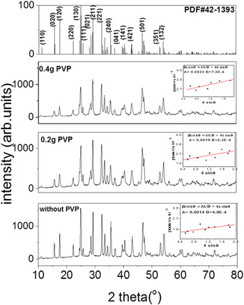
Figure 1 shows the XRD patterns of the Sb2S3 NPs. The reference pattern of orthorhombic mineral stibnite is also included in the figure. For all the samples, the observed XRD peaks could be well indexed to orthorhombic stibnite structure (PDF# 42–1392). It is important to note that the synthesized samples were highly pure as no peaks corresponding to any other phases were observed. The broadening of XRD peaks with the addition of PVP indicates the reduction of crystallite size. The use of PVP for the synthesis of nanomaterials with narrow size distribution has been previously reported, especially for the transition metal colloids. It is an uncharged, water soluble, polar organic polymer that imparts stabilization to colloids through steric effect. Reference Wagner and Köhler19 In the present case we assume that the random polymer chains of PVP attach to the surface of Sb2S3 nuclei during the growth stage suppressing the mean crystallite size.
X-ray diffraction pattern of Sb2S3 particles obtained with different amounts of PVP.

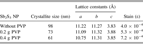
The lattice parameters calculated from the XRD data using Eq. (1) are given in Table I.
 $${d_{hkl}} = {1 \over {\sqrt {{{{h^2}} \over {{a^2}}} + {{{k^2}} \over {{b^2}}} + {{{l^2}} \over {{c^2}}}} }}\quad .$$
Estimated crystallite size, lattice constants and strain of Sb2S3 particles.

The derived lattice parameters of Sb2S3 particles, particularly of PVP-stabilized samples are deviated to some extent from the standard values: a = 11.239 Å, b = 11.313 Å, and c = 3.841 Å [PDF# 42-1392].
From the XRD profile, the crystallite size (D) and the micro strain (ε) of samples can be calculated using the Williamson–Hall method. The full-width of the diffraction line at half maximum intensity (FWHM) and the micro strain are related by the equation:
 $$\beta \cos \theta = {{k\lambda } \over D} + 4\varepsilon \sin \theta \quad .$$
A plot of β cos θ versus sin θ gives a straight line graph; the strain (ε) and the crystallite size (D) are obtained from the slope and intercept of the best straight-line fit respectively. The calculated crystallite size and the micro strain are given in Table I. It can be seen that the crystallite size decreased from 98 nm to 61 nm with the addition of 0.4 g PVP. The increase of micro strain with the addition of PVP is an indication of increased lattice imperfections which has been reflected further in lattice parameter values of PVP-stabilized Sb2S3.
B. Raman study
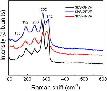
Raman scattering spectroscopy has been recognized as one of the most sensitive techniques to study the structural properties of the material at the atomic scale. Figure 2 shows the Raman spectra of Sb2S3 nanostructures obtained with and without PVP in the reaction medium. Sb2S3 crystallizes in the Pnma space group (#62) with orthorhombic crystal structure which is centrosymmetric. Therefore, the vibrational modes of Sb2S3 cannot be Raman active and IR active at the same time. Reference Liu, Ting, Chua, Sum and Gan20 Theoretically, there are 30 Raman active optical phonon modes at the Γ point of the Brillouin zone:
 $${\Gamma _{{\rm{Raman}}}} = 10{{\rm{A}}_{\rm{g}}} + 5{{\rm{B}}_{{\rm{1g}}}} + 10{{\rm{B}}_{{\rm{2g}}}} + 5{{\rm{B}}_{{\rm{3g}}}}\quad .$$
Raman spectra of Sb2S3 nanostructures confirming the orthorhombic phase.

However, only a few of them have been observed experimentally. Reference Shuai and Shen4,Reference Pilapong, Thongtem and Thongtem21 In the present work, the stibnite phase of Sb2S3 was confirmed in all three samples by the appearance of two strong peaks at 280 and 305 cm−1, the relatively weak peaks at 192 and 238 cm−1 and a weak shoulder at 155 cm−1. Reference Makreski, Petrusevski, Ugarkovic, Jovanovski and Jovanovski22 The Raman spectra of the samples confirm that the PVP does not affect the orthorhombic phase purity of Sb2S3. In comparison to Sb2S3 obtained without using PVP, the samples grown with PVP showed a slight broadening in the Raman bands suggesting the reduction in crystallite size which is in accordance with XRD results. For the samples synthesized with PVP there is a red shift in the Raman peak, which is very pronounced for the sample prepared with 0.4 g PVP; the shift is from 312 cm −1 to 303 cm−1. The peak position most frequently reported in the literature for stibnite Sb2S3 phase is 305 cm−1. The shift can be due to the fact that Sb2S3 prepared with PVP has the stoichiometry more proximate to the ideal value as can be seen in the EDS results.
C. SEM analysis
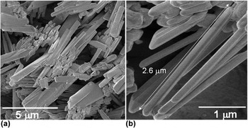
Figure 3 displays FESEM images of Sb2S3 nanorods synthesized without PVP. As shown in Fig. 3(a), long nanorods of high aspect ratio were obtained. The coexistence of some short nanorods can also be seen in the micrograph. The higher magnification of SEM images [Fig. 3(b)] shows that the longer nanorods possess a length of ∼2.5 µm and diameter of ∼70–80 nm of an aspect ratio of ∼35.
SEM image of Sb2S3 nanorods obtained at different magnification; (a) at 5000× revealing the formation of nanorods with low as well as high aspect ratio, and (b) at 30,000× showing the nanorods with an aspect ratio close to 35.

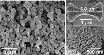
Figure 4 shows the morphology of Sb2S3 particles synthesized using 0.2 g of PVP as the surfactant. Interestingly, the morphology has been significantly changed upon using PVP under otherwise same reaction condition. The formation of dumbbell-shaped bundles can be observed in the low-magnification FESEM image [Fig. 4(a)]. Figure 4(b) displays a higher magnification SEM image of a single dumbbell-shaped architecture which seems to consist of a highly ordered assembly of hundreds of uniform nanorods as the building blocks. The length of the bundles is found to be close to 3 µm [Fig. 4(b)] and the diameter of the bell at each end is around 2 µm. By close inspection of the ends of dumbbell structure it is clear that such shape is formed by the assemblage of many individual rods; the diameter of each rod lies close to 70 nm [Fig. 4(c)].
SEM images of dumbbell shaped Sb2S3 structures captured at different magnifications; (a) shows the general morphology of the product (5000× magnification); (b) shows an individual dumbbell-shaped Sb2S3 particle indicating that this specific structure was formed by oriented attachment of hundreds of individual nanorods (30,000×), and (c) shows the tip/end of the structure formed by uniform nanorods; the diameter of an individual nanorod is approximately 70 nm.

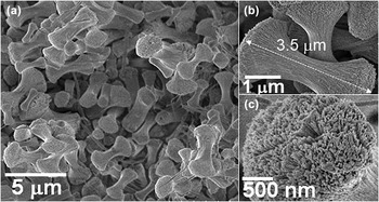
Figure 5 shows typical SEM images of Sb2S3 particles obtained using 0.4 g of PVP. The morphology is very similar to that obtained using 0.2 g of surfactant. A large quantity of uniform, dumbbell shaped and well dispersed nanorod bundles can be observed [Fig. 5(a)]. The high magnification SEM images show that [Figs. 5(b) and 5(c)] the dumbbell shaped structure is truly made of a large number of individual nanorods tightly aligned with each other. The length of such bundle structures is about 3.5 µm and the diameter of the bell at each end is in the range of 1.7–2.0 µm [Fig. 5(c)]. The SEM analysis showed that the addition of PVP contributed a significant change in the morphology of the Sb2S3 particles while the amount of PVP merely has an effect in changing the shape or size of the product. In an attempt to disperse the bundles we sonicated the samples for 3 h, however the SEM images showed that the bundles are intact.
SEM images of Sb2S3 structures synthesized using 0.4 g of PVP: (a) showing an overall distribution of the dumbbell shaped morphology. (b) & (c) High-magnification SEM images showing that each individual dumbbell structure consists of a bundle of nanorods.

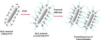
The SEM results suggest that PVP plays the key role in the formation of dumbbell shaped Sb2S3. Similar type of observations were reported by Senthil et al. for the preparation of ball/dumbbell shaped Sb2S3 particles where they added both citric acid and PVP in the reaction medium and the processing temperature was 200 °C 24 h. Reference Senthil, Muthukumarasamy and Kang8 In their work because of the simultaneous addition of both PVP and citric acid it was not clear which of the two agents were truly involved in constructing such a fascinating Sb2S3 architecture. Zhang et al. studied catalytic properties of dumbbell-shaped Sb2S3 nanowire bundles synthesized by PVP assisted hydrothermal processing. Reference Zhang, Ge, Yang, Zhou, Chen, Li and Liu7 However, the synthetic approach is not complete as it did not contain any insight about the morphology aspect of the product without using PVP. In the present work we have performed the synthesis in the absence of PVP and with different amounts of it. Without PVP in the reaction medium we obtained nanorods while the addition of PVP transforms the morphology into dumbbell shape which is the consequence of self-assembly of Sb2S3 nanorods in an ordered fashion. The structural properties of Sb2S3 were not found to be affected by the different amount of PVP. The formation of dumbbell shaped nanorod-bundles can be explained as follows: the hydrothermally synthesized Sb2S3 nanorods are covered with hydroxyl groups making the surface hydrophilic. Reference Wu, He and Jiang23 We speculate that in the presence of PVP the hydrophilic end of PVP (pyrrolidone part) attaches to the surface of Sb2S3 nuclei while the hydrophobic end (polyvinyl part) remains extended facing toward outside which further becomes adhered with more and more PVP-covered nanorods via hydrophobic and van der waals forces giving rise to the formation of nanorod bundles. Reference Seo, Lee, Sun, Suzuki, Mann, Liu, Terashima, Yang, McConnell, Nishimura and Dai24 The proposed PVP induced shape transition of Sb2S3 nanorods is illustrated schematically in Fig. 6.
Schematic representation for the formation of dumbbell shaped architectures starting from the Sb2S3 nanorods as the building blocks.

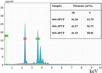
To verify the chemical composition of the samples energy dispersive x-ray spectroscopic (EDXS) study was performed. Figure 6 shows a representative EDXS spectrum of PVP-protected Sb2S3 nanostructures where the emission peaks of Sb and S can be clearly seen. The EDXS composition analysis shown as the inset of Fig. 7 reveals that the stoichiometry of the material has been improved with the addition of PVP approaching the Sb:S atomic ratio close to 2:3.
A typical EDXS spectrum of PVP-protected Sb2S3 particles; inset shows the chemical composition analysis of different powder samples.

D. XPS analysis
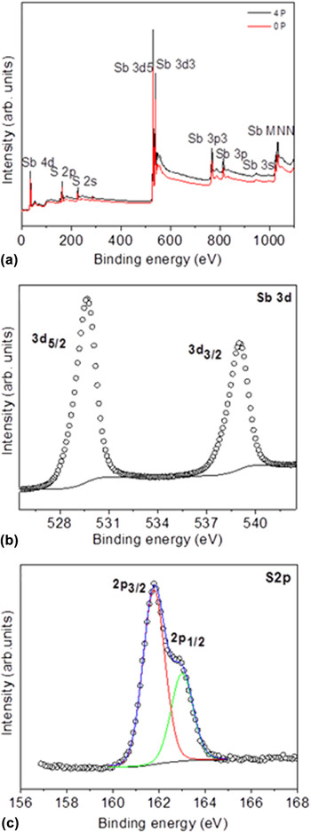
The XPS spectra were obtained to identify the chemical nature of Sb2S3 nanoparticles developed with and without PVP. The spectra were recorded after etching with 3 keV Ar+ ions for 150 s. Figure 8(a) shows the survey scan of Sb2S3 nanoparticles synthesized with and without PVP. The peaks from Sb 4d, 3d, 3p, 3s, and Sb Auger and S 2p and 2s are clearly seen in the spectrum. The high resolution spectra of Sb 3d and S 2p from Sb2S3 without PVP (0P) are shown in Figs. 8(b) and 8(c) respectively. The XPS results obtained from Sb2S3 nanoparticle synthesized with PVP were identical and hence not presented. The two peaks of the Sb core level spectrum at binding energy (BE) values 529.6 eV and 539 eV with a separation of 9.4 eV corresponds to Sb 3d 5/2 and 3d 3/2, respectively [Fig. 8(b)]. The BE values and the separation by spin orbit coupling coincide well with the reported values of Sb3+ in Sb2S3. Reference Xiao, Xu, Ye, Liu, Fu, Li, Chen and Su25 The high resolution spectrum of sulfur core level shows two peaks at 162.98 eV and 161.78 eV which correspond to 2p 1/2 and 2p 3/2 of S2− respectively. The intensity ratio (1:2) and separation (1.2 eV) of the peaks are in good agreement with sulfur ion (S2−). Reference Ota and Srivastava26 The BE values and the spectral characteristics observed for our samples agree well with the reported values of Sb2S3. Reference Zakaznova-Herzog, Harmer, Nesbitt, Bancroft, Flemming and Pratt27 The XRD and XPS results confirm the phase purity of the synthesized Sb2S3 nanoparticles. The atomic ratio of Sb to S calculated was very close to the theoretical ratio of 2:3 for Sb2S3.
XPS spectra of the Sb2S3 nanoparticles: (a) survey scan, (b) high resolution spectra of Sb 3d core level, (c) S 2p core level.

E. BET measurement
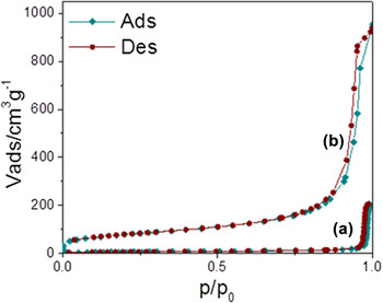
Nitrogen adsorption–desorption isotherms were measured to determine the specific surface area of Sb2S3 nanorods and dumbbell-shaped nanorod bundles and the results are presented in Fig. 9. The isotherm shows a typical type IV curve with a hysteresis loop at relative pressure (P/P o) between 0.5 and 1. BET surface area was increased from 19 m2/g in nanorods to 178 m2/g in the case of nanorod-bundles. The larger surface area can be attributed to the existence of nanopores among the self-assembled individual nanorods.
Adsorption–desorption isotherms for Sb2S3 (a) nanorods and (b) nanorod-bundles.

F. Optical studies
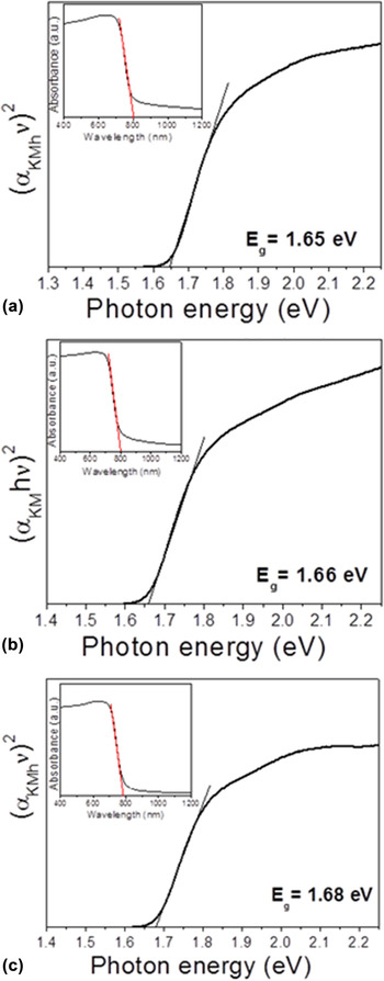
The UV–Vis absorption spectra of Sb2S3 nanorods and dumbbell shaped nanorod bundles were recorded at room temperature in diffuse reflectance mode and are given in the insets of Fig. 10. In all three cases the Sb2S3 particles absorb strongly in the region between 700 and 800 nm with a slight red shift in the absorption edge for the PVP protected dumbbell shaped particles. The reflectance spectra were analyzed and converted into Kubelka–Munk function (αKM) using the following relation:
 $${\alpha _{{\rm{KM}}}} = {{{{\left( {1 - {R_\alpha }} \right)}^2}} \mathord{\left/ {\vphantom {{{{\left( {1 - {R_\alpha }} \right)}^2}} {2{R_\alpha }}}} \right. \kern-\nulldelimiterspace} {2{R_\alpha }}}\quad ,$$
 $${R_\alpha } = {{{R_{{\rm{sample}}}}} \mathord{\left/ {\vphantom {{{R_{{\rm{sample}}}}} {{R_{{\rm{ref}}}}}}} \right. \kern-\nulldelimiterspace} {{R_{{\rm{ref}}}}}}\quad ,$$
where R α is the reflectance of an infinitely thick sample with respect to a reference at each wave length. Band gap energies (E g) of the samples were estimated from the variation of Kubelka–Munk function with photon energy using Tauc’s formula for direct band gap semiconductors:
 $${\left( {{\alpha _{{\rm{KM}}}}h\nu } \right)^n} = A\left( {h\nu - {E_{\rm{g}}}} \right)\quad ,$$
where A is a constant, (hν) is photon energy, and n = 2 for allowed direct transition. The plot of (αKM hν)2 versus (hν) for the powder samples are displayed in Fig. 10. By extrapolating the linear portion of the graph to photon energy axis gives the band gap. The E g values were estimated as 1.65, 1.66, and 1.68 eV for Sb2S3 particles prepared with 0, 0.2, and 0.4 g of PVP respectively. The estimated band gap values lie in the red part of solar spectrum implying that they may be good candidates as absorber materials for solar photovoltaics.
UV–Vis absorption spectra of Sb2S3 powder samples: (a) without PVP, (b) 0.2 g PVP and (c) 0.4 g PVP. The corresponding Kubelka–Munk plots for each sample were inserted as inset to determine the band gap energies associated with direct transition.

IV. CONCLUSIONS
Highly crystalline Sb2S3 nanorods and dumbbell shaped nanorod bundles have been synthesized by hydrothermal process at temperature as low as 150 °C. The use of PVP as surfactant is found to play a crucial role in causing the assembly of Sb2S3 nanorods into well-ordered Sb2S3 bundles. The possible mechanism behind the PVP assisted shape transition has been outlined. The Sb2S3 nanorods have an average diameter and length in the range of 70–80 nm and 2.5–2.6 µm respectively while the uniformly sized dumbbell like bundles constituted by Sb2S3 nanorods have a radial length of ∼3.5 µm. The well-defined XPS peaks at 529.6 and 539 eV corresponding to Sb 3d 5/2 and Sb 3d 3/2 states confirm the +3 oxidation state of Sb in Sb2S3 indicating the phase purity of our samples. The strong light absorption capacity of Sb2S3 particles was observed in the red part of solar spectrum.
ACKNOWLEDGMENTS
The authors wish to thank P.E. Altuzar Coello for the XRD analysis. Authors also acknowledge the projects CeMIE-Sol 207450/28 and PAPIIT IN107815 for supporting with experimental facilities in nanomaterial synthesis. MP acknowledges the projects PRODEP DSA/103.5/15/7449, PROFOCIE-2015, and VIEP 2016 for the financial support in acquiring some experimental setup. Thanks to Dr. Umapada Pal from Instituto de Fisica of BUAP for extending the facilities of BET measurements. This work has collateral application in project PAPIIT IN 113214.