Introduction
Tetralogy of Fallot is the most common type of cyanotic CHD, accounting for 5–10% of CHD. Tetralogy of Fallot belongs to the category of conotruncal malformation. It includes four kinds of malformations: generalised right ventricular outflow tract stenosis, unrestricted ventricular septal defect, aortic saddle (saddle range ≤50%), and secondary right ventricular hypertrophy. Reference Schulte, Miller and Bhat1 A total of 50% of patients without repaired Tetralogy of Fallot will die within 3 years, and most of them will not live to 30 years old. The radical operation of Tetralogy of Fallot includes ventricular septal defect repair and right ventricular outflow tract dredging and reconstruction to restore the normal physiological function of the right heart. Reference Huishan and Shoujun2 In 1955, Lillehei et al. Reference LILLEHEI, COHEN and WARDEN3 performed the radical operation for Tetralogy of Fallot for the first time, and created the pioneer of radical operation for Tetralogy of Fallot. Early single-stage repair surgery can prevent multi-organ dysfunction resulting from prolonged hypoxia, improve cardiac function, promote pulmonary artery and alveolar tissue development, and achieve favorable clinical outcomes. In recent years, the mainstream view in the world advocates early intervention. With the rich experience of surgeons and the improvement of surgical techniques, the early mortality after Tetralogy of Fallot repair is significantly reduced. Cuypers et al. Reference Cuypers, Menting and Konings4 found that the survival rates of 10, 20, 30, and 40 years after Tetralogy of Fallot radical surgery were 98, 96, 92, and 86%, respectively. However, patients with Tetralogy of Fallot may have some mid- and long-term complications after radical repair, such as pulmonary valve regurgitation, tricuspid regurgitation, aortic root dilatation, heart failure, arrhythmia, and sudden death, which increase the mortality of patients significantly, and affect the quality of life of patients to varying degrees. Long-term follow-up has found that pulmonary valve regurgitation has become the most common late complication after Tetralogy of Fallot surgery, and 40–85% of children have moderate to severe pulmonary valve regurgitation 5 to 10 years after surgery, and the incidence increases with the extension of postoperative time. Reference Menachem and Opotowsky5 Early pulmonary valve regurgitation after repaired Tetralogy of Fallot can be asymptomatic, and even survive asymptomatic for a long time, especially in young patients, which was previously considered to be a benign disease. However, a large number of studies have found that long-term pulmonary valve regurgitation and chronic right ventricular volume overload can lead to decreased exercise tolerance, decreased right ventricular ejection fraction, arrhythmia, and sudden death, which are the main causes of long-term complications. Reference Bokma, Geva and Sleeper6 The pathophysiological mechanism, surgical timing, and surgical methods of long-term pulmonary valve regurgitation after repaired Tetralogy of Fallot are still controversial in clinical practice, and there is a lack of detailed clinical guidelines. This review mainly summarises the current pathophysiological mechanism, diagnosis and treatment progress of pulmonary valve regurgitation after radical repair of repaired Tetralogy of Fallot, discusses the clinical examination methods for severity grading of pulmonary valve regurgitation, surgical indications and surgical timing to guide postoperative follow-up and timely intervention, compares the advantages and disadvantages of different intervention methods, and summarises a better clinical diagnosis and treatment strategy.
Pathophysiological mechanisms of pulmonary valve regurgitation after repaired Tetralogy of Fallot
At present, it is generally believed that the transannular patch in Tetralogy of Fallot repair can cause significant damage to the pulmonary valve function, which is an important factor leading to pulmonary valve regurgitation. For children with severe pulmonary valve hypoplasia, transannular patch is often needed, which will destroy the structure of the valve annulus and leaflet and further aggravate pulmonary valve insufficiency. Reference Wei, Li and Ling7 However, the study by d’Udekem et al. Reference D’Udekem, Ovaert and Grandjean8 showed that the incidence of postoperative pulmonary valve regurgitation and the degree of right ventricular enlargement were the same after transannular patch compared with isolated right ventricular patch. Another theory is that during the process of Tetralogy of Fallot radical surgery to relieve the stenosis of the pulmonary artery and right ventricular outflow tract, the surgical approach of right ventricular incision and the resection of part of the hypertrophic muscle bundle and abnormal wall bundle may cause the instability of the muscle column supporting the pulmonary valve, resulting in the impairment of the pulmonary valve function and the dilatation of the right ventricular infundibulum. Reference Ylitalo, Lehmonen, Lauerma, Holmstrom, Pitkanen-Argillander and Jokinen9 HOASHI et al. Reference Hoashi, Kagisaki and Meng10 also found that there was no significant difference in the degree of pulmonary valve regurgitation between patients with transannular patch and those without transannular patch, suggesting that the “pulmonary valve preservation” technique could not guarantee the function of the pulmonary valve. Even if the pulmonary valve annulus was preserved, only 35.7% of patients did not develop moderate to severe pulmonary valve regurgitation 20 years after surgery. In addition, some studies have shown that for patients with transannular patch and valved conduit implantation in right ventricular outflow tract, valve failure, calcification, and loss of function will occur in the long term, eventually leading to pulmonary valve regurgitation. Reference Siyuan, Zihao, Xiangming and Xucong11 Factors such as pulmonary regurgitation area, right ventricular compliance, main pulmonary artery-right ventricular diastolic pressure gradient, pulmonary artery volume, and diastolic duration can affect the degree of pulmonary valve regurgitation. Residual distal pulmonary artery stenosis or pulmonary hypertension can also increase the degree of pulmonary valve regurgitation. The adaptation of the right ventricle to pulmonary valve regurgitation-induced volume load depends on the degree, duration of the regurgitation, and properties of the right ventricular and pulmonary arteries. Moderate to severe chronic pulmonary valve regurgitation can lead to right ventricular volume overload and progressive deterioration of myocardial function. The tolerance of postoperative pulmonary valve regurgitation is poor in adults undergoing radical repair of Tetralogy of Fallot in the late stage (which may be related to the poor right ventricular dilatation and compliance due to preoperative anatomical abnormalities in adults with Tetralogy of Fallot), but the mechanism of right ventricular adaptation and remodelling is still unclear. A study of 124 patients by Frigiola et al. Reference Frigiola, Redington, Cullen and Vogel12 showed that the degree of pulmonary valve regurgitation was associated with a decrease in myocardial systolic function. The progressive dilatation of the right ventricle can lead to the dilatation of the tricuspid annulus, resulting in tricuspid regurgitation. At the same time, tricuspid regurgitation will further lead to the dilatation of the right atrium and right ventricle. In addition, right ventricular dilatation slows the conduction between the ventricles and also produces abnormal reentry loops, which are easy to induce sustained ventricular tachycardia. Reference Li, Yu, So, Wong and Cheung13 Gatzoulis et al. Reference Gatzoulis, Balaji and Webber14 followed 793 patients with Tetralogy of Fallot for 21 years and found that postoperative pulmonary valve regurgitation was the most important pathological change causing sustained ventricular tachycardia, and pulmonary valve regurgitation increased the risk of atrial arrhythmia and sudden cardiac death. The study conducted by Harrison et al. Reference Harrison, Harris and Siu15 found that moderate or greater pulmonary valve regurgitation after repaired Tetralogy of Fallot was associated with ventricular bigeminy and non-paroxysmal ventricular tachycardia. Patients with repaired Tetralogy of Fallot may have no obvious symptoms in the early stage of pulmonary valve regurgitation and can tolerate pulmonary valve regurgitation for a long time. It has been shown that even when the function of the pulmonary valve is completely lost, the pulmonary valve regurgitation fraction rarely exceeds 40%, because during ventricular systole, blood flow passes through the low-resistance pulmonary artery into the alveolar capillary network, which acts as a unidirectional valve to prevent retrograde flow back to the ventricle. Reference Bouzas, Kilner and Gatzoulis16 At the same time, children’s heart rate is faster, diastolic period is shorter, and right ventricular compliance is better, but in adulthood, patients’ heart rate is decreased, diastolic period is prolonged, and the compliance of right ventricle and pulmonary artery is decreased. Persistent pulmonary valve regurgitation leads to right ventricular preload overload, ventricular muscle function decline, myocardial fibrosis, and damage to biventricular function. When symptoms occur, right ventricular dysfunction is usually present and may become irreversible, mainly manifested as exercise intolerance, congestive heart failure, atrial or ventricular arrhythmias, and sudden cardiac death. Reference Dennis, Moore, Kotchetkova, Pressley, Cordina and Celermajer17
Preoperative assessment and clinical diagnostic criteria for pulmonary valve regurgitation after repaired Tetralogy of Fallot
Echocardiography makes it difficult to evaluate the degree of pulmonary valve regurgitation and right ventricular function, and depends on the experience of the examiner, while cardiac magnetic resonance is gradually becoming the standard method to evaluate right ventricular function and pulmonary valve regurgitation after Tetralogy of Fallot. Reference Bansal, Gupta, Joshi, Zerin and Aggarwal18,Reference Stout, Daniels and Aboulhosn19 Pulmonary valve regurgitation fraction is often used as an important index to evaluate the severity of pulmonary valve regurgitation in cardiac magnetic resonance. Pulmonary valve regurgitation fraction ≤ 20% is mild regurgitation; moderate regurgitation was defined as 20% < pulmonary valve regurgitation fraction ≤ 40%; pulmonary valve regurgitation fraction > 40% is severe regurgitation. Reference Koestenberger, Nagel, Halb and Gamillscheg20 In addition, right ventricular end-diastolic volume index and right ventricular end-systolic volume index are two important parameters for cardiac magnetic resonance assessment of right ventricular dilatation. When right ventricular end-diastolic volume index > 160mL/m2 and right ventricular end-systolic volume index ≥ 80 mL/m2, patients can be considered to have severe progressive right ventricular dilatation. Reference Holmes21 Patients with Tetralogy of Fallot undergoing radical surgery may have stenosis of right ventricular outflow tract, especially the pulmonary artery branches, which leads to the uneven distribution of pulmonary artery blood flow. Cardiac magnetic resonance can display the geometry of right ventricular outflow tract and quantitatively assess the blood flow into the left and right lungs at the level of pulmonary artery branches, which is better than echocardiography. In addition, cardiac magnetic resonance is also valuable in identifying right ventricular fibrosis, which is associated with poor prognosis of Tetralogy of Fallot radical surgery, including right ventricular dilatation, increased risk of arrhythmia, and reduced exercise capacity. Reference Yamamura, Yuen and Hickey22 Late gadolinium enhancement imaging technology can show segmental myocardial fibrosis and irreversible myocardial damage.
Due to the characteristics of non-invasiveness, easy operation, low cost, strong comfort, and no effect on the metal in the implanted body, at present, echocardiography is still the main examination method for follow-up of patients with repaired Tetralogy of Fallot, and it is also a reliable way to assess the long-term complications of patients with repaired Tetralogy of Fallot. Echocardiography can be used to diagnose the severity of pulmonary valve regurgitation, residual ventricular horizontal shunt and its size, right ventricular outflow tract obstruction and its degree, left/right ventricular size, function, and motion, severity of tricuspid regurgitation, aortic dilatation, and so on. The degree of pulmonary valve regurgitation after Tetralogy of Fallot radical repair was evaluated by a semi-quantitative method, and the location of retrograde diastolic blood flow in the pulmonary artery lumen was used as an evaluation index for severity classification. Reference Mercer-Rosa, Yang, Kutty, Rychik, Fogel and Goldmuntz23 Retrograde diastolic flow was limited to the proximal segment of the main pulmonary artery, which indicated mild regurgitation. Retrograde diastolic flow occurred in the distal segment of the main pulmonary artery, but did not exceed the level of the pulmonary artery branches, which indicated moderate regurgitation. Retrograde diastolic flow was observed at both the main and branch levels of the pulmonary artery, indicating severe regurgitation.
Due to the risk of ionising radiation, CT is not currently the preferred examination method. CT can be used to examine coronary artery conditions, the degree of calcification of the right ventricular-pulmonary conduit, and aortopulmonary collateral arteries. At the same time, cardiac CT angiography can also be used to examine the coronary artery of the patient’s heart and understand the blood supply of each part of the heart. In addition, compared with cardiac magnetic resonance, CT can more clearly display the morphology of trachea and bronchus, and better evaluate the development of lung. Reference Lapierre, Dubois, Rypens, Raboisson and Dery24 When echocardiography cannot provide relatively objective data indicators for patients with pulmonary valve regurgitation, and the patient has contraindications to cardiac magnetic resonance, such as artificial pacemaker implantation, cardiac CT angiography can be selected to evaluate the patient’s right ventricular function and pulmonary valve regurgitation degree. Reference Huishan and Shoujun2
Prevention of pulmonary valve regurgitation after repaired Tetralogy of Fallot
Transannular patch surgery increases the risk of postoperative pulmonary regurgitation, which in turn aggravates the decline of right ventricular function and left ventricular function. Postoperative preservation of mild pulmonary valve stenosis (right ventricular systolic pressure of 60–70 mmHg, transvalvular pressure gradient of 15–30 mmHg) can significantly reduce long-term pulmonary valve regurgitation and the risk of later pulmonary valve replacement, Reference Park, Lee, Lim, Kim and Kim25,Reference Wang, Li, Ding, Zhu, Li and Yi26 while too high or too low right ventricular pressure will aggravate the decline of right ventricular function. The risk of reoperation in patients with postoperative right ventricular outflow tract gradient >36 mmHg is 10 times higher than that in patients with lower transvalvular gradient, and one third of these patients will need reoperation within 5 years after surgery due to right ventricular outflow tract stenosis progression. Reference Tan, Soquet, Brizard and D’Udekem27 Data from Japan and Korea point to progressive right ventricular outflow tract stenosis (31.7%) or even higher than pulmonary valve regurgitation (29%), suggesting that the right ventricular outflow tract transvalvular pressure gradient measured immediately after Tetralogy of Fallot repair should not be>26.8 mmHg. Reference Yamamura, Sakamoto and Morihana28,Reference Kim, Song, Huh, Kang, Yang and Jun29
Selection of the timing for pulmonary valve regurgitation intervention after repaired Tetralogy of Fallot
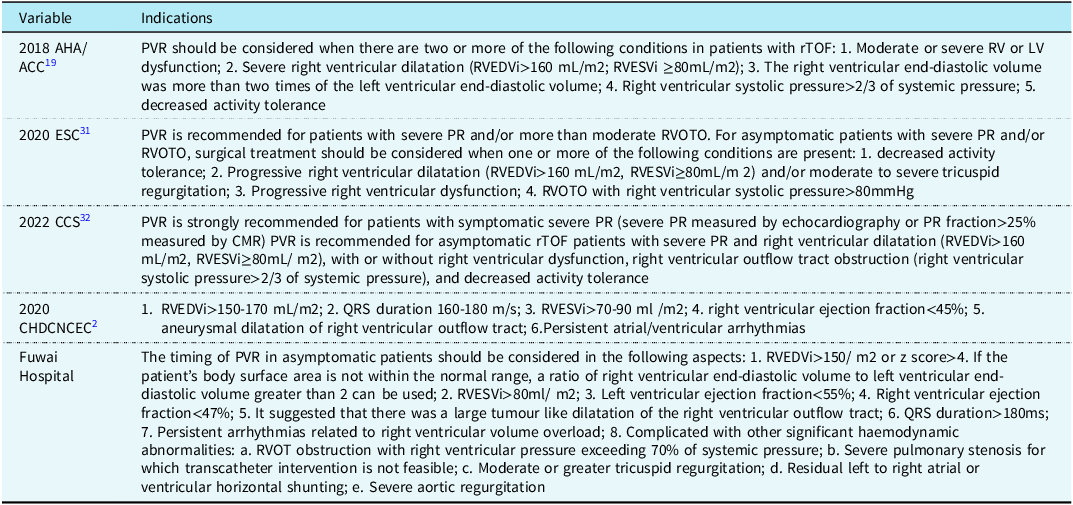
Persistent pulmonary valve regurgitation after repaired Tetralogy of Fallot is the core factor causing long-term complications. Pulmonary valve replacement is the standard procedure for the treatment of symptomatic pulmonary valve regurgitation after Tetralogy of Fallot radical surgery. According to clinical statistics, 37.8% of patients need pulmonary valve replacement after repaired Tetralogy of Fallot. Reference Hoashi, Kagisaki and Meng10 The goal of pulmonary valve replacement is not only to perform it before postoperative complications produce irreversible right ventricular dysfunction, malignant arrhythmia, and sudden death tendency, but also to avoid long-term multiple replacements caused by premature surgery as much as possible. Reference Bokma, Geva and Sleeper6 However, there is no clear and unified standard for the best time to intervene. CESNJEVAR et al. Reference Cesnjevar, Harig and Raber30 proposed the concept of “critical point,” that is, after the “critical point” is missed and pulmonary valve replacement is performed, the right ventricular function will still be irreversibly affected, even affecting the left ventricular function. But the “critical point” for the timing of pulmonary valve replacement is a moving target and may be different for individual patients. The 2018 American Heart Association/American College of Cardiology, the 2020 European Society of Cardiology, and the 2022 Canadian Cardiovascular Society have published guidelines for the management of CHD in adults, respectively Reference Stout, Daniels and Aboulhosn19,Reference Baumgartner, De Backer and Babu-Narayan31,Reference Marelli, Beauchesne and Colman32 (Table 1). All these guidelines agree that the management of pulmonary valve regurgitation after radical repair of Tetralogy of Fallot in adults is closely related to the severity of pulmonary valve regurgitation, right ventricular function, and clinical manifestations. It is necessary to perform pulmonary valve replacement in patients with decreased exercise capacity, arrhythmia attack, and right ventricular dysfunction. If the right ventricular reverse remodelling and clinical symptoms are not improved in these patients after surgery, it indicates that the patient has missed the best opportunity for surgery. In order to better reverse the right ventricular outcome and improve the prognosis, many researchers have focused on the study of early intervention timing for patients with asymptomatic pulmonary regurgitation. The timing of surgery for asymptomatic pulmonary regurgitation is controversial. The indications recommended by 2020 Chinese Congenital Heart Disease Committee of National Cardiovascular Experts Committee Reference Huishan and Shoujun2 mainly include: right ventricular end-diastolic volume index >150–170 mL/m2, QRS duration 160–180 m/s, right ventricular end-systolic volume index>70–90 ml /m2, right ventricular ejection fraction <45%, aneurysmal dilatation of right ventricular outflow tract, persistent atrial or ventricular arrhythmias, etc (Table 1).
Different PVR indications for pulmonary valve regurgitation after rTOF repair

At present, there is no clear consensus on the timing of surgical intervention for minors with pulmonary valve regurgitation after Tetralogy of Fallot radical repair. However, regardless of whether the patient is an adult or a minor, if there are obvious symptoms or no symptoms related to pulmonary valve regurgitation, and clinical examination shows significant enlargement of the right ventricular volume or right ventricular dysfunction, new or progressive arrhythmia, or progressive tricuspid regurgitation, intervention should be carried out in a timely manner before irreversible changes occur in the right ventricular volume and function, regardless of the patient’s age. The argument in favour of early intervention is that pulmonary valve replacement reduces the duration of right ventricular volume overload, delays irreversible right ventricular dysfunction, and improves the patient’s long-term quality of life. Based on international research and guidelines, the timing of surgery for asymptomatic patients in our hospital is mainly considered from the following aspects: 1. right ventricular end-diastolic volume index > 150/ m2 or z score > 4. If the patient’s body surface area is not within the normal range, a ratio of right ventricular end-diastolic volume to left ventricular end-diastolic volume greater than 2 can be used; 2. Right ventricular end-systolic volume index > 80ml/ m2; 3. Left ventricular ejection fraction < 55%; 4. Right ventricular ejection fraction < 47%; 5. It suggested that there was a large tumour-like dilatation of the right ventricular outflow tract; 6. QRS duration > 180ms; 7. Persistent arrhythmias related to right ventricular volume overload; and 8. Complicated with other significant haemodynamic abnormalities: a. right ventricular outflow tract obstruction with right ventricular pressure exceeding 70% of systemic pressure; b. Severe pulmonary stenosis for which transcatheter intervention is not feasible; c. Moderate or greater tricuspid regurgitation; d. Residual left-to-right atrial or ventricular horizontal shunting; and e. Severe aortic regurgitation.
Selection of different strategies for pulmonary valve regurgitation intervention after repaired Tetralogy of Fallot
After determining the timing of pulmonary valve regurgitation after Tetralogy of Fallot radical repair, the appropriate treatment should be selected according to the patient’s own condition. Pulmonary valvuloplasty can be selected for very few pulmonary valve regurgitation patients with preserved intrinsic pulmonary valve leaflets and annulus structures. The surgical opportunity depends on the protection of intrinsic pulmonary valve leaflets during Tetralogy of Fallot repair and the growth and function maintenance of pulmonary valve leaflets after operation in pulmonary valve regurgitation patients. Reference Adamson, McElhinney and Lui33 Most patients with TAO (transannular patch) or without pulmonary valve leaflet preservation can only choose pulmonary valve replacement, including surgical pulmonary valve replacement, percutaneous pulmonary valve intervention, and hybrid pulmonary valve replacement.
Surgical pulmonary valve replacement
Previous studies have shown that surgical pulmonary valve replacement is the first choice for pulmonary valve replacement in pulmonary valve regurgitation patients, and it is an objective and effective surgical procedure with mature technology. There are two main methods: isolated pulmonary valve replacement and pulmonary valved conduit replacement. Reference Cleveland and Wells34 Isolated pulmonary valve replacement mainly includes artificial bioprosthetic valve and mechanical prosthetic valve. The choice of bioprosthetic valve and mechanical prosthetic valve should be comprehensively considered according to the patient’s condition, age, general condition, medical condition, and patient’s preference. Mechanical prosthetic valve has a long service life, but it is easy to form thrombosis, which can cause valve stenosis and thromboembolism, so most scholars do not recommend the use of mechanical prosthetic valve. However, Haas et al. Reference Haas, Schreiber, Horer, Kostolny, Holper and Lange35 believe that mechanical prosthetic valve should be the first choice when patients are given good anticoagulation and surgical indications are strictly controlled. Some scholars also believe that mechanical prosthetic valve might not be superior to bioprosthetic valve in terms of prosthesis failure or reintervention. Dong-Hee Kim et al. Reference Kim, Choi and Kwon36 reported the results of 131 patients with repaired Tetralogy of Fallot at their institution who underwent pulmonary valve replacement for the first time using mechanical prosthetic valve or bioprosthetic valve. The median age at pulmonary valve replacement was 19 years. Bioprosthetic valve and mechanical prosthetic valve were used in 88 (67.2%) and 43 (32.8%) patients, respectively. The median follow-up duration was 7.4 years, and the 10-year survival rate was 96.4%. Risk factors for prosthesis failure were smaller body surface area and smaller prosthesis size. Risk factors for prosthesis reintervention were smaller body surface area and prosthesis size. Probability of prosthesis failure and reintervention at 10 years was 24.6% (19.5% in bioprosthetic valve versus 34.8% in mechanical prosthetic valve, P = 0.34) and 7.8% (5.6% in bioprosthetic valve versus 11.9% in mechanical prosthetic valve, P = 0.079), respectively. Bioprosthetic valve is the most commonly used valve in adult clinical practice, and bovine pericardial stent valves are the most commonly used. Reference Kwon and Baird37 The short-term results are good without the need for lifelong anticoagulation, and it has been found that bovine pericardial valves have better flow characteristics and durability compared with conventional porcine pericardial valves. A retrospective study of 611 patients at Boston Children’s Hospital Reference Nomoto, Sleeper and Borisuk38 showed that bioprosthetic valve had good short-term results, but age at surgery and valve type were risk factors for reoperation. The risk of reoperation for bioprosthetic valve in children was 5 times higher than that in adults, and the risk of reoperation decreased by 10% for every 1 year of age at surgery. Moreover, valve calcification and other problems will still occur in the long term. McKenzie et al. Reference McKenzie, Khan and Dietzman39 also reached a similar conclusion: when the age at surgery was less than 13 years old (P = 0.03) and the diameter of the valve annulus was small (P = 0.025), the risk of reoperation would increase. We believe that bioprosthetic valve should be prioritised over mechanical prosthetic valve for paediatric patients in the era of transcatheter pulmonary valve replacement.
Pulmonary valved conduits mainly include valved homograft conduits, valved xenogeneic conduits, artificial valved conduits, and tissue-engineering valved conduits. Reference Wang, Ma and Li40 Valved homograft conduits mainly use aortic or pulmonary valved conduits, which have good replacement effect, but the source is limited, and the freedom from intervention rate reaches 70% after 15 years. Reference Li, Wang and Jin41 However, the results of different studies vary greatly. The valved heterogenous conduit mainly uses bovine jugular vein conduit, which is convenient to obtain materials, but the long-term effect is not as good as the valved homograft conduit, and the risk of long-term conduit calcification, obstruction, and regurgitation is high. Reference Nomoto, Sleeper and Borisuk38 The valved homograft conduit is more consistent with the haemodynamic characteristics of the pulmonary valve, with a lower pressure gradient, and can also provide a suitable release site for subsequent percutaneous pulmonary valve replacement. Dittrich et al. Reference Dittrich, Alexi-Meskishvili and Yankah42 showed in their mid-term follow-up study after pulmonary valve replacement that valved homograft conduits were more prone to calcification than valved heterogenous conduits, while valved heterogenous conduits had a higher probability of stenosis. The incidence of conduit stenosis was 12% in valved homograft conduit and 66% in valved heterogenous conduits. Valved heterogenous conduit stenosis often occurs at the distal anastomosis due to intimal hyperplasia. About 46% of patients with pulmonary valve replacement using valved heterogenous conduits require re-intervention, and the incidence of postoperative complications is higher. The most commonly used artificial valve conduit is polytetrafluoroethylene conduit, which can be made according to different ages and weights. This conduit can be used to solve a variety of cardiac vascular anatomical problems such as pulmonary artery stenosis and aneurysm. Reference Wang, Ma and Li40,Reference Allen, El-Zein, Cuneo, Cava, Barth and Ilbawi43 Polytetrafluoroethylene conduits are considered by many heart centres to be the next best option to valved homograft conduits because of their simple construction, cost-effectiveness, easy access, and good long-term haemodynamic results. Compared with conventional Dacron conduits, patients who underwent replacement of polytetrafluoroethylene conduits had significantly lower rates of pseudointima and early obstruction. Reference Allen, El-Zein, Cuneo, Cava, Barth and Ilbawi43 The short-term effect of polytetrafluoroethylene conduit is positive, but long-term follow-up data are still needed to verify. The emerging tissue engineering technology is expected to use recipient-derived endothelial cells to implant on decellularised valved homograft conduits or degradable synthetic materials to construct recipient-specific tissue engineering valved conduits. This progress offers great potential for ideal biological graft materials and pulmonary valve replacement in CHD. The problems of immune rejection and failure of catheter growth to adapt to the body growth of children will be completely solved. Reference Nakatsuji, Yamagishi and Maeda44 In addition, the study found that the prognosis of surgical pulmonary valve replacement is significantly related to the replacement material of pulmonary valve. The 5-year freedom from valve failure rate after surgical pulmonary valve replacement in children with valved homograft conduit or bioprosthetic valve is similar, but the long-term effect of valved homograft conduit is significantly better than that of bioprosthetic valve. Moreover, the long-term effect of pulmonary valved homograft conduit is significantly better than that of aortic valved homograft conduit. Reference Dong, Keming and Shoujun45 Therefore, we believe that compared with other materials, valved homograft conduit is more in line with physiological characteristics, better haemodynamics, easy to suture, good haemostasis, anti-calcification, anti-infection, and no lifelong anticoagulation. It is the best material for reconstructing right ventricular outflow tract, followed by bioprosthetic valve.
Percutaneous pulmonary valve intervention
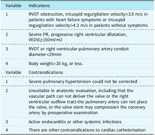
In recent years, with the development of interventional technology, percutaneous pulmonary valve intervention has developed rapidly due to its advantages of no need for general anaesthesia, short operation time, less trauma and rapid recovery, and can partially replace surgical pulmonary valve replacement. Reference Ribeiro, Teixeira, Lopes, Costa, Pires and Goncalves46 In 2000, Bonhoeffer et al. Reference Bonhoeffer, Boudjemline and Saliba47 performed the world’s first percutaneous pulmonary valve intervention on a 12-year-old boy with right ventricular outflow tract obstruction. After the operation, the degree of pulmonary valve regurgitation was significantly reduced, the right ventricular systolic pressure and transvalvular pressure gradient were significantly reduced, and the right ventricular function was significantly improved, which provided a new treatment for pulmonary valve regurgitation. Compared with surgical pulmonary valve replacement, percutaneous pulmonary valve intervention has higher requirements for the shape and size of right ventricular outflow tract, which is particularly important for patients who use transannular patch during Tetralogy of Fallot radical surgery. Percutaneous pulmonary valve intervention can be preferred as long as the anatomy of the right ventricular outflow tract allows. Reference Siyuan, Zihao, Xiangming and Xucong11 The indications for percutaneous pulmonary valve intervention in Munich Heart Center, Germany Reference Georgiev, Ewert and Tanase48 included: (1) right ventricular outflow tract obstruction, tricuspid regurgitation velocity >3.5 m/s in patients with heart failure symptoms or tricuspid regurgitation velocity >4.3 m/s in patients without symptoms; (2) severe pulmonary valve regurgitation, progressive right ventricular dilatation, VEDVi ≥150ml/m2; (3) right ventricular outflow tract or right ventricular-pulmonary artery conduit diameter <29mm; and (4) body weight >20 kg. Percutaneous pulmonary valve intervention has low risk and small trauma, which can effectively improve cardiac function and symptoms, reduce the risk of sudden death in some patients, and improve the prognosis and quality of life of patients. However, it is difficult to operate and is only suitable for patients with appropriate right ventricular outflow tract anatomy. There are also a series of contraindications: (1) It cannot correct severe pulmonary hypertension; (2) Inappropriate anatomic evaluation, including that the vascular path can not deliver the valve or the right ventricular outflow tract-the pulmonary artery can not place the valve, or the valve stent may compress the coronary artery by preoperative examination; (3) Active endocarditis or other systemic infections; (4) There are other contraindications to cardiac catheterisation. Details of the indications and contraindications for percutaneous pulmonary valve intervention are provided (Table 2).
Indications and contraindications of PPVI for pulmonary valve regurgitation after rTOF

At present, Melody valve (Medtronic), SAPIEN valve (Edwards), and Venus-P valve (China) are the most common valve stents used for percutaneous pulmonary valve intervention in the market. Studies have shown that the freedom from reintervention rate and reimplantation rate within 5 years after percutaneous pulmonary valve intervention are 76 and 92%, respectively. Common complications include stent fracture, coronary artery compression, infective endocarditis, right ventricular outflow tract rupture or dissection, and stent migration or embolism. Reference Cheatham, Hellenbrand and Zahn49 Early stent fracture is the most common complication, occurring in 20 to 43% of patients after valve stenting alone. In 2011, Nordemeyer et al. Reference Nordmeyer, Lurz and Khambadkone50 proposed that right ventricular outflow tract should be preprocessed before percutaneous pulmonary valve intervention, and bare stent protection with valved stent should be placed, which can reduce the incidence of stent fracture by 65%. Therefore, the two commonly used valve systems require pre-implantation of a bare stent in the right ventricular outflow tract, which greatly reduces the incidence of stent fracture. Compared with SAPIEN valve, Melody valve is more prone to stent fracture. Relevant meta-analysis showed that the incidence of stent fracture of Melody valve reached 12.4%, and patients using Melody valve have a higher probability of postoperative infective endocarditis. Reference Virk, Liou, Chandrakumar, Gupta and Cao51 In an early trial by Bonhoeffer, the weight threshold for percutaneous pulmonary valve intervention was 20 kg. However, Martin et al. Reference Martin, Shahanavaz and Peng52 reported 43 cases of successful percutaneous pulmonary valve intervention with body weight less than 20 kg. In 2018, Shahanavaz et al. Reference Shahanavaz, Qureshi and Levi53 reported 117 cases of successful percutaneous pulmonary valve intervention with right ventricular outflow tract<16 mm and non-guideline recommended indications. The continuous innovation of valve materials and delivery systems will further expand the indications of percutaneous pulmonary valve intervention, which has profound significance for the popularisation and promotion of percutaneous pulmonary valve intervention. Venus-P valve is a newly developed pulmonary valve in our country. The 1-year follow-up results were good: all patients had remission of pulmonary regurgitation, and the average right ventricular end-diastolic volume index decreased from 139ml/m2 before surgery to 78 ml/m2 after surgery. Reference Garay, Pan, Zhang, Wang and Springmuller54 In recent years, with the development and maturity of different methods of pulmonary valve replacement, the age of percutaneous pulmonary valve intervention intervention and perioperative mortality of pulmonary valve regurgitation patients are decreased. Reference Egbe, Vallabhajosyula and Connolly55
Hybrid pulmonary valve replacement
Because percutaneous pulmonary valve intervention is limited by age, weight, and right ventricular outflow tract anatomy, pulmonary valve replacement requires thoracotomy under cardiopulmonary bypass, which is more traumatic, so some patients cannot tolerate it. Hybrid pulmonary valve replacement combined with surgery and interventional therapy can make up for these shortcomings to a certain extent. Compared with surgical pulmonary valve replacement, the operation time of hybrid pulmonary valve replacement is significantly shorter, and the surgical trauma is relatively small. However, hybrid pulmonary valve replacement still requires median sternotomy, which is more traumatic than percutaneous pulmonary valve intervention, and hybrid pulmonary valve replacement leaves a puncture incision in the anterior wall of the right ventricle, which may cause early complications such as local dyskinesia and arrhythmia of the right ventricle. However, no obvious complications have been found in patients after hybrid pulmonary valve replacement. In 2007, Schreiber et al. Reference Schreiber, Horer and Vogt56 performed hybrid pulmonary valve replacement for the first time on six patients with right ventricular outflow tract diameter>22 mm and severe pulmonary valve regurgitation after Tetralogy of Fallot repair, who underwent off-pump thoracotomy and valved-stent placement through the right ventricle. Except for one case with severe enlargement of right ventricular outflow tract, the remaining five cases had good surgical results with only mild pulmonary valve regurgitation. Subsequently, in 2008, Dittrich et al. Reference Dittrich, Gloeckler and Arnold57 reported a case of hybrid pulmonary valve replacement under cardiopulmonary bypass, with an 8-year-old child weighing 35 kg and a right ventricular outflow tract width of about 28 mm. Surgery was performed through the third intercostal space incision to reach the pulmonary artery, through which a valve stent was placed, avoiding injury to the median sternotomy and myocardium. In 2013, Chen et al. Reference Chen, Turner, Caputo, Stoica, Marianeschi and Parry58 performed six cases of off-pump hybrid pulmonary valve replacement. The operation was performed through a median sternal incision, and the pulmonary artery with a diameter of >29 mm was folded and sutured, and then a purse was sutured at the proximal end of the trans-ring patch in the right ventricular outflow tract of the anterior wall of the right ventricle. It can avoid the complications caused by cardiopulmonary bypass and prevent myocardial damage. Holoshitz et al. Reference Holoshitz, Ilbawi and Amin59 reported a case of hybrid pulmonary valve replacement in a 3-year-old child weighing 12 kg. The sheath tube was delivered through the subxiphoid incision, and the valve was placed through the right ventricle. This surgical method preserves the complete sternum, which is less invasive than the surgical method of median sternotomy, and brings light to the hybrid treatment of children with low age and weight.
Comparison of different treatment strategies for pulmonary valve regurgitation intervention after repaired Tetralogy of Fallot
Surgical pulmonary valve replacement and percutaneous pulmonary valve intervention have their own advantages and disadvantages, and the selection of intervention methods should consider the individual condition and the prognosis of children, and try to reduce the risk of reoperation. At present, there is no consensus on the choice of long-term pulmonary valve regurgitation intervention methods after repaired Tetralogy of Fallot. In a retrospective cohort study of surgical pulmonary valve replacement and percutaneous pulmonary valve intervention surgery conducted by Hribernik I et al. Reference Hribernik, Thomson and Ho60 in the United Kingdom from 1998 to 2020, patients undergoing pulmonary valve replacement surgery were divided into two groups: surgical pulmonary valve replacement group (365 cases) and percutaneous pulmonary valve intervention group (128 cases). The postoperative complication rate of surgical pulmonary valve replacement and percutaneous pulmonary valve intervention was 5.5% and 5.2%, the perioperative mortality rate was 1.1 and 0%, the 5-year survival rate was 96.8 and 92%, the 10-year survival rate was 95.1 and 85.8%, and the reintervention rate was 14.8 and 12.5%, respectively. Therefore, they concluded that surgical pulmonary valve replacement, as compared with percutaneous pulmonary valve intervention, has similar rates of perioperative mortality, mid-term mortality, and postoperative reintervention. Some scholars have also conducted long-term studies on the two surgical methods, showing that the right ventricular volume is basically normal and the heart function is significantly improved after surgical pulmonary valve replacement. However, the right ventricular volume decreased after percutaneous pulmonary valve intervention, but the improvement of right ventricular function was not obvious, indicating that the postoperative effect of surgical pulmonary valve replacement was better than that of percutaneous pulmonary valve intervention. Reference Zablah, Misra, Gruber, Kholwadwala and Epstein61 Among them, the risk factors affecting the postoperative effect of surgical pulmonary valve replacement include: surgical age ≥28 years old, preoperative right ventricular mass: volume ≥0.45g/ml, and right ventricular ejection fraction <40%. Reference Geva, Mulder and Gauvreau62 Other studies have reported that patients older than 35 years old at the time of surgical pulmonary valve replacement will have a 5.6-fold increase in 10-year mortality. Reference Dorobantu, Sharabiani and Taliotis63 Then some people put forward different views. In 2018, Li et al. Reference Li, Pollard and Karimi64 conducted a comparative study on 32 cases of percutaneous pulmonary valve intervention and 30 cases of pulmonary valve replacement, and found that for patients younger than 17 years old, the pulmonary valve function after percutaneous pulmonary valve intervention was better than that after pulmonary valve replacement. In adults, the incidence of pulmonary valve dysfunction is low in the two groups, and there is no difference in postoperative effect. Therefore, percutaneous pulmonary valve intervention is recommended as the first choice for children and adolescents. When reintervention of pulmonary valve regurgitation after repaired Tetralogy of Fallot is performed, the choice of reintervention methods needs further research and discussion.
Our institution proposed a systematic strategy based on the synthesis of published research views at home and abroad and our own experience during clinical practice. The decision of which intervention to use was made during the preoperative multidisciplinary team meeting and finalised according to the intraoperative evaluation (Figure 1). We believe that pulmonary valvuloplasty can be considered for pulmonary valve regurgitation patients after Tetralogy of Fallot with normal pulmonary valve leaflet function. Pulmonary valve replacement is required for patients with appropriate anatomical conditions who have clinical examination results of exercise capacity decline, arrhythmia attack, right ventricular dysfunction, or asymptomatic but moderate to severe pulmonary regurgitation, severe tricuspid regurgitation, severe progressive right ventricular dilatation, or moderate to severe right ventricular dysfunction after TOF. Percutaneous pulmonary valve intervention is recommended for patients with no stenosis or obstruction of the catheter access from the vascular approach to the right ventricular outflow tract and pulmonary artery, no deformity of the anatomical morphology of the pulmonary artery root, valves can be placed at the right ventricular outflow tract and pulmonary artery, and there is no compression of the coronary artery. For patients with unsuitable cardiac and vascular anatomy, complicated with other cardiac anatomical problems, or younger age, surgical pulmonary valve replacement is recommended. If the patient cannot accept the trauma caused by surgical pulmonary valve replacement, hybrid pulmonary valve replacement is recommended.
Pulmonary valve surgery decision-making process.

Perspectives
In recent years, the long-term pulmonary valve regurgitation after Tetralogy of Fallot repair has gradually attracted the attention of scholars at home and abroad, and the choice of intervention time and the best treatment method is particularly important. In addition, with the development of materials science and the in-depth research of tissue engineering in the cardiovascular field, some scholars have made new explorations on valve and patch materials, such as self-cell tissue engineering valves and pedicled autologous pericardial valve with growth characteristics, which provide a new direction for the prevention and treatment of pulmonary valve regurgitation although the long-term prognosis needs further observation. Reference Wang, Ma and Li40 In the future, with the improvement of diagnostic methods, the refinement of surgical indications, the improvement of surgical equipment, and other medical fields, the long-term pulmonary valve regurgitation problem after repaired Tetralogy of Fallot is expected to be solved satisfactorily.
Conclusions
With the improvement of Tetralogy of Fallot diagnosis, treatment, and nursing, the number of Tetralogy of Fallot patients undergoing radical repair is increasing, and the long-term pulmonary valve regurgitation is increasing accordingly. How to provide standardised, scientific, and individualised treatment strategies and life-cycle health services for repaired Tetralogy of Fallot patients with long-term pulmonary valve regurgitation has become a challenge for CHD surgeons around the world. Therefore, by understanding the pathophysiological mechanism of pulmonary regurgitation after repaired Tetralogy of Fallot, clarifying the severity of pulmonary valve regurgitation grading diagnosis, paying attention to prevent the occurrence of pulmonary valve regurgitation during the initial repair, identifying the intervention time of severe pulmonary valve regurgitation, and selecting the optimal intervention method, we can better improve the long-term prognosis and quality of life of patients with pulmonary valve regurgitation.
Data availability statement
All data are incorporated into the article, and the data underlying this article are available in the article.
Acknowledgements
We acknowledge the roles of our colleagues, perfusionists, nurses, and others involved in the care of the study participants.
Author contributions
Zhangwei Wang: Conceptualisation; Data curation; Formal analysis; Investigation; Methodology; Software; Validation; Visualisation; Writing—original draft. Honghao Fu: Writing—review & editing. Shoujun Li: Writing—review & editing.
Financial support
No funding was received for this work.
Competing interests
None declared.
Ethical standard
Not applicable.
Consent for publication
The manuscript is not currently under consideration elsewhere, and the work reported will not be submitted for publication elsewhere until a final decision has been made as to its acceptability by the journal.