Introduction
Delusions are a difficult-to-manage feature of Alzheimer's disease (AD), most often emerging during the middle stages of the disease, and causing considerable distress to individuals with AD and their caregivers, and frequently leading to institutionalization (Magni et al., Reference Magni, Binetti, Bianchetti and Trabucchi1996; Kloszewska, Reference Kloszewska1998). Delusions in AD often have paranoid features, involving ideas that others are trying to harm the individual (persecution), are stealing from them, or are being unfaithful (Burns et al., Reference Burns1996; Kloszewska, Reference Kloszewska1998). Other types of delusions, more specific to the dementias than psychotic illness, include delusional misidentification of loved ones or one's surroundings (Burns et al., Reference Burns1996). Clinical management of delusions and other psychotic symptoms is difficult in AD; antipsychotics have historically been the mainstay of management of psychotic symptoms in nursing homes, but are not without risk in this group (Schneider et al., Reference Schneider, Dagerman and Insel2005). Cholinesterase inhibitors have shown efficacy in reducing delusions and other psychotic symptoms, though data is currently limited (Fischer et al., Reference Fischer, Bozanovic, Atkins and Rourke2006). Understanding the pathophysiology of delusions in AD is important, as this may provide clues to specific and targeted treatments to manage these distressing symptoms.
Neuroimaging approaches to understanding delusions in AD have included seeking structural correlates, using magnetic resonance imaging (MRI), as well as regional under or overactivity in the resting state, using positron emission tomography (PET) and single photon emission computed tomography (SPECT). Heterogeneity of groups studied in terms of disease stage and types of delusions measured has led to variable results, though the existing body of literature points consistently to disturbances in frontal and temporal regions. Structural imaging studies have found greater atrophy of frontal regions (Bruen et al., Reference Bruen, McGeown, Shanks and Venneri2008) as well as increased frontal hyperintensities and white matter changes (Binetti et al., Reference Binetti1995), and greater left temporal atrophy and white matter changes in those with delusions (Geroldi et al., Reference Geroldi, Bresciani, Zanetti and Frisoni2002; Lee et al., Reference Lee2006). In addition, some subcortical changes have been reported to occur in those with delusions (Bruen et al., Reference Bruen, McGeown, Shanks and Venneri2008). Similarly, PET and SPECT studies find mainly right hemisphere disturbances (largely hypoactivity) in prefrontal regions, namely frontopolar regions (Sultzer et al., Reference Sultzer, Brown, Mandelkern, Mahler, Chen and Cummings2003), orbitofrontal cortex (Mentis et al., Reference Mentis1995; Mega et al., Reference Mega, Lee, Dinov, Mishkin, Toga and Cummings2000; Sultzer et al., Reference Sultzer, Brown, Mandelkern, Mahler, Chen and Cummings2003), lateral prefrontal and dorsolateral prefrontal cortex (Mega et al., Reference Mega, Lee, Dinov, Mishkin, Toga and Cummings2000; Sultzer et al., Reference Sultzer, Brown, Mandelkern, Mahler, Chen and Cummings2003; Nakano et al., Reference Nakano, Yamashita, Matsuda, Kodama and Yamada2006; Moran et al., Reference Moran, Becker, Satlin, Lyoo, Fischman and Johnson2008), and anterior cingulate cortex (Mega et al., Reference Mega, Lee, Dinov, Mishkin, Toga and Cummings2000; Nakano et al., Reference Nakano, Yamashita, Matsuda, Kodama and Yamada2006). Changes in superior, middle and inferior temporal cortices have also been associated with delusions, representing hypoactivity in some studies, and hyperactivity in others (Mentis et al., Reference Mentis1995; Nakano et al., Reference Nakano, Yamashita, Matsuda, Kodama and Yamada2006; Moran et al., Reference Moran, Becker, Satlin, Lyoo, Fischman and Johnson2008). Less consistent findings include reduced activity in the basal ganglia (Mentis et al., Reference Mentis1995; Mega et al., Reference Mega, Lee, Dinov, Mishkin, Toga and Cummings2000) and the parietal lobe (Mega et al., Reference Mega, Lee, Dinov, Mishkin, Toga and Cummings2000; Fukuhara et al., Reference Fukuhara2001; Nakano et al., Reference Nakano, Yamashita, Matsuda, Kodama and Yamada2006). Two studies have suggested that mechanisms underlying delusions in men and women with AD may differ; delusions have been associated with cortical hypoactivity in frontal and temporal regions in women (Moran et al., Reference Moran, Becker, Satlin, Lyoo, Fischman and Johnson2008), and with hyperactivity (Moran et al., Reference Moran, Becker, Satlin, Lyoo, Fischman and Johnson2008) and increased D2/3 receptor availability (Reeves et al., Reference Reeves, Brown, Howard and Grasby2009) in striatal regions in men.
Attempts to distinguish neural correlates of different kinds of delusions have highlighted a role for secondary visual centers in delusional misidentification syndromes, including parieto-occipital regions (Lee et al., Reference Lee2006) and medial occipital cortex (Nakano et al., Reference Nakano, Yamashita, Matsuda, Kodama and Yamada2006), as well as frontotemporal disturbances. However, most studies have included individuals with paranoid and misidentification delusions in a single delusions group. Frontotemporal dysfunction may represent a common mechanism for delusions of any kind and is a putative pathophysiological model of delusions in wider psychotic illness and schizophrenia (Blackwood et al., Reference Blackwood, Howard, Bentall and Murray2001). In schizophrenia, aberrant connectivity between frontal and temporal regions is thought to underlie psychotic phenomena, whereas in dementia there is also evidence of atrophy occurring in these regions (Bruen et al., Reference Bruen, McGeown, Shanks and Venneri2008). Greater specificity in examining the pathophysiology of delusions in AD may be achieved by considering specific types of delusions (Fukuhara et al., Reference Fukuhara2001; Nakano et al., Reference Nakano, Yamashita, Matsuda, Kodama and Yamada2006) as well as examining males and females separately (Moran et al., Reference Moran, Becker, Satlin, Lyoo, Fischman and Johnson2008). Paranoid delusions are likely to involve executive and attentional centers as well as those which are involved in representations of others’ intentions and behaviors, such as the frontal and temporal regions (Blackwood et al., Reference Blackwood, Howard, Bentall and Murray2001).
The present study will examine the association of one subtype of delusions, namely paranoid delusions, with regional cortical thickness and subcortical volume in AD. Following previous studies, we hypothesize reduction in cortical thickness in orbitofrontal and dorsolateral prefrontal regions, as well as temporal regions associated with paranoid delusions. Subcortical regions of interest include the striatum and pallidus, and also the nucleus accumbens, as part of the extended frontolimbic network.
Methods
Subjects
One-hundred-and-thirteen participants with mild to moderate AD were recruited as part of AddNeuroMed, a multi-centre European study of AD biomarkers (Lovestone et al., Reference Lovestone, Francis and Strandgaard2007). The six participating clinical centers included university hospitals in Kuopio (Finland), Perugia (Italy), Lodz (Poland), Thessaloniki (Greece), Toulouse (France) and London (UK). Participants were individuals with a diagnosis of AD, attending local dementia clinics. All subjects also met the following inclusion criteria: (1) ADRDA/NINCDS and DSM-IV criteria for possible or probable AD; and (2) Mini-Mental State Examination score > 12. Exclusion criteria for the study included: (1) significant neurological or psychiatric illness other than AD; and (2) significant unstable systematic illness or organ failure. Informed consent was obtained for all participants, and protocols and procedures were approved by the relevant local ethical committees at each site.
Clinical data
A comprehensive semi-structured evaluation was conducted with all patients and their caregivers to assess clinical, cognitive and functional status. Disease severity was assessed using the Global Deterioration Scale (GDS; Reisberg et al., Reference Reisberg, Ferris, Deleon and Crook1982), and the Clinical Dementia Rating Scale (CDR; Morris, Reference Morris1993). Global cognitive status was assessed using the Alzheimer's Disease Assessment Scale – Cognitive subscale (ADAS-Cog; Rosen et al., Reference Rosen, Mohs and Davis1984) and the Mini-Mental State Examination (MMSE; Folstein et al., Reference Folstein, Folstein and McHugh1975), completed by the participant. Delusions and other neuropsychiatric symptoms were assessed using the Neuropsychiatric Inventory, rated by the caregiver/informant (NPI; Cummings, Reference Cummings1997). The NPI provides information on history of delusions, types of delusions experienced, frequency of delusions and their severity in terms of distress and disruption experienced over the previous month. For the purposes of the present study, however, participants were asked if they had experienced neuropsychiatric symptoms at any point in time since cognitive decline began. Since experience of psychotic symptoms may fluctuate (Reisberg et al., Reference Reisberg, Ferris, Deleon and Crook1982) over the course of the disease and against a background of progressive cortical atrophy, we felt that history of delusions rather than recent experience was a more suitable variable for analysis. Therefore, those with a history of paranoid delusions (belief that others are planning to harm him/her, belief that other are stealing from him/her, belief that his/her spouse is unfaithful) were assigned to the delusions group, and those with no history of delusions of any kind to the non-delusions group. No participants with delusional misidentification only were included in the analysis.
Magnetic resonance imaging
Data were acquired using six 1.5T MR systems (4 General Electric, 1 Siemens and 1 Picker). Following a three plane localizer, high resolution T1-weighted sagittal 3D MP-RAGE volume and a dual echo fast spin echo scans were obtained, providing proton density and T2-weighted images. Full brain and skull coverage was required for both of the latter datasets and detailed quality control carried out on all MR images (see Simmons et al., Reference Simmons2011). Image quality control took place immediately after the images had been acquired at each site. All MR images were given a clinical check by an on-site radiologist in order to exclude any subjects with non-AD related pathologies.
Image analysis
A highly automated structural MRI image processing pipeline developed by Fischl and Dale was utilized for data analysis (Fischl and Dale, Reference Fischl and Dale2000; Fischl et al., Reference Fischl2004), which produced regional cortical thickness and volume measures. Cortical reconstruction included removal of non-brain tissue using a hybrid watershed/surface deformation procedure, automated Talairach transformation, intensity normalization, segmentation of the subcortical white matter and deep grey matter volumetric structures (including hippocampus, amygdala, caudate, putamen, ventricles), tessellation of the gray/white matter boundary, automated topology correction, and surface deformation following intensity gradients to optimally place the gray/white and gray/cerebrospinal fluid borders at the location where the greatest shift in intensity defines the transition to the other tissue class. Surface inflation was followed by registration to a spherical atlas which utilized individual cortical folding patterns to match cortical geometry across subjects and parcellation of the cerebral cortex into units based on gyral and sulcal structure (Fischl et al., Reference Fischl2004). All volumes were normalized by the subject's intracranial volume. The regional cortical thickness was measured from 34 areas (entorhinal cortex, parahippocampal gyrus, isthmus of cingulate cortex, posterior cingulate cortex, fusiform gyrus, temporal pole, inferior temporal gyrus, middle temporal gyrus, superior temporal gyrus, transverse temporal cortex, banks of superior temporal sulcus, medial orbitofrontal cortex, lateral orbitofrontal cortex, orbital operculum, frontal pole, superior frontal gyrus, frontal operculum, pars triangularis, rostral middle frontal gyrus, caudal middle frontal gyrus, precentral gyrus, superior parietal gyrus, inferior parietal gyrus, post central gyrus, paracentral cortex, supramarginal gyrus, pericalcarine gyrus, cuneus cortex, precuneus cortex, lateral occipital cortex, lingual gyrus, corpus callosum, caudal anterior cingulate cortex and anterior cingulate cortex) and the regional cortical volume was measured from 22 areas (right and left hippocampus, right and left amygdala, right and left caudate, right and left accumbens, right and left putamen, right and left thalamus, right and left cerebellum cortex, brainstem, right and left pallidum, posterior corpus callosum, anterior corpus callosum, central corpus callosum, mid-anterior corpus callosum, mid-posterior corpus callosum).
Specified regions of interest for the analysis, according to our hypotheses, were frontal pole, lateral orbitofrontal, medial orbitofrontal, superior frontal, rostral middle frontal, temporal pole, superior temporal, middle temporal, inferior temporal and subcortical regions (caudate, putamen, nucleus accumbens and pallidum).
Statistical analysis
To investigate the relationship between delusions and MRI measures, a group comparisons method was used (paranoid delusions vs no delusions). Means obtained for the frequency × severity (F×S) score on the delusions subscale of the NPI were low and the data heavily skewed towards zero, and therefore a regression approach using the F×S score was not possible. Group comparisons for clinical variables were conducted using t-tests and χ2 comparisons for ordinal and categorical variables respectively. Regions of interest were compared for paranoid delusions and no delusions groups using ANCOVA. Clinical covariates for ANCOVA group comparisons were on the basis of t-test comparisons between the groups. For the exploratory analysis of cortical regions, ANCOVA comparisons between the groups were corrected for multiple comparisons (n = 50), and only those significant at the p < 0.001 level were deemed to have survived correction.
Results
Delusions were a relatively infrequent neuropsychiatric symptom within this cohort, affecting 20.4% of participants at some point during the course of their illness. Within the spectrum of delusory thought, paranoid ideation (ideas of others threatening harm or theft and of spousal infidelity) was the most common, and delusional misidentification less common. Frequency and severity of current delusions was low (mean frequency × severity score 2.7, ±2.6, in the delusions group). The present results therefore consider the association of a history of paranoid delusions with regional brain atrophy.
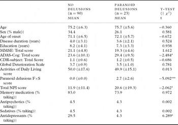
Table 1 compares demographic and clinical variables of those with and without delusions. There were no significant differences in age, sex, education, age of onset or disease duration between the groups. Those with delusions displayed lower scores on the ADAS-Cog assessment of cognitive status, although global dementia ratings (CDRS and GDS) did not differ significantly. Those with paranoid delusions also experienced a greater overall burden of neuropsychiatric symptoms, suggesting that delusions tend to occur in the context of pre-existing neuropsychiatric disturbance. Those with delusions were no more likely to be taking antipsychotic, sedative, or cognitive enhancing medication, but were less likely to be taking antidepressants. Cognitive status, measured by the ADAS-Cog, was selected as a covariate in analyses of regional brain atrophy associated with paranoid delusions.
Clinical variables in those with and without paranoid delusions

* p < 0.05; ** p < 0.01; *** p < 0.001.
Notes: † Dichotonous variable, χ2 comparisons; Memory medications included acetylcholinesterase inhibitors, NMDA agonists and nootropics.
Antidepressants % refers to the percentage of patients taking at least one type of antidepressant. MMSE = Mini-Mental State Examination; ADAS-Cog = Alzheimer's Disease Assessment Scale – Cognitive subscale; CDR = Clinical Dementia Rating scale; NPI = Neuropsychiatric Inventory.
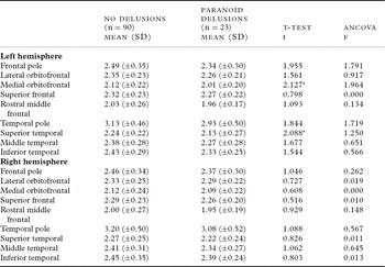
Table 2 compares the MRI measures of cortical thickness in those with and without paranoid delusions. Left medial orbitofrontal and superior temporal cortices were significantly thinner in those with paranoid delusions, though this difference was not significant when ADAS-Cog was included as a covariate. Of the subcortical regions of interest, only the left putamen showed a trend for greater atrophy in the sample (t = 1.816, p = 0.072), though the trend remained following covariation of the cognitive status score (F = 2.973, p = 0.087).
Regional cortical thickness in those with and without paranoid delusions

* p < 0.05; ** p < 0.01; *** p < 0.001.
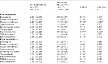
A further analysis considered female participants only, following previous findings of gender specific associations with regional atrophy and delusions. Table 3 shows a stronger effect for widespread frontal and temporal atrophy in the left hemisphere of female participants with paranoid delusions. Three areas, specifically left medial orbitofrontal, and left superior temporal and temporal pole, were significantly thinner in the female delusions group even after cognitive status was included as a covariate. There were no differences in volumetric measures of subcortical regions of interest in female participants. Male participants were treated as a separate analysis, and showed no associations between paranoid delusions and cortical thickness, or subcortical regional volume (data not shown). However, the sample size of males in this cohort was small (31 males without delusions, 6 males with delusions).
Regional cortical thickness in females with and without paranoid delusions

* p < 0.05; ** p < 0.01; *** p < 0.001.
Additional exploratory analyses on the remaining cortical regions required a statistical significance of p < 0.001 to correct for multiple comparisons. The strongest effect was found for greater thinning of the insula cortex. Across the whole sample, this reached the required statistical significance corrected for multiple comparisons in the left insula (t = 3.030, p = 0.003), and for both left (L) and right (R) insula in the female group alone (L: t = 3.627, p = 0.001; R: t = 3.341, p = 0.001). However, this was not significant following covariation of the cognitive status score, though the strongest effect remained for increased cortical thinning in the left insula in the female group (F = 7.156, p = 0.009).
Discussion
The present study examined associations between regional structural brain atrophy and delusions in Alzheimer's disease. Those with paranoid delusions showed greater impairment on a measure of cognitive ability, and this variable was chosen as a covariate for group comparisons. Across the whole sample, those with paranoid delusions showed significantly thinner left medial orbitofrontal and superior temporal cortex than those without, though this was not significantly different after controlling for cognitive status. Of the subcortical regions of interest, none differed significantly between groups, though there was a trend for smaller volume of the left putamen in the delusions groups. Female participants with paranoid delusions showed a consistent regional reduction in frontal and temporal cortical thickness, with significantly thinner left medial orbitofrontal, superior temporal, and temporal pole cortex, even after controlling for cognitive status, compared to those females without delusions. An exploratory analysis of regions outside those hypothesized to show differences demonstrated the strongest effect for the left insula in female participants. This finding was independent of cognitive status, but fell short of the statistical level required following multiple comparisons. Male participants showed no significant differences on any of the MRI measures hypothesized as relating to delusion status.
These results are broadly consistent with previous studies, highlighting a role of atrophy of both frontal, in particular orbitofrontal, and temporal regions in the experience of delusions as a whole (Mentis et al., Reference Mentis1995; Mega et al., Reference Mega, Lee, Dinov, Mishkin, Toga and Cummings2000; Geroldi et al., Reference Geroldi, Bresciani, Zanetti and Frisoni2002; Sultzer et al., Reference Sultzer, Brown, Mandelkern, Mahler, Chen and Cummings2003, Bruen et al., Reference Bruen, McGeown, Shanks and Venneri2008; Moran et al., Reference Moran, Becker, Satlin, Lyoo, Fischman and Johnson2008). Our study also replicates findings of specific pathophysiological-neuropsychiatric associations dependant on gender in AD (Moran et al., Reference Moran, Becker, Satlin, Lyoo, Fischman and Johnson2008). We found that atrophy of frontal and temporal cortical regions was associated with delusions in women with AD, although we did not find any specific associations for the male group, possibly due to the small number of men with delusions in this cohort. As such, theories proposing that delusions in men are subcortically driven were not supported by our study. A trend for greater atrophy of the left putamen associated with delusions was noted across the entire sample. One key difference between the present study and the previous literature is an emphasis on the role of right hemisphere dysfunction associated with delusions in other previous studies, and our finding of left hemisphere changes. The significance of this discrepancy is not clear; frontal and temporal regions contribute bilaterally to the cognitive processes thought to be aberrant in individuals with delusions, namely executive and attentional function and language processing.
In the context of the present study, orbitofrontal and temporal regions show greater atrophy associated specifically with a specific subgroup of “paranoid” delusions. Our results emphasize the role of ventral regions in paranoid delusions, compared to findings of posterior involvement in studies focusing on, or including, a large number of participants with delusional misidentification (Nakano et al., Reference Nakano, Yamashita, Matsuda, Kodama and Yamada2006; Lee et al., Reference Lee2006). This suggests that disturbance of frontotemporal regions may be the core mechanism for delusions as a whole, and that wider involvement of more posterior regions is associated with perceptually driven delusions, namely delusional misidentification syndromes (DMS). Specific regions associated with paranoid features, which fundamentally involve social processing, are less clear. Only two studies have examined neuroanatomical correlates of specific types of paranoid delusions, though results were not consistent; delusions of theft have been associated with bilateral anterior cingulate hypoactivity (Nakano et al., Reference Nakano, Yamashita, Matsuda, Kodama and Yamada2006) and with right posterior parietal region hypoactivity (Fukuhara et al., Reference Fukuhara2001).
Blackwood et al. (Reference Blackwood, Howard, Bentall and Murray2001) provide a cognitive neuroanatomical framework to describe the cognitive biases and pathophysiology associated with persecutory delusions in psychotic illness. Dysfunction in a number of everyday cognitive processes may lead to feelings of persecution; over-attention to threatening stimuli (Phillips et al., Reference Phillips, Senior and David2000); misinterpretation of the behaviors, emotions and facial expressions of others (Whittaker et al., Reference Whittaker, Deakin and Tomenson2001); tendency to jump to conclusions (Young and Bentall, Reference Young and Bentall1997); and misattribution of others’ mental states (Drury et al., Reference Drury, Robinson and Birchwood1998). Blackwood et al. (Reference Blackwood, Howard, Bentall and Murray2001) draw together evidence from neuroimaging studies of individuals with delusions, as well as studies of the brain regions involved in these processes. They highlight the role of prefrontal, orbitofrontal, ventral striatal and superior temporal regions in the pathology of persecutory delusions. Within this framework, our findings of greater medial orbitofrontal atrophy suggest that delusions may be driven by failures in social reasoning centers, wrongly assigning emotional or social significance to everyday non-threatening social stimuli. Pathology of the superior temporal cortex, which controls representations of others’ behavior, could also lead to misidentifying everyday behavior as being threatening or emotionally significant. Though the role of the insula is not considered in Blackwood's framework, the insula is also a key center for processing social and emotional information. With extensive innervation from the amygdala, the brain's fear processing center, disturbance to the insula or its connection from the amygdala, may lead to greater salience and a greater degree of cortical processing being given to potentially threat-related emotional and social stimuli. Our findings are consistent with this framework of heightened attention to threat and misattribution of other's intentions or behaviors, and, unlike the previous literature in AD, imaging studies of delusions in psychotic illness emphasize the role of the left hemisphere over the right (Blackwood et al., Reference Blackwood, Howard, Bentall and Murray2001).
The present study has some limitations which may impact on the generalizability of the findings. First, the mean MMSE score for the sample was 20.2 (±5.0), and therefore this sample represents a group of mild to moderately impaired AD patients. Examining patients with a broader range of disease severity may have revealed different associations with specific regions, or may have found a statistically greater effect for regions of interest. The frequency of delusions in the wider AD population may therefore have been underrepresented in this sample. Secondly, the nature of the frequency × severity score derived by the NPI means that low frequency symptoms will be skewed toward zero. This skewing limited the nature of the analyses conducted, and a larger sample, or one with a broader range of delusion severity may have allowed us to examine associations with delusion severity using a regression design, rather than a comparison of those with or without a history of delusions. Third, our sample included only a small number of males and therefore we can describe a pattern of associations in the female participants, but cannot draw conclusions about the presence of a sex difference. Lastly, we used caregiver rated NPI to assess the presence and type of delusions, which is likely to be less reliable than a full clinical interview, particularly in those cases where the informant was a professional carer, which may have resulted in an underestimation of delusion frequency.
Strengths of the present study include its size, which is comparable to the largest existing neuroimaging studies of delusions in AD. This relatively large sample also allowed us to examine male and female associations separately, suggesting the possibility of different mechanisms for delusions in the two groups. The selection of a specific subtype of delusion for analysis also increases the specificity of findings and removes confounders that may result from mixing individuals with only visually mediated delusional misidentification and those with only paranoid delusions. Some authors consider DMS to be a “disorder of perception” more akin to hallucinations than delusions or “disordered thought” (Burns, Reference Burns1996). Finally, use of cognitive status as a key covariate has allowed us to examine regional structural differences independently of cognitive decline, giving greater specificity to brain regions associated with neuropsychiatric as opposed to cognitive symptoms.
Conclusions
Our study found that paranoid delusions are associated with reduced cortical thickness of orbitofrontal and superior temporal regions in women with AD, but not in men. Frontotemporal atrophy is a putative model for delusions in AD, affecting key structures involved in social and emotional processing. At present, risperidone and donepezil have shown the greatest effect in the management of delusions (Fischer et al., Reference Fischer, Bozanovic, Atkins and Rourke2006), though differential drug effects on different types of delusions have not been examined. The association of orbitofrontal atrophy with delusions in the present study may underlie the beneficial effects of atypical antipsychotics on delusions, which impact on extrastriatal or corticostriatal domapine pathways. The possibility of differential involvement of key structures between sexes warrants further investigation in a larger sample, and comparison of different classes of delusions (i.e. paranoid vs misidentification) may be fruitful in delineating specific mechanisms and further targeting pharmacological treatments.
Conflict of interest
None.
Description of authors' roles
D. Whitehead analyzed the above data, and wrote the paper. C. Tunnard collected data, assisted with analysis and contributed to the writing. C. Hurt assisted with statistical analysis and contributed to the writing. D. Cromb assisted with image processing. L. O. Wahlund, P. Mecocci, M. Tsolaki, B. Vellas, C. Spenger, I. Kłoszewska, H. Soininen, D. Cromb, S. Lovestone and A. Simmons designed and implemented the study. In addition, S. Lovestone contributed to writing the paper, and A. Simmons oversaw MRI data collection and processing, assisted with analysis and contributed to writing.
Acknowledgments
The AddNeuroMed study is funded through EU FP7 as part of InnoMed and by funding from the NIHR Biomedical Research Centre for Mental Health at the South London and Maudsley NHS Foundation Trust and Institute of Psychiatry, Kings College London.