Introduction
At birth, the serum of the lamb is virtually void of immunoglobulin G (IgG; O’Doherty and Crosby, Reference O’Doherty and Crosby1997a) as the ovine placenta prevents the in utero transfer of maternal Ig’s to the foetus (Gilbert et al., Reference Gilbert, Gaskins, Hillers, Parker and McGuire1988). The lamb is reliant on the uptake of IgG, the main Ig in ovine colostrum (Smith et al., Reference Smith, Dawson, Wells and Burrells1975), from colostrum to confer disease resistance in early life. This is similar to the situation with vitamin E, an important antioxidant (Buckley et al., Reference Buckley, Morrissey and Gray1995), which is also involved in maximising immunocompetence (Puls, Reference Puls1994). Blood tocopherol levels are much lower in the newborn than those of the mother (Njeru et al., Reference Njeru, McDowell, Wilkinson, Linda and William1994) as placental transfer of α-tocopherol is very limited in sheep (Pehrson et al., Reference Pehrson, Hakkarainen and Blomgren1990; Njeru et al., Reference Njeru, McDowell, Wilkinson, Linda and William1994). Vitamin E and IgG are present in colostrum at much higher concentrations than in normal milk in order to facilitate the rapid uptake by the newborn in the immediate post partum period. Early ingestion of colostrum is essential (Campbell et al., Reference Campbell, Siegel and Knowlton1977) as the lamb’s ability to absorb intact maternal globulins decrease rapidly between 24 and 48 h after birth (Gilbert et al., Reference Gilbert, Gaskins, Hillers, Parker and McGuire1988) due to gut closure.
A number of studies have shown that high dietary intakes of some mineral supplementations by ewes in late pregnancy results in their progeny having decreased serum IgG concentrations with reduced efficiency of colostral IgG absorption, despite the fact that they had adequate intakes of colostrum (O’Doherty, 1994; Boland et al., Reference Boland, Brophy, Callan, Quinn, Nowakowski and Crosby2004; Crosby et al., Reference Crosby, Boland, Brophy, Quinn, Callan and Joyce2004). Recent studies have shown that vitamin E absorption is also reduced as a result of high-level mineral supplementation (Boland et al., Reference Boland, Callan, Brophy, Quinn and Crosby2006). Iodine has been identified as the specific element responsible for this reduced efficiency of IgG absorption from colostrum. Furthermore, iodine supplementation of the pregnant ewe reduced IgG absorption from a colostrum supplement where lambs had no access to maternal colostrum during the first 24 h of life (Rose et al., Reference Rose, Wolf and Haresign2007). Minerals other than iodine have also demonstrated a role in altering lamb serum IgG levels following pre partum maternal supplementation. The progeny of ewes supplemented with Se during pregnancy had increased absorption of IgG (Rock et al., Reference Rock, Kincaid and Carstens2001). In another study, it was reported that high levels of cobalt may also have a negative effect on IgG absorption (Boland et al., Reference Boland, Brophy, Callan, Quinn, Nowakowski and Crosby2005a).
The key role of iodine in the body is in the synthesis of the thyroid hormones, thyroxine (T4) and tri-iodothyronine (T3) produced in the thyroid gland. These hormones regulate the metabolic pattern of most cells and play a vital role in the process of cellular differentiation, growth and development in the foetus and neonate (Stanbury, Reference Stanbury1996; Sethi and Kapil, Reference Sethi and Kapil2004) probably mediated by effects on gene expression (Brody, Reference Brody1999). Earlier work by Cabello indicated a relationship between the concentration of thyroid hormones during the immediate pre partum period and the period of transmission of IgG in the newborn (Cabello and Levieux, Reference Cabello and Levieux1982; Cabello et al., Reference Cabello, Levieux, Girardeau and Lefaivre1983).
The objectives of this experiment were to determine the effects of offering a high level of supplementary iodine to the ewe on lamb serum IgG, vitamin E, T3 and T4 concentrations at 1 h post partum and the effects of offering supplementary cobalt for the final 3 weeks of pregnancy or supplementary iodine for either the final 3 weeks or final week of pregnancy on lamb serum vitamin E and IgG concentrations at 24 and 72 h post partum.
Material and methods
Animals and management
Sixty twin-bearing ewes were selected following pregnancy scanning in week 14 of pregnancy. The ewes were weighed and body condition was scored using a scale of 1 to 5 (Jeffries, Reference Jeffries1961) and allocated to one of four treatments (n = 15), which were balanced for breed, age (3.9 ± 0.95 year), body condition score (3.1 ± 0.33) and live weight (80 ± 11 kg). On day 119 of pregnancy, the ewes were individually housed on timber-slatted floors, and following an acclimatisation period of 1 week experimental dietary supplements were introduced on day 126 of pregnancy. All ewes were offered a basal diet of grass silage ad libitum supplemented with 800 g/day of a 190 g/kg crude protein (CP) concentrate from day 126 of gestation until lambing. The silage was harvested in early June 2004 using a precision-chop harvester following a 24-h wilt. No additive was used in the preservation of the mainly perennial ryegrass sward. The ewes received either a control diet with no mineral supplement or received one of three mineral supplements denoted I-3, I-1 or Co-3 (Table 1).
Iodine and cobalt inclusion levels, expressed in terms of mg per ewe per day of active ingredient and mg per ewe per day of source material

†I-3 = ewes offered 26.6 mg iodine for 3 weeks; I-1 = ewes offered 26.6 mg iodine for 1 week; Co-3 = ewes offered 20.0 mg cobalt for 3 weeks.
‡The 26.6 mg level of iodine equates to a mineral intake of 34 g which we have previously shown to have a negative effect on IgG and vitamin E absorption (Boland et al., Reference Boland, Guinan, Brophy, Callan, Quinn, Nowakowski and Crosby2005b).
§The level of 20 mg of cobalt equates to a mineral intake of 48 g per ewe per day, which has been shown to negatively affect IgG absorption efficiency (Boland et al., Reference Boland, Brophy, Callan, Quinn, Nowakowski and Crosby2004).
Feeding
Fresh silage and water was available to ewes at all times with uneaten silage being removed at 0730 h each day, immediately following which fresh silage was offered at 1.1 times the previous day’s intake. The silage was split-fed with ewes receiving their second daily silage compliment at 1700 h. The concentrate was also split-fed in two equal portions offered at 0900 h and 1730 h. Supplements (calcium iodate and cobalt carbonate) were manually mixed into the concentrate on the day prior to feeding. Before being added to the concentrate, the daily allowance was first added to 20 g (fresh weight) of the concentrate, which was used as a carrier. Prior to the mineral inclusion, this carrier was dried using forced air circulation at 55°C for 72 h, following which it was then ground through a 0.8-mm screen using a Christy & Norris hammer mill (Christy & Norris Ltd, Ipswich, UK). The daily concentrate allowances in treatments I-3, I-1 and Co-3 were adjusted to take account of the carrier contribution. The average daily intakes of cobalt and iodine are presented in Table 2. Mineral intakes from concentrates are based on book values for the individual concentrated ingredients (Sauvant et al., Reference Sauvant, Perez and Tran2004) and the composition of the mineral supplement in the mineral premix (Inform Nutrition, Cork, Ireland). The levels of iodine and cobalt in the forages are based on the values of Rogers and Murphy (1999). As with previous reports the levels of supplementary minerals greatly exceeded that present in the basal diet (Boland et al., Reference Boland, Brophy, Callan, Quinn, Nowakowski and Crosby2004).
Dietary intakes of iodine (I) and cobalt (Co) based on recorded daily feed intakes of pregnant ewes (least-square means ± s.e.)

†C = no supplement; I-3 = ewes offered 26.6 mg iodine for 3 weeks; I-1 = ewes offered 26.6 mg iodine for 1 week; Co-3 = ewes offered 20.0 mg cobalt for 3 weeks.
Lambing
All ewes lambed in their individual pens and remained there until 24 h post partum. They were then moved to a straw-bedded group pen for a further 48 h. Immediately after birth, all lambs received navel treatment with tincture of iodine to prevent joint ill, lambs were weighed, tagged and their sex recorded. An apron was placed over the ewe’s udder to prevent the lamb from suckling and removed after the lambs were blood sampled at 24 h post partum.
Ten lambs (five sets of twin mates) were selected at birth from each of C and I-3 treatments and these lambs were blood sampled using jugular venipuncture and non-heparinised vacutainers. All blood samples were stored at room temperature for 1 h and then refrigerated at 4°C for 24 h, when they were centrifuged at 1700 × g for 15 min and the sera collected were stored at −20°C until required to be analysed for IgG, vitamin E, T3 and T4 concentrations.
Milking
All ewes were hand milked at 1, 10 and 18 h post partum. Prior to the commencement of milking, 10 IU of oxytocin (Oxytocin-S; Intervet (Irl.) Ltd, Magna Business Park, Dublin, Ireland) was administrated intramuscularly to facilitate complete milk let-down (Doney et al., Reference Doney, Peart and Smith1979). After 3 min, both sides of the udder were completely milked out by hand. At each milking, colostrum yield was recorded and a 5 ml colostrum sample was collected and frozen at −20°C until required for IgG analysis.
Following each milking, the lambs were fed measured quantities of colostrum via stomach tube at a rate of between 20 and 50 ml/kg birth weight (BW) depending on colostrum yield. The quantity of colostrum fed to each lamb was recorded. All lambs received colostrum from their own dam except when colostrum yield was insufficient to supply 20 ml/kg BW. These lambs were fed substitute colostrum and excluded from further IgG studies. A 10 ml blood sample was taken from all lambs at 24 and 72 h post partum as described above.
Chemical analysis
Colostrum IgG concentration in the 1, 10 and 18 h samples were measured using the method of Fahey and McKelvey (Reference Fahey and McKelvey1965), using single radial immunodiffusion (RID) kits (Bethyl Laboratories, Montgomery, TX, USA). In order to obtain a reading on a standard curve, the colostrum samples were diluted by a factor of 20, 10 and 5 for the 1, 10 and 18 h samples, respectively. The RID kits were designed to measure colostral IgG concentration only.
Total serum Ig content was measured using the zinc sulphate turbidity (ZST) test (McEwan et al., Reference McEwan, Fisher, Selman and Penhale1970). The serum IgG concentration was estimated by reducing the Ig concentration value by 0.09 to equate with the IgG only measured in colostrum (Larson et al., Reference Larson, Ward, Frederiksen, Ardrey and Frank1974). When calculating the efficiency of IgG absorption it was assumed that in lambs, as in calves, 0.075 of BW is equivalent to blood plasma volume (Quigley et al., Reference Quigley, Hammer, Russel and Polo2005). The following equation was used to calculate the IgG absorption efficiency:

Serum T4 and T3
Serum T4 and T3 concentrations were determined by solid phase time-resolved fluoroimmunoassay using AutoDELFIA kits (Wallac Oy, Turku, Finland). Free serum T4 and T3 concentrations were analysed using the following protocol: anti-T4/T3 monoclonal antibody was first reacted with a solid phase coated with anti-mouse IgG. The sample T4/T3 was then added and reacted with the T4/T3 antibody. Following the second incubation, buffer and serum were washed away and in the third incubation step, europium-labelled T4/T3 was added and occupied the remaining empty binding sites on the anti-T4/T3. An enhancement solution was added to dissociate the europium ions from the labelled T4/T3, forming highly fluorescent chelates with components of the enhancement solution. The fluorescent was then measured by a time-resolved fluorometer, with the amount of resulting fluorescence inversely proportional to the amount of free T4 and T3 in the sample.
Serum α-tocopherol
A modification of the method of Bieri et al. (Reference Bieri, Tolliver and Catignani1979) was used to extract α-tocopherol from serum samples. In a 2-ml screw cap micro tube (ref. 72.694.006, Sarstedt, Germany), 500 μl of serum was combined with 500 μl of ethanol containing the internal standard (50 μg/ml dl-α-tocopherylacetate). The contents were then vortexed for 45 s, and following the addition of 500 μl of hexane the samples were vortexed for a further 45 s following which they were then centrifuged at 7800 × g for 5 min. The upper hexane layer was then transferred to a 1.5 ml Eppendorf tube, evaporated to dryness and redissolved in 200 μl ethanol.
High-pressure liquid chromatography (HPLC) separation of samples was carried out on a Varian Prostar 230 solvent delivery module (Varian Ltd, Surrey, UK), equipped with a 20 μl loop. Detection was achieved with a Varian Prostar 310 AV vis-detector (Varian Ltd) set at 292 nm. The HPLC was fitted with a Gemini 5u C18 110A (150 × 4.6 mm i.d.) column (Phenomenex Ltd, Cheshire, UK) and a Gemini C18 guard cartridge (AJO-7597; Phenomenex Ltd) was eluted with methanol/water (99 : 1) at a flow rate of 2.0 ml/min.
Percentage recovery was determined by the addition of known quantities of α-tocopherol to the serum samples prior to extraction. Following extraction, the resulting peak areas and chromatograms were compared with those obtained by direct injection onto the column of standard α-tocopherol solutions.
Statistical analysis
The data were analysed using the general linear model procedure (PROC GLM) of the Statistical Analysis Systems Institute (Cary, NC, USA) (1999–2001) (SAS, Version 8). The experiment was analysed as a randomised-block design. All data were subjected to analysis of variance providing treatment means with standard errors of the mean ± s.e. The model included mineral supplementation and ewe BW as a covariate.
Results
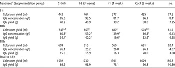
The effect of treatment on colostrum and IgG yield and IgG concentration at 1, 10 and 18 h post partum and on total colostrum and IgG yields to 18 h post partum are presented in Table 3. Seven ewes yielded insufficient colostrum at 1 h to provide the minimum colostrum requirement of 20 ml/kg BW for their lambs at the 1 h feeding (one ewe from each of C, I-3 and Co-3 and four ewes from I-1). At 10 h, four ewes yielded insufficient colostrum (one from each of I-3 and Co-3 and two from I-1) while only one ewe from I-1 yielded insufficient colostrum at 18 h. Treatment had a significant effect on colostrum yield at 10 h post partum (P < 0.05) but not at 1 or 18 h or on total colostrum yield to 18 h (P > 0.05). I-3 ewes had a significantly higher colostrum yield at 10 h post partum than I-1 ewes (653 v. 480 ml; P < 0.05). While it did not reach significance there was a tendency (P = 0.12) for a lower colostrum yield to 18 h post partum in the I-1 group with more ewes from this treatment failing to yield sufficient colostrum at the 10 h milking to feed both lambs with at least 20 ml/kg BW.
Effects of treatment on colostrum yield, concentration and yield of IgG at 1, 10 and 18 h post partum and the total yield of IgG to 18 h (least-square means ± s.e.)

†C = no supplement; I-3 = ewes offered 26.6 mg iodine for 3 weeks; I-1 = ewes offered 26.6 mg iodine for 1 week; Co-3 = ewes offered 20.0 mg cobalt for 3 weeks; s.e. = standard error; IgG = immunoglobulin G.
Means with different superscripts are significantly different: a,b(P < 0.05); x,y(P < 0.01).
As with colostrum yield, there were large variations in IgG concentrations and IgG yield at 1, 10 and 18 h post partum. Colostral IgG concentration decreased from the first to the third milking with mean concentrations of 86.7, 55.0 and 26.0 g/l for 1, 10 and 18 h colostrum, respectively, similar to previous results (O’Doherty and Crosby, Reference O’Doherty and Crosby1997a; Boland et al., Reference Boland, Brophy, Callan, Quinn, Nowakowski and Crosby2004; Guinan et al., Reference Guinan, Harrison, Boland and Crosby2005). Treatment had a significant effect on colostral IgG concentration and yield at 10 h post partum, with I-1 ewes having a significantly lower IgG concentration (P < 0.05) and yield (P < 0.01) at 10 h post partum than any other treatment. Treatment had no effect on colostral IgG concentration and yield at 1 or 18 h post partum or on total IgG yield to 18 h post partum (P > 0.05).
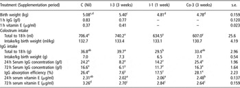
The effect of treatment on lamb BW, serum IgG and vitamin E levels at 1 h post partum, colostrum intake, IgG consumption, lamb serum IgG and vitamin E concentrations and the efficiency of colostral IgG absorption are presented in Table 4. Treatment had a significant effect on lamb BW (P < 0.01). The progeny of I-3 ewes had higher BWs than the progeny of the I-1 and Co-3 ewes (P < 0.01). Iodine supplementation had no effect on 1 h serum IgG or vitamin E concentration (P > 0.05). The progeny of Co-3 and I-1 ewes had lower colostrum intakes on an absolute basis to 18 h than lambs born to C or I-3 ewes (P < 0.001). Lambs born to I-3 ewes had a higher intake of colostral IgG to 18 h than lambs from I-1 ewes (P < 0.05), reflecting the lower BW of I-1 lambs. Treatment had no effect on the volume of colostrum or on the quantity of IgG fed per kg lamb BW over the first 18 h post partum (P > 0.05).
Effect of treatment on lamb birth weight, colostrum and IgG intakes, lamb serum IgG and vitamin E concentrations and IgG absorption efficiency (least-square mean ± s.e.)

†C = no supplement; I-3 = ewes offered 26.6 mg iodine for 3 weeks; I-1 = ewes offered 26.6 mg iodine for 1 week; Co-3 = ewes offered 20.0 mg cobalt for 3 weeks; s.e. = standard error; IgG = immunoglobulin G.
Means with different superscripts are significantly different: a,b(P < 0.05); c,d(P < 0.01); x,y,z(P < 0.001).
There was a major effect of treatment on lamb serum IgG values at both 24 and 72 h post partum. Lambs born to C and Co-3 ewes had significantly higher serum IgG values at both 24 (P < 0.001) and 72 h post partum than lambs born to iodine-supplemented ewes (I-1, P < 0.05 and I-3, P < 0.001). The progeny of the I-3 ewes had lower serum IgG concentrations at 24 and 72 h post partum than the progeny of I-1 ewes (P < 0.05). At 24 h post partum, Co-3 lambs had higher serum vitamin E values (P < 0.05) than lambs born to I-1 or I-3 ewes, while at 72 h post partum C lambs had a higher serum vitamin E concentration than those from all other treatments (P < 0.01). Serum vitamin E concentrations for the progeny of ewes offered supplementary iodine (I-3) tended to be lower than lambs born to the control animals at 24 h post partum, but the difference failed to reach significance (P > 0.05).
The C and Co-3 lambs had a higher IgG absorption efficiency than the I-1 and I-3 progeny (P < 0.01 and 0.001, respectively). The period of iodine supplementation also had a significant effect on lamb IgG absorption efficiency with lambs from I-3 ewes having lower absorption efficiency than I-1 lambs (P < 0.01).
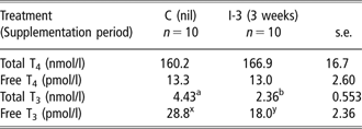
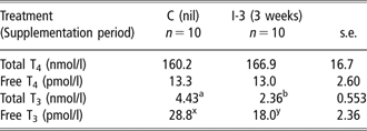
The effect of iodine supplementation on total T4 concentration, free T4 concentration, total T3 concentration and free T3 concentration at 1 h post partum are presented in Table 5. Iodine supplementation had no effect on total and free T4 concentration (P > 0.05). Treatment had a significant effect on total and free T3 concentrations at 1 h post partum. Total T3 concentration (nmol) was lower in the progeny of the I-3 ewes compared with C lambs (2.36 v. 4.44 nmol; s.e. 0.553; P < 0.05). Free T3 concentration of lamb serum was also affected by iodine supplementation, as C lambs had higher free T3 concentrations than the I-3 lambs (28.8 v. 18.0 pmol; s.e. 2.36; P < 0.01).
Effect of iodine supplementation on lamb total T4 concentration (nmol/l), free T4 concentration (pmol/l), total T3 concentration (nmol/l) and free T3 concentration (pmol/l) at 1 h post partum (least-square means ± s.e.)
†C = no supplement; I-3 = ewes offered 26.6 mg iodine for 3 weeks; s.e. = standard error.
Means with different superscripts are significantly different: a,b(P < 0.05); x,y(P < 0.01).
Discussion
The minimum dietary iodine requirements of farm animals cannot be given with any accuracy (Underwood, Reference Underwood1981), but requirements of 0.1 to 0.8 mg/kg dry matter (DM) have been reported for sheep (National Research Council, 1985). Thus in the current study ewes in the C and Co-3 treatments received adequate levels of iodine to meet requirements. However, dietary iodine intakes in these two treatments were lower than the iodine requirements of 0.7 mg per ewe per day as outlined by Boland et al. (Reference Boland, Brophy, Callan, Quinn, Nowakowski and Crosby2004) while ewes in the I-1 and I-3 treatments received iodine at levels in excess of requirements for the final week and 3 weeks of pregnancy, respectively. However, these levels were considerably lower than the published toxicity levels of 50 mg per ewe per day. The cobalt requirements of sheep are 0.07 mg/kg DM per day (Agricultural Research Council (ARC), 1980) and the concentrations fed in the current study were higher than the requirements of 0.13 mg per ewe per day as outlined by Boland et al. (Reference Boland, Brophy, Callan, Quinn, Nowakowski and Crosby2004). The levels of mineral supplementation offered in this study did not reach the current quoted toxicity levels (ARC, 1980) but the findings of this study and recent work (Boland et al., Reference Boland, Brophy, Callan, Quinn, Nowakowski and Crosby2005a, Reference Boland, Guinan, Brophy, Callan, Quinn, Nowakowski and Crosby2005b and Reference Boland, Callan, Brophy, Quinn and Crosby2006) show that high levels of iodine inclusion can have negative effects on animal performance even at rates below the quoted toxicity level. Based on these findings, the indications are that the toxicity level for iodine in the diet of the pregnant ewe should be lowered to 20 mg per ewe per day, equivalent to 40% of its current level.
There was wide variation in colostrum yield between ewes at 1, 10 and 18 h post partum, which is in agreement with the results previously reported (Pattinson et al., Reference Pattinson, Davies and Winter1995; O’Doherty and Crosby, Reference O’Doherty and Crosby1997a; Boland et al., Reference Boland, Brophy, Callan, Quinn, Nowakowski and Crosby2004). Mineral supplementation has been shown to have inconsistent affects on colostrum yield, with yield at individual time points being altered but the overall yield to 18 h remaining unaffected (Boland et al., Reference Boland, Brophy, Callan, Quinn, Nowakowski and Crosby2004; Guinan et al., Reference Guinan, Harrison, Boland and Crosby2005). In the current study, there was no effect of treatment on colostrum yield at 1 or 18 h post partum or on total colostrum yield to 18 h. However, colostrum yield at 10 h post partum was significantly lower for ewes in the I-1 v. I-3 treatment. It is unlikely that this lower colostrum yield can be fully attributable to iodine supplementation as a similar level of iodine has been offered previously with no effect on colostrum yield (Boland et al., Reference Boland, Guinan, Brophy, Callan, Quinn, Nowakowski and Crosby2005b). O’Doherty and Crosby (Reference O’Doherty and Crosby1997b) reported a negative linear correlation between underfeeding in late pregnancy and colostrum production during the first 18 h post partum and data collected on feed intake from day 135 to day 139 of gestation in the current study indicate that I-1 ewes had lower dietary intakes than all other groups (data not shown). This may also partly explain the lowered colostrum production by this group at 10 h. However, as supplementation of this group only began on day 138 it is unlikely that the treatment employed was responsible for this reduced intake.
Similar to the variation in colostrum yield, there was wide variation in colostral IgG concentration and IgG yield at 1, 10 and 18 h post partum. The decrease in colostral IgG concentration from 1 to 18 h post partum coincides with a reduction in the ability of the lamb’s small intestine to absorb IgG in the first 24 h after birth (Campbell et al., Reference Campbell, Siegel and Knowlton1977) as a result of gut maturation (Parker and Nicol, Reference Parker and Nicol1990). The lower IgG concentrations in the I-1 treatment at 10 h post partum than all other treatments can be linked to their significantly lower DM intake, which consequently resulted in lower CP and it has been demonstrated that inadequate dietary protein intake has a negative impact on colostral IgG concentration (O’Doherty and Crosby, Reference O’Doherty and Crosby1997a).
The lower BW of the I-1 and Co-3 lambs was reflected in differences in total colostrum and IgG intakes to 18 h post partum, as all lambs were fed colostrum and IgG proportional to BW. The higher total IgG intake by the I-3 lambs compared with the I-1 lambs (39.7 v. 29.5 g; P < 0.05) was as a result of a tendency for a higher total colostrum and IgG yield to 18 h post partum and a higher lamb BW in the I-3 treatment. Colostrum and IgG intake per kg BW were unaffected by treatment. Lambs require between 180 and 210 ml of colostrum per kg BW within the first 18 h of life to provide sufficient fuel for heat production (Mellor and Murray, Reference Mellor and Murray1986). However, O’Doherty (1994) reported a level of 112 ml/kg BW as sufficient in an indoor lambing system. Following this recommendation, all lambs received adequate colostrum in the first 24 h of life on the assumption that IgG absorption efficiency would be within normal limits.
The ovine placenta acts as a major barrier to the in utero transfer of IgG (Gilbert et al., Reference Gilbert, Gaskins, Hillers, Parker and McGuire1988) and vitamin E (Njeru et al., Reference Njeru, McDowell, Wilkinson, Linda and William1994). Thus, the newborn is virtually void of IgG and vitamin E at birth (Campbell et al., Reference Campbell, Siegel and Knowlton1977; Njeru et al., Reference Njeru, McDowell, Wilkinson, Linda and William1994; O’Doherty and Crosby, Reference O’Doherty and Crosby1997a), and therefore the transfer of IgG and vitamin E via the colostrum is of critical importance. Recent results have shown that mineral supplementation for just 2 weeks immediately prior to lambing has a deleterious effect on IgG absorption in the neonate (Guinan et al., Reference Guinan, Harrison, Boland and Crosby2005). In addition, iodine has been indicated as the element responsible (Boland et al., Reference Boland, Brophy, Callan, Quinn, Nowakowski and Crosby2005a). Furthermore, when iodine was supplemented to pregnant ewes at around one-third of the current published toxicity levels (ARC, 1980), their progeny were still severely compromised, in relation to the acquirement of passive immunity from the colostrum (Boland et al., Reference Boland, Brophy, Callan, Quinn, Nowakowski and Crosby2005a and Reference Boland, Guinan, Brophy, Callan, Quinn, Nowakowski and Crosby2005b; Guinan et al., Reference Guinan, Harrison, Boland and Crosby2005).
In the current study, iodine supplementation had a major effect on lamb serum IgG concentrations at 24 and 72 h post partum and on colostral IgG absorption efficiency at 24 h after birth. The progeny of ewes receiving supplementary iodine for either 3 or 1 week (I-3, I-1) prior to lambing had lower 24 and 72 h serum IgG concentrations than C or Co-3 progeny, confirming that iodine supplementation at a level of 26.6 mg per ewe per day for a little as 1 week prior to lambing is sufficient to reduce lamb serum IgG concentration below the normal levels (23.0 g/l) as previously recorded for the progeny of ewes offered silage ad libitum and concentrates in late pregnancy (Boland et al., Reference Boland, Brophy, Callan, Quinn, Nowakowski and Crosby2004). This finding, that supplementation with a relatively high level of iodine for as short a period as 1 week immediately prior to parturition can seriously compromise the lamb’s attainment of passive immunity has not been previously reported in the literature and is only half the duration of our earlier of 2 weeks (Guinan et al., Reference Guinan, Harrison, Boland and Crosby2005). Recent results have shown that although lambs may appear to receive an adequate supply of colostrum containing sufficient levels of IgG, their serum IgG concentrations can be low, indicating a poor IgG absorption efficiency which leaves the lambs extremely susceptible to infections (Boland et al., Reference Boland, Brophy, Callan, Quinn, Nowakowski and Crosby2004; Crosby et al., Reference Crosby, Boland, Brophy, Quinn, Callan and Joyce2004; Guinan et al., Reference Guinan, Harrison, Boland and Crosby2005). A negative linear relationship has been demonstrated between iodine level and serum IgG concentration (Boland et al., Reference Boland, Guinan, Brophy, Callan, Quinn, Nowakowski and Crosby2005b) and these new findings suggest that there may also be a similar relationship between the period of supplementation and serum IgG concentration. Although the serum IgG concentration at 24 h in the I-1 lambs was significantly lower than in the C lambs, it was intermediate between the C and I-3 lambs, indicating that a supplementation period of more than 1 week is required to see the complete negative effects of high levels of iodine supplementation on lamb serum IgG concentration. The absorptive mechanism internally in the lamb does function at some reduced level, as despite serum IgG levels at 24 h being only a fraction of normal levels, they are much greater than levels recorded in lambs prior to colostrum feeding in this study and elsewhere (Hunter et al., Reference Hunter, Reneau and Williams1977; O’Doherty and Crosby, Reference O’Doherty and Crosby1997a). In all lambs, serum IgG levels declined from 24 to 72 h post partum, indicating that no further absorption of IgG from the colostrum occurred and in fact IgG clearance from the blood stream is already well underway at this stage. This is in agreement with various authors, who stated that the lamb’s small intestine becomes incapable of absorbing intact maternal globulins between 24 and 48 h after birth (Campbell et al., Reference Campbell, Siegel and Knowlton1977; Gilbert et al., Reference Gilbert, Gaskins, Hillers, Parker and McGuire1988). These results support the findings of Guinan et al. (Reference Guinan, Harrison, Boland and Crosby2005) and reinforce the importance of the correct mineral balance in final 2 weeks immediately prior to parturition. The addition of supplementary cobalt for the final 3 weeks of progeny had no effect on lamb serum IgG concentrations or colostral IgG absorption efficiency.
Boland et al. (Reference Boland, Callan, Brophy, Quinn and Crosby2006) when supplementing iodine at a level of 26.6 mg per ewe per day, equal to the supplementation level in the current experiment, reported a significant reduction in lamb serum vitamin E concentration at 24 h post partum compared with that of the control treatment (P < 0.001). In the above study, however, supplementary iodine was offered for the final 6 weeks of pregnancy as opposed to the final 3 weeks in the current study. It is therefore possible that the period of supplementation in the current experiment was insufficient to elicit an equally negative effect as reported by Boland et al. (Reference Boland, Callan, Brophy, Quinn and Crosby2006). Lamb serum vitamin E concentrations increased from 24 to 72 h post partum, indicating that the uptake of vitamin E from colostrum is a more gradual process than the uptake of IgG. Furthermore, at 72 h post partum, the negative effects of iodine supplementation on lamb serum vitamin E concentration were apparent. These results are in broad agreement with our previous findings indicating that the transfer of vitamin E is affected in a manner similar to that of IgG (Boland et al., Reference Boland, Callan, Brophy, Quinn and Crosby2006). The finding that high-level cobalt supplementation during the final 3 weeks of pregnancy will have a negative effect on serum vitamin E concentration at 72 h post partum is a new and significant finding and has not previously been reported in the literature. This lends support to our previous findings that the exclusion of cobalt from mineral supplements for the pregnant ewe may have beneficial effects for the uptake of essential nutrients from the colostrum by the newborn lamb (Boland et al., Reference Boland, Brophy, Callan, Quinn, Nowakowski and Crosby2005a). The mechanism behind this reduction merits further investigation.
Lamb serum vitamin E concentrations at 1 h post partum were 0.37 and 0.41 μg/ml for lambs born to C and I-3 ewes, respectively, and were similar to that of 0.35 μg/ml reported by Njeru et al. (Reference Njeru, McDowell, Wilkinson, Linda and William1994) while Pehrson et al. (Reference Pehrson, Hakkarainen and Blomgren1990) found lamb serum vitamin E concentrations in the range of 0.08 to 0.22 mg/l prior to colostrum feeding. The latter authors noted a significantly higher serum vitamin E concentration in lambs born to ewes given a single dose of vitamin E in late pregnancy (0.22 v. 0.10 mg/l) indicating the possibility to increase vitamin E transfer across the placenta by supplementing vitamin E in late pregnancy. However, the results in the current study are in agreement with several authors confirming that the newborn lamb is virtually void of IgG and vitamin E at birth and is thus dependant upon the transfer of IgG and vitamin E via the colostrum (Campbell et al., Reference Campbell, Siegel and Knowlton1977; Njeru et al., Reference Njeru, McDowell, Wilkinson, Linda and William1994; O’Doherty and Crosby, Reference O’Doherty and Crosby1997a) for protection from disease and increased survival.
Several studies have been carried out on the transfer of thyroid hormones, T3 and T4 from the mother to foetus in humans, rats and sheep. Similar to human and rats, maternal thyroid metabolism in sheep is important for foetal development during early pregnancy. However, in contrast to man and rats, the ovine placental transfer of thyroid hormones is seen to be absent or minimal in the second half of gestation (Piosik et al., Reference Piosik, Van Groenigen, Van Doorn, Baas and Vijlder1997). Choksi et al. (Reference Choksi, Jahnke, Hilaire and Shelby2003) stated that thyroid development in sheep appears mostly to occur in utero during the latter two-thirds of pregnancy. A direct link exists between the levels of thyroid hormones in the circulatory system of newborn ruminants and the efficiency with which colostral IgG is absorbed, as plasma T4 levels are negatively related to maximal plasma IgG1 levels in both pre-term and full-term bottle-fed lambs (Cabello and Levieux, Reference Cabello and Levieux1982), confirming the importance of thyroid function for the acquisition and maintenance of passive immunity.
Plasma thyroid hormone levels, particularly T4, increase in the lamb prior to parturition before falling rapidly within the first few days of life (Slebodzinski, Reference Slebodzinski1965; Nathanielsz et al., Reference Nathanielsz, Comline, Silver and Thomas1973). The total and free T4 concentrations for control and iodine-treated lambs observed in the current study are in agreement with published values (Davicco et al., Reference Davicco, Lefaivre and Barlet1982; Kececi, Reference Kececi2003). Davicco et al. (Reference Davicco, Lefaivre and Barlet1982) recorded lamb serum T4 and T3 concentrations of 185 and 5.54 nmol/l, respectively, at 6 h post partum, while Kececi (Reference Kececi2003) reported thyroid hormone levels at 2 h post partum of 137.45 nmol/l, 3.36 nmol/l, 27.15 pmol/l and 8.47 pmol/l for total T4, total T3, free T4 and free T3, respectively. A positive relationship has been reported between BW and serum T3 and T4 concentrations in newborn lambs (Wrutniak and Cabello, Reference Wrutniak and Cabello1987) and this most likely contributed to the higher thyroid hormone levels in the current study compared with those reported by Kececi (Reference Kececi2003) where lamb BW was only 2.77 kg compared with 4.7 and 5.2 kg for C and I-3 lambs, respectively, in the current experiment. Also, contributing to the higher levels of thyroid hormones reported by Davicco et al. (Reference Davicco, Lefaivre and Barlet1982) would be later sampling time at 6 h post partum, as it has been reported that plasma thyroid hormone levels increase significantly during the first 8 h post partum following which, they decrease rapidly (Cabello and Wrutniak, Reference Cabello and Wrutniak1986).
There is a strong relationship between total and free hormonal fractions, indicating that the amount of total T3 is a major determinant of free T3 concentration (Cabello and Wrutniak, Reference Cabello and Wrutniak1986) while the neonatal rise in free T4 levels is more important than total T4 levels as free T4 is the direct substrate for T3 production (Wrutniak and Cabello, Reference Wrutniak and Cabello1987; Cabello and Wrutniak, Reference Cabello and Wrutniak1989). The lower total and free T3 levels suggest a decline in the peripheral T4 to T3 conversion in the I-3 lambs. Thyroxine is converted to T3 by a selenium (Se)-containing deiodinating enzyme, type I iodothyronine 5′-deiodinase (5′D-I; Kohrle, Reference Kohrle1994) which is present in the thyroid, liver and kidneys and plays a major role in converting T4 to T3, accounting for more than 80% of T3 production with only 15% originating directly in the thyroid (Beckett et al., Reference Beckett, Russell, Nicol, Sahu, Wolf and Arthur1992; Naziroglu et al., Reference Naziroglu, Cay, Tahan, Bal and Delibas1998, Kelly, Reference Kelly2000). Thus Se has a critical role to play in the synthesis and homoeostatic control of the thyroid hormones. Se deficiency has been shown to affect thyroid metabolism by impairing the conversion of T4 to T3 and Donald et al. (Reference Donald, Langlands, Bowles and Smith1994) reported lower levels of circulating T3 concentration and a greater T4/T3 ratio in Se-deficient sheep. Beckett et al. (Reference Beckett, Russell, Nicol, Sahu, Wolf and Arthur1992) when feeding rats a Se-deficient diet for 6 weeks reported inhibited production of both T3 from T4 and also the catabolism of T3 to 5-deiodination in liver homogenates. In the current experiment, ewes in both treatments received 0.32 mg per ewe per day Se from their mineral supplement, a level above requirements, and it is therefore unlikely that Se deficiency was a contributory factor in the decreased T3 concentrations. However, supplementation of iodine to angora goats has been shown to increase plasma T4 concentration while decreasing T3 concentration (Wichtel et al., Reference Wichtel, Thompson, Craigie and Williamson1996). An increase in the conversion of T4 to the biologically inactive reverse T3 (rT3) or a decrease in the ability to degrade rT3 could result in decreased serum T3 concentrations (Kelly, Reference Kelly2000). It is possible given the similar T4 levels but reduced T3 levels in the I-3 lambs that there was an increase in the conversion of T4 to rT3 in this group of lambs. It is not possible at this stage to indicate the mode of action for the reduced T3 concentrations in lambs born to ewes offered high levels of supplementary iodine in late pregnancy, as the mechanisms involved in thyroid homoeostasis are complex. There is a strong association between free T3 and free T4 levels and mortality and thus any factors disturbing normal thyroid hormone levels will seriously compromise the health of the neonate.
The role of T3 in colostral Ig absorption has previously been reported in the calf with postnatal administration of T3 but not T4, exerting a positive effect on the transfer of immunolactoglobulins in the neonatal calf intestine (Slebodzinski, Reference Slebodzinski1995). The results of the current study also suggest a positive relationship between T3 levels in lamb serum and IgG absorption from colostrum as the progeny with the lower T3 levels had lower IgG absorption efficiency. However, this is not in agreement with the report of Rose et al. (Reference Rose, Wolf and Haresign2007) where high-level iodine supplementation of the pregnant ewe increased plasma T4 levels at birth but plasma T3 levels were unaffected. The exact mechanism through which iodine reduces IgG and vitamin E uptake from colostrum is still unclear.
From the findings of this experiment we conclude that offering 26.6 mg of iodine for 1 week prior to lambing will have negative effects on the absorption of colostral IgG to 24 and 72 h post partum and on serum vitamin E concentration at 72 h post partum. This period of supplementation is much shorter than previously reported in the literature. There is no measurable absorption of IgG between 24 and 72 h post partum. Offering cobalt at a rate of 20 mg per ewe per day also leads to a breakdown in the transfer of vitamin E from colostrum to the serum of the newborn lamb. This is a completely novel finding and merits confirmation and further investigation. The reduced T3 levels in the serum of lambs fed with high levels of iodine offers a link between iodine supplementation and reduced lamb serum IgG levels. There is a need to identify the mechanism that brings about this reduction in T3 levels and also if and how this reduced T3 level results in a reduced IgG and vitamin E absorption efficiency.
Acknowledgements
The authors express their thanks to Mr Stephen Lott his assistance throughout the experiment and to Ms Bernie Flynn for her laboratory assistance.