




No CrossRef data available.
Article contents
Successful management of fetal hydrops due to congenitally complete atrioventricular block
Published online by Cambridge University Press: 24 May 2005
Abstract
At 32 weeks, a fetus was found to be hydropic with congenitally complete heart block in an asymptomatic mother who was positive to anti-Ro antibodies. Maternal therapy with oral salbutamol was successful in prolonging gestation for a period of 3 weeks so that preterm delivery was delayed until the 35th week of gestation. Following treatment with inotropes and diuretics, a permanent transvenous pacemaker was implanted at 8 weeks of age. The child is now thriving at 20 months of age.
Information
- Type
- Brief Report
- Information
- Copyright
- © 2003 Cambridge University Press
There is a high fetal and early neonatal mortality when congenitally complete heart block is associated with hydrops. Reported mortalities range from 73%1 to 100%.2 Premature delivery and early implantation of a pacemaker has been advocated in the management of this condition. Severe respiratory distress, and infective complications, may accompany delivery of a hydropic neonate at an early gestation leading to significant morbidity and mortality. Delayed delivery may, however, result in intrauterine death. We present our recent experience with a fetus having this combination, outlining our strategy that maximised fetal maturation before delivery.
Case report
A 26-year-old woman, seropositive for anti-Ro antibodies, and without any symptoms of a connective tissue disease, was referred at 32 weeks gestation in her second pregnancy, with an irregular fetal heartbeat and poor fetal movements. Fetal ultrasound revealed hydrops, with oedema, bilateral pleural effusions, and ascites. There was complete atrioventricular block, with a ventricular rate of 43 beats per min, and poor biventricular function (Table 1). The mother was administered two doses of dexamethasone to accelerate pulmonary maturity to prepare for an imminent delivery. She was also started simultaneously on slow-release salbutamol 8 mg twice daily. Four days later, the fetal heart rate was 50 beats per min, with improved contractility. The salbutamol was increased 2 days later to 8 mg three times daily, with an increase in the fetal heart rate to 55 beats per min. Three weeks after starting salbutamol, the ascites began to increase further, although the fetal movements remained good.
Table 1. Fetal parameters following maternal administration of salbutamol.

At 35 weeks gestation, a caesarean section was performed. A female baby was delivered weighing 2.21 kg. Apgar scores were 6 and 8, at 1 and 5 min, respectively. She rapidly developed respiratory distress, requiring intubation and positive pressure ventilation, and was also treated with surfactant. To increase her heart rate from a baseline of 30 beats per min, an intravenous infusion of isoprenaline was commenced at 1.0 mg/kg/min. This achieved a heart rate of 55 to 60 beats per min. Dopamine and dobutamine were commenced at 5 μg/kg/min and 10 μg/kg/min respectively for hypotension. Bilateral pleural effusions were present, and a pleural tap drained 20 ml of straw-coloured fluid (Fig. 1a). An echocardiogram revealed a structurally normal heart with a patent oval foramen and a patent arterial duct. No pericardial fluid was noted. Renal and cranial ultrasonic scans were normal.

Figure 1. The chest X-ray after delivery (a) showing bilateral pleural effusions, hepatomegaly and ascites. Subsequent to implantation of the pacemaker (b), an electrode is seen in the right ventricle, with a loop of the lead in the right atrium to allow for growth.
On the 3rd day, bilateral chest drains were inserted, draining a total of 55 ml. She now weighed 1.805 kg, representing a position between the 0.4th and the 2nd centiles. The baby received positive pressure ventilation until the 7th day of life, and was weaned via nasal continuous positive airway pressure to head box oxygen by the 9th day, and into room air by the 12th day. Dopamine and dobutamine had been discontinued by the 5th day. She was weaned from isoprenaline by the 12th day, and subsequently maintained a heart rate of around 45 to 50 beats per min. Total parenteral nutrition was needed from the 2nd until the 8th day of life. She was then established on enteral nutrition with diuretics to maintain fluid balance. Physiological jaundice was treated with phototherapy until the 8th day.
The electrocardiogram confirmed complete heart block, with an atrial rate of 150 beats per min, a ventricular rate of 48 beats per min, normal QRS complexes and a normal QT interval. A 24-hour recording revealed a maximum heart rate of 63 beats per min, and a minimum heart rate of 34 beats per min.
A transvenous endocardial pacemaker was implanted at the age of 8 weeks, when she weighed 2.95 kg, and feeding had been established. Through a left subclavicular incision and subclavian venous puncture, a 46 cm Tendril Bipolar Active Fixation Endocardial Lead (St Jude Medical, Solna, Sweden) was placed in the right ventricle with a threshold of 0.4 V and attached to a Microny II VVI pacemaker placed subcutaneously (Fig. 1b). The rate was set at 90 beats per min. She was discharged five days later having discontinued diuretics, immediately after the insertion of the pacemaker.
At follow-up one month later, the pacemaker was functioning normally. At the age of 18 months she is thriving, and now weighs 10.9 kg, above the 25th centile.
Discussion
Congenitally complete heart block has an incidence of approximately 1 in 20,000.2 In the absence of structural heart disease, a strong association has been established between maternal anti-Ro and anti-La antibodies and congenitally complete heart block in the fetus. These antibodies cross the placenta and, after binding to their respective antigens, are thought to produce inflammation and fibrosis of the conducting system in the region of the atrioventricular node.3 Transfer of antibodies across the placenta occurs prior to the 20th week of gestation, and heart block is most often detected between the 20th and 30th weeks of gestation.4 The majority of these fetuses progress normally through pregnancy and are delivered without complications.
Fetal hydrops, apparent as pleural and pericardial effusions, oedema of the tissues and ascites, may appear in as many as one-quarter of involved pregnancies.1, 5 It is thought that bradycardia and a low cardiac output result in an increased cardiac volume, with an inability of the fetal heart to adapt. This, in turn, leads to congestive cardiac failure, with a high fetal mortality.1, 2 In addition, an inflammatory myocarditis has been demonstrated. This, too, may contribute to development of hydrops by depressing myocardial function.
Treatment for this situation is not standardized. With increasing fetal hydrops and signs of fetal distress, early delivery is often undertaken, but is accompanied by a high mortality due to complications of prematurity even if effective pacing is commenced.1 Fetal management has included administration of steroids to reduce inflammation from the concomitant myocarditis, sympathomimetics to improve heart rate and contractility, paracentesis of effusions, injection of digoxin and diuretics directly into the fetus, direct transabdominal and transthoracic ventricular pacing, and even hysterotomy for implantation of a pacemaker, with subsequent return of the fetus to the uterus.1, 2, 5–8 Of these, sympathomimetics and steroids would appear to have the most promise. Premature delivery is indicated once the fetus is compromised.
As far as we know, there has been no success with direct fetal transabdominal and transthoracic ventricular pacing.7 While effective pacing was initiated, this was not maintained, and the fetus died shortly after. Potential complications include the introduction of sepsis and dislodgement of the pacemaker with fetal movement. Permanent implantation has been attempted in one fetus by hysterotomy and exteriorizing the fetus, but again the fetus died shortly thereafter.8
Sympathomimetic therapy has the potential to increase the heart rate and myocardial contractility, and thereby to reverse fetal hydrops. In a trial conducted by Groves et al.,6 intravenous salbutamol and isoprenaline were compared for their efficacy in increasing fetal heart rate. Salbutamol was shown to produce a better inotropic and chronotropic fetal response compared to isoprenaline, due to better transfer of the former across the placenta. In one case, administration of intravenous, and then oral, salbutamol resulted in resolution of the fetal hydrops.6 In our patient, oral salbutamol administered to the mother successfully increased the fetal heart rate and contractility. It is not clear whether tachyphylaxis to the salbutamol occurred, as the highest heart rate achieved was not maintained.9 Nevertheless, gestation was prolonged by three weeks, thereby allowing further maturation of the fetus, so that the complications of delivery of a hydropic neonate at the early gestation of 32 weeks were avoided. Maternal hypokalaemia is a potential problem when high doses of salbutamol are given and the serum potassium needs monitoring.
When administered maternally, dexamethasone remains unmetabolised before reaching the fetus, and has been shown to resolve hydrops.5 Small pericardial effusions alone do not indicate a poor prognosis.1 In the current report, dexamethasone was administered twice prior to delivery with the intent of hastening fetal lung maturity, and not with the specific aim of reducing the fetal hydrops. Longer-term administration may lead to immune suppression and corticosteroid dependency.
Subsequent to delivery, the hydropic premature neonate is at risk of pulmonary and infective complications of prematurity, with a high morbidity and mortality.1 Staged pacing has been undertaken successfully.10 Temporary transvenous pacing in this setting poses the risks of sepsis, thrombosis, and perforation of the heart, while infection of temporary epicardial leads may preclude permanent epicardial pacing. We avoided the need for temporary pacing by providing chronotropic and inotropic support with isoprenaline, dopamine and dobutamine while the hyaline membrane disease and hydropic state resolved. Subsequently, treatment with diuretics allowed permanent implantation of a pacemaker at greatly reduced risk.
The successful outcome in our case illustrates the advantage of close collaboration between the obstetric, fetal, and paediatric cardiologic teams, allowing for optimum monitoring and management of the mother and the unborn fetus. While administration of sympathomimetics to the mother only delayed delivery by 3 weeks, this was enough to allow significant growth, which together with steroids to hasten lung maturation, contributed to avoiding the major complications of prematurity. This allowed for a smoother course immediately after delivery, and permitted implantation of a permanent pacemaker under stable, controlled conditions.
References
Groves A, Allan LD, Rosenthal E. Outcome of isolated congenital complete heart block diagnosed in utero. Heart 1996; 75: 190–194.Google Scholar
Schmidt K, Ulmer H, Silverman N, Kleinman C, Copel J. Perinatal outcome of fetal complete atrioventricular block: a multicenter exeperience. J Am Coll Cardiol 1991; 17: 1360–1366.Google Scholar
Ho SY, Esscher E, Anderson RH, Michaëlsson M. Anatomy of congenital complete heart block and relation to maternal anti-Ro antibodies. Am J Cardiol 1986; 58: 291–294.Google Scholar
Waltuck J, Buyon J. Autoantibody-associated congenital heart block: outcome in mothers and children. Ann Intern Med 1994; 120: 544–551.Google Scholar
Richards DS, Wagman AJ, Cabaniss ML. Ascites not due to congestive heart failure in a fetus with lupus-induced heart block. Obstet Gynecol 1990; 76: 957–959.Google Scholar
Groves A, Allan L, Rosenthal E. Therapeutic trial of sympathomimetics in three cases of complete heart block in the fetus. Circulation 1995; 92: 3394–3396.Google Scholar
Carpenter R, Strasburger J, Garson A, Smith R, Deter R, Engelhardt HT. Fetal ventricular pacing for hydrops secondary to complete atrioventricular block. J Am Coll Cardiol 1986; 8: 1434–1436.Google Scholar
Silverman MH, Kohl TE, Harrison MR, Hanley FL. Experimental surgery in the animal model and in the human fetus. In: Proceedings of the second world congress of paediatric cardiology and cardiac surgery, Honolulu, Hawaii, 1997; 106.
Robinson BV, Ettedgui JA, Sherman RS. Use of terbutaline in the treatment of complete heart block in the fetus. Cardiol Young 2001; 11: 683–686.Google Scholar
Weindling S, Saul JP, Triedman J, et al. Staged pacing therapy for congenital complete heart block in premature infants. Am J Cardiol 1994; 74: 412–413.Google Scholar

Table 1.
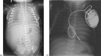
The chest X-ray after delivery (a) showing bilateral pleural effusions, hepatomegaly and ascites. Subsequent to implantation of the pacemaker (b), an electrode is seen in the right ventricle, with a loop of the lead in the right atrium to allow for growth.
 You have
Access
- 14
- Cited by