

Introduction
The use of the endoscope for middle-ear surgery was first described in the late 1960s.Reference Mer, Derbyshire, Brushenko and Pontarelli1 Initially, the endoscope was used as an adjunct to microscopic techniques for diagnostic purposes.Reference Nomura2 Early studies focused on delineating the anatomy of the middle ear. Endoscopes were soon found to be useful in second-look cholesteatoma surgery, to identify residual or recurrent disease.Reference Yung3, Reference Haberkamp and Tanyer4 It is only in the last 15 years that endoscopes have been increasingly used as the sole instrument in middle-ear surgery.
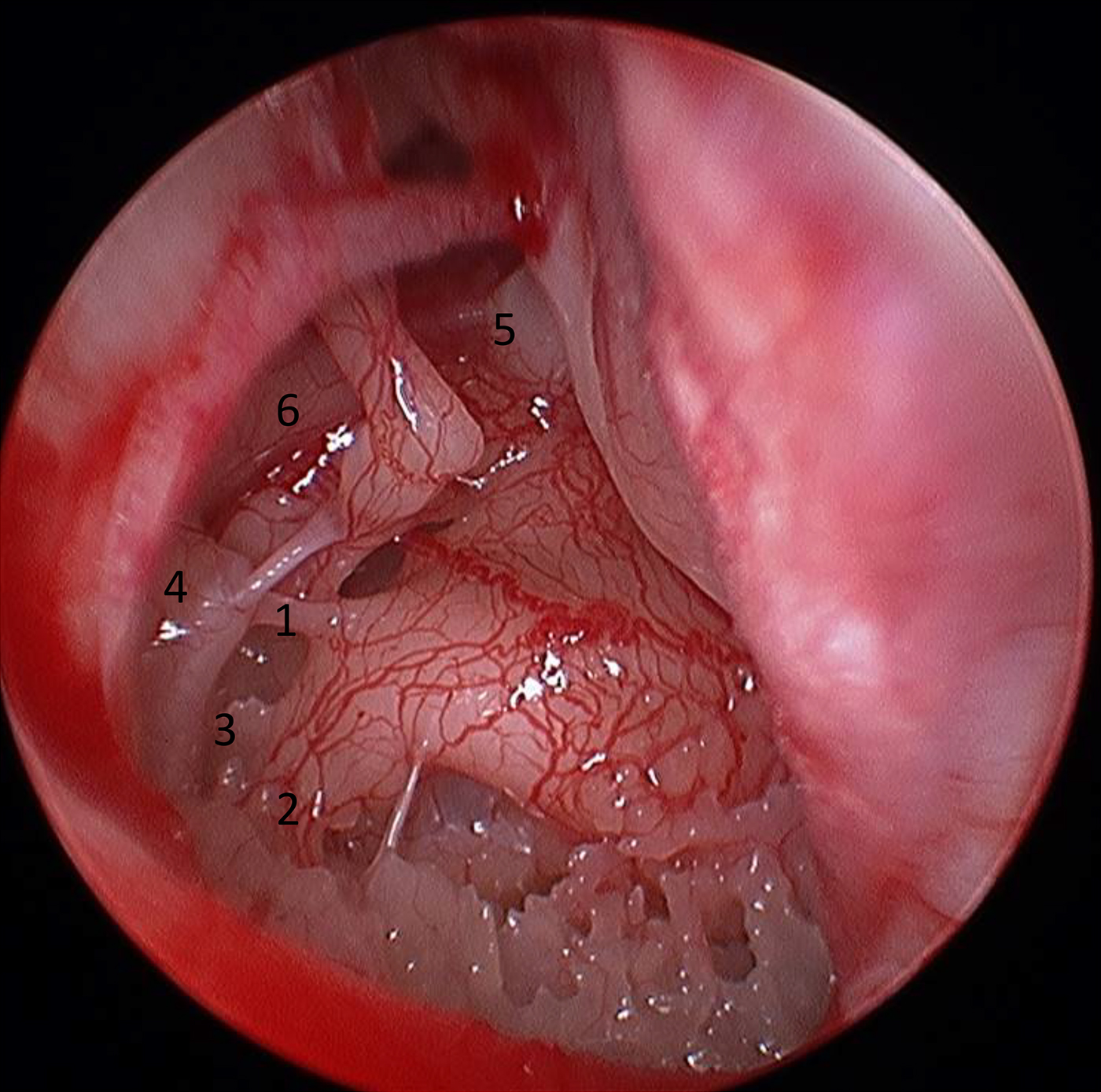
The advantages of the endoscopic approach include higher magnification and resolution, alongside the wider field of view. It allows visualisation into deeper recesses of the middle ear, and improves access to difficult to reach areas, such as the sinus tympani, anterior epitympanum and facial recess (Figures 1 and 2).Reference Tarabichi, Ayache, Nogueira, Al Qahtani and Pothier5 As the light source is attached to the tip of the endoscope, adequate light can reach the middle ear without the need for soft tissue retraction and bony drilling, which are frequently required during a microscopic approach. This, coupled with the fact that the external auditory canal is used as the surgical port, minimises the morbidity associated with external wounds and drilling.
Endoscopic overview of the middle ear. 1 = chorda tympani; 2 = head of stapes; 3 = Eustachian tube; 4 = ridge of tensor tympani muscle; 5 = anterior attic (supratubal recess); 6 = attic

Endoscopic view of the retrotympanum. 1 = ponticulus; 2 = subiculum; 3 = sinus tympani; 4 = pyramid; 5 = tensor tympani; 6 = facial nerve
The main disadvantage of endoscopic middle-ear surgery is that one hand is occupied in controlling the endoscope, leaving only one hand to operate. This can be challenging, particularly when there is blood in the operating field. Another disadvantage is the loss of binocular vision provided by traditional microscopes. There is also concern about the heat generated by extended static application of the endoscope.Reference Tarabichi6
To date, very few studies have systematically reviewed the outcomes of procedures using the endoscope as an operative tool. They have included only a small number of patients, and operations were performed by a select group of experts.Reference Tarabichi, Ayache, Nogueira, Al Qahtani and Pothier5–Reference Kozin, Lehmann, Carter, Hight, Cohen and Nakajima7 Here, we present the preliminary outcomes of the first 103 cases performed in our centre, which, to our knowledge, is the largest case series in the UK.
Materials and methods
A prospective review of the first 103 consecutive patients undergoing totally (transcanal) endoscopic ear surgery or endoscope-assisted microsurgery was conducted. Totally endoscopic ear surgery was defined as a procedure where the entire operation was carried out using an endoscope, while endoscope-assisted microsurgery denoted those procedures where conventional microscopic techniques were required at a later stage. This was invariably because of disease extending beyond the reach of the endoscopic instruments, which was defined as crossing the midway point of the lateral semi-circular canal.
All procedures were carried out in a single centre (NHS Lanarkshire) by one operator. The data were collected using the Common Otology Audit tool,8 with supplementary information obtained from the patients’ case notes. Procedures were carried out between January 2014 and August 2016. Complete information was available for 97 patients, with 6 being lost to follow up.
All patients were reviewed at three weeks post-operatively and the further minimum review period was three months. Tympanic membrane healing was assessed using otoscopy and out-patient microscopy.
Audiograms were carried out pre-operatively and three months’ post-operatively. The mean of 0.5, 1, 2 and 3 kHz was used to calculate a four-tone pure-tone average, as per guidelines.8 The status of the chorda tympani was assessed intra-operatively. Statistical analysis was conducted using Excel spreadsheet software (Microsoft, Redmond, Washington, USA).
Results
Basic demographics
There was an even number of men and women. The mean age was 43 years, with a range of 6–66 years. Of the 103 procedures performed, 78 were completed totally endoscopically, while 25 were endoscopically assisted. The different types of procedures performed, as well as the number of each, can be found in Table I.
Summary of different types of surgery performed

Tympanic membrane healing
The 29 patients undergoing stapedectomy were removed from the analysis as they all had intact tympanic membranes pre-operatively and at 3 months post-operatively. Of the remaining 74 patients, 4 were not grafted at the time of surgery. In all cases, this was because of the extent of the disease. Four patients were lost to follow up. The tympanic membrane healing rate of the remaining 66 patients was 89 per cent (59 patients). Of the seven patients who had residual perforations, two were ‘pinhole’ perforations and three were revision procedures. One patient had previously undergone radiotherapy for nasopharyngeal carcinoma and two had documented post-operative infection. In those patients with perforations, the material used to reconstruct the tympanic membrane was as follows: four patients had perichondrial and cartilage grafts, two had bio-engineered grafts, and one had a fascial graft.
Audiological data
Of the 29 patients undergoing stapedectomy, the chorda tympani was displaced in 26 and lacerated in 2 patients. Only in one case was it necessary to cut the chorda tympani to gain adequate access. This corresponds to the overall complications, with only one patient complaining of post-operative dysgeusia; however, their chorda tympani was not cut.
The mean air–bone gap across all procedures at three months’ follow up was 15.498 dB, with a range of 2.5–50 dB. The mean air–bone gaps for the different procedures are outlined in Table II. Detailed audiological data for the patients undergoing stapedectomy are published elsewhere.Reference Sproat, Yiannakis and Iyer9
Air–bone gap at three months’ follow up

ABG = air–bone gap
Complications
Fourteen of the 103 patients had documented complications, as seen in Table III. Of these, seven patients were treated for infection, two of whom required hospital admission. The patient with facial nerve palsy presented 9 days post-operatively with a House–Brackmann III palsy. This was treated with topical and systemic antimicrobials and steroids, with removal of aural packing. The weakness had completely resolved by three weeks. Five patients reported two simultaneous complications: two with vertigo and infection, one with reduced hearing and infection, one with pain and vertigo, and one with temporary facial nerve palsy and infection. With the exception of the patients with chronic pain, all complications resolved within three months.
Reported complications

Discussion
Safety
Our preliminary results with regard to complications and patient-reported morbidity support those previously published in the literature, and indicate that endoscopic middle-ear surgery is safe. The overall complication rate was low. The only complication that had not resolved by three months was patient-reported chronic pain. Interestingly, this patient complained of bilateral otalgia, with the worse ear being the side that had been operated on using microscopic techniques.
Improved visualisation effect on outcomes
One of the principle advantages of using the endoscope is better visualisation, particularly of middle-ear structures and the sinus tympani. However, research is limited as to whether this translates into better outcomes. With regard to tympanic membrane healing, our results were comparable to the established literature. Although our results represent, to our knowledge, the largest patient group from the UK, the number of individual procedures, such as myringoplasties or ossiculoplasties, is small. Therefore, it is difficult to establish whether there is a statistically significant difference in tympanic membrane healing rates compared with the same surgeon's outcomes for conventional surgery and published data. A further question would be whether the type of graft and packing material had an impact on healing rates.
Our audiological outcomes were noted to be similar to the benchmark, as well as the same surgeon's outcomes using microscopic techniques. Although our sample size for patients undergoing stapedectomy is too small to allow statistical analysis, our results are promising.
Our secondary outcome results, regarding injury to the chorda tympani, are also encouraging. This confirms previous proposals that the improved visualisation of the middle ear provided by an endoscopic technique results in less intentional or accidental damage to the chorda tympani.Reference Migirov and Wolf10
Future research
Whether the improved visualisation using endoscopic techniques as opposed to traditional microscopic techniques translates into reduced rates of residual or recurrent cholesteatoma remains to be seen. The average time to recurrence is approximately 12–18 months. Consequently, although no evidence of recurrence has been identified in our patient series to date, it is not possible to draw conclusions with regard to this. The advantage of our prospective series is that we can actively collect these data alongside tympanic membrane healing rates and audiological data.
• Endoscopic middle-ear surgery is emerging as an alternative to microscopic techniques
• Both tympanic membrane healing rates and audiological outcomes are comparable to conventional techniques
• Preliminary results indicate that endoscopic middle-ear surgery is safe, with low overall complication rates
The two principal problems with this study are the small sample sizes for individual procedures and the length of follow up. The sample sizes make statistical analysis unfeasible, while the length of follow up limits the conclusions that can be made regarding long-term outcomes. The study does not take into account the impact of the learning curve for individual surgeons. This has implications on both the success of the procedures, and the financial and time costs.
Conclusion
Our results confirm that endoscopic middle-ear surgery is safe, with low complication rates. They indicate that short-term outcomes are comparable to those of conventional microscopic techniques. However, it is difficult to draw a statistically significant conclusion given the small sample sizes for individual procedures.
Long-term data are essential, particularly with regard to cholesteatoma recurrence, where the average time to recurrence is approximately 18 months. Despite these limitations, we feel that endoscopic middle-ear surgery provides a promising addition to standard techniques.
Competing interests
None declared



