Introduction
Amino acids have a major effect on the control of protein metabolism. Provision of the different essential amino acids is a prerequisite for maintenance of optimal rates of protein synthesis, and this is an unquestionable major concept in protein nutrition. In other words, amino acids are substrates for protein synthesis and the lack of even a single essential amino acid causes a decrease in protein synthesis. Amino acid availability affects not only protein synthesis rates, but also proteolysis and amino acid oxidation, thus modulating the different pathways involved in protein metabolism. Moreover, studies conducted during the last ten years indicate that amino acids act as regulators of metabolic pathways, with for instance an effect targeted on mRNA translation into proteins.
Amino acids exert their action in various tissues and organs, and in particular they are able to regulate muscle development. The regulation of protein metabolism by amino acids has been studied extensively in skeletal muscle in order to improve muscle growth and meat quality in animal production, and to reduce muscle wasting in some physiological (e.g. early lactation) and physiopathological situations (e.g. ageing, infections, etc). Indeed, due to the differences between the amino acid composition of acute-phase proteins synthesized during catabolic states and that of muscle proteins, a considerable amount of muscle protein has to be degraded to provide the amino acids used in the acute-phase response (Reeds et al., Reference REEDS, FJELD and JAHOOR1994). This review aims to address the important question of how protein nutrition, i.e. provision of amino acids, may contribute to the optimisation of metabolic efficiency and product quality. It also describes the general effects of amino acids with implications for body composition and meat quality. It also highlights the role of amino acids as nutrient signals, which appears to be important for cell functions and metabolic pathways. The last part of this review focuses on sulphur amino acids and their role in controlling oxidative status and the methylation process.
Body composition and muscle quality
Amino acids are known as anabolic factors, which induce protein gain by stimulating protein synthesis while inhibiting proteolysis. These effects on protein turnover have been clearly demonstrated both in mammals and in avian species (Hocquette et al., Reference HOCQUETTE, TESSERAUD, CASSAR-MALEK, CHILLIARD and ORTIGUES-MARTY2007; Muramatsu, Reference MURAMATSU1990; Tesseraud et al., Reference TESSERAUD, ABBAS, DUCHENE, BIGOT, VAUDIN and DUPONT2006 for reviews). Protein-deficient diets cause inhibition of protein synthesis, whereas the effect on proteolysis seems to depend on the severity of the deficiency. Hyperaminoacidemia induced by intravenous infusion of amino acids increases muscle protein synthesis, at least when measurements are performed using the constant infusion of labelled amino acids (Watt et al., Reference WATT, CORBETT and RENNIE1992; Tesseraud et al., Reference TESSERAUD, GRIZARD, DEBRAS, PAPET, BONNET, BAYLE and CHAMPREDON1993). Amino acids are essential for postprandial stimulation of protein synthesis, as demonstrated in the rat (Yoshizawa et al., Reference YOSHIZAWA, KIMBALL, VARY and JEFFERSON1998) and the chicken (Yaman et al., Reference YAMAN, KITA and OKUMURA2000). Stimulation of protein synthesis by amino acids is associated with an increase in translational efficiency with, in particular, stimulation of the initiation phase which corresponds to the sequential binding of the 40S and 60S ribosomal sub-units on the mRNA molecule. Because re-feeding elicits a rise in plasma concentrations of insulin and amino acids, the role of insulin has also to be considered. Interestingly, some studies have suggested that a minimum level of insulin is required for the stimulation of protein synthesis by re-feeding or amino acids (see Kimball et al., Reference KIMBALL, FARRELL and JEFFERSON2002 for a review). Insulin may thus have a permissive effect for the amino acid-induced stimulation of protein synthesis.
Evidence of the action of amino acids in protein turnover has been provided using diets supplemented with or deprived of one or more amino acids. Methionine is for example the first limiting factor in classical diets used for growing chickens because of a high requirement for sulphur amino acids for the synthesis of feathers, whereas poultry diets based on corn and soybean meal are deficient in sulphur amino acids without supplementation (Baker, Reference BAKER2006). Decreased growth in chickens fed methionine- and cysteine-free diets is mainly caused by lower rates of whole-body protein synthesis associated with lower RNA efficiency, suggesting translational regulation. Methionine supplementation enhances muscle growth, and the addition of methionine to a methionine-deficient diet, otherwise balanced in terms of other amino acids, increases accretion and synthesis of protein in the Gastrocnemius and Pectoralis major muscles in chickens (Barnes et al., Reference BARNES, CALVERT and KLASING1995). Similarly, lysine supplementation to a lysine-deficient diet otherwise balanced in terms of other amino acids increases the amounts of protein synthesized and gained in chicken skeletal muscle (Tesseraud et al., Reference TESSERAUD, PERESSON, LOPES and CHAGNEAU1996a, Reference TESSERAUD, TEMIM, LE BIHAN-DUVAL and CHAGNEAU2001).
Studies performed using lysine-deficient diets have revealed the possibility of a major effect of dietary amino acid levels on protein degradation: the fractional rates of proteolysis (values expressed as % per day) measured in the Pectoralis major muscle of growing chickens are always higher in lysine-deprived animals, irrespective of age or genotype (Tesseraud et al., Reference TESSERAUD, PERESSON, LOPES and CHAGNEAU1996a, Reference TESSERAUD, TEMIM, LE BIHAN-DUVAL and CHAGNEAU2001). Amino acid deficiency stimulates autophagic (lysosomal degradation) and proteasome-mediated proteolysis (Hamel et al., Reference HAMEL, FAWCETT, BENNETT and DUCKWORTH2004; Bechet et al., Reference BECHET, TASSA, COMBARET, TAILLANDIER and ATTAIX2005). In chicken muscle, daily variations in dietary lysine content affect the expression of two important genes controlling the ubiquitin-proteasome proteolytic pathway, i.e. E3 ubiquitin ligases Muscle Atrophy F box (MAFbx, also called atrogin-1) and Muscle Ring Finger-1 (MuRF1) (Tesseraud et al., Reference TESSERAUD, BOUVAREL, COLLIN, AUDOUIN, CROCHET, SEILIEZ and LETERRIER2009a).
It should be noted that lysine is one amino acid that probably exerts the most specific effects on carcass composition and muscle growth. A particularly drastic effect has been observed on breast muscle development in chickens (Tesseraud et al., Reference TESSERAUD, PERESSON, LOPES and CHAGNEAU1996a). It is probable that the breast muscle of chickens represents a major reservoir of lysine that can be mobilised in deficiency states. Increased proteolysis in such conditions provides free amino acids that are used for protein synthesis. It is of note that neither threonine nor valine exhibits an effect on body composition as pronounced as that of lysine, as observed in an experiment in which these three amino acids were studied together in similar conditions (Leclercq, Reference LECLERCQ1998). The specific effect of lysine on breast muscle development may be due to the fact that lysine is the most abundant essential amino acid in skeletal muscle protein. Interestingly, a recent study has also revealed that increasing lysine can improve chicken breast muscle quality by increasing its ultimate pH and water holding capacity (Berri et al., Reference BERRI, BESNARD and RELANDEAU2008), but the underlying mechanisms have yet to be defined.
Based on findings reported in piglets (Conde-Aguilera et al., Reference CONDE-AGUILERA, BAREA, LE FLOC'H, LEFAUCHEUR and VAN MILGEN2010), it appears that amino acids modify the chemical composition of tissues with a potential effect on meat quality. In particular, a deficient supply of methionine and/or cysteine affects lipid content and amino acid composition of proteins in muscles.
Nutrient signalling
Several reviews have reported that amino acids are signal mediators acting on the same intracellular protein kinases as certain hormones (e.g. insulin) (Kimball and Jefferson, Reference KIMBALL and JEFFERSON2006a; Dann and Thomas, Reference DANN and THOMAS2006; Tesseraud et al., Reference TESSERAUD, ABBAS, DUCHENE, BIGOT, VAUDIN and DUPONT2006; Métayer et al., Reference MÉTAYER, SEILIEZ, COLLIN, DUCHÊNE, MERCIER, GERAERT and TESSERAUD S.2008). Thus, amino acids, in particular the branched-chain amino acid leucine in skeletal muscle, affect the activity of several proteins involved in the control of mRNA translation, such as 4E binding protein (4E-BP1) and 70 kDa ribosomal protein S6 kinase (p70S6K, also called S6K1) (Figure 1). S6K1 and, similarly, 4E-BP1 are phosphorylated in response to insulin via a signal transduction pathway involving phosphatidylinositol-3’ kinase (PI3K) and the mammalian target of rapamycin (mTOR) in mammals and avian species (Proud, Reference PROUD2006; Tesseraud et al., Reference TESSERAUD, MÉTAYER, DUCHÊNE, BIGOT, GRIZARD and DUPONT2007a; Dupont et al., Reference DUPONT, TESSERAUD and SIMON2009). Amino acid signalling also originates from mTOR, and activates S6K1 and 4E-BP1 (Meijer and Dubbelhuis, Reference MEIJER and DUBBELHUIS2004; Tesseraud et al., Reference TESSERAUD, ABBAS, DUCHENE, BIGOT, VAUDIN and DUPONT2006). Despite the increased understanding of the roles of amino acids, many questions still remain and the underlying mechanisms need to be elucidated. One of the key issues relates to the specificity of amino acids acting as ‘nutrient signals’. The effects of branched-chain amino acids on the mTOR/S6K1 pathway have been clearly demonstrated (Kimball and Jefferson, Reference KIMBALL and JEFFERSON2006b; Yoshizawa, Reference YOSHIZAWA2004). However, other amino acids may also exert an effect on cell signalling. For example, arginine regulates S6K1 and 4E-BP1 in intestinal cells (Ban et al., Reference BAN, SHIGEMITSU, YAMATSUJI, HAISA, NAKAJO, TAKAOKA, NOBUHISA, GUNDUZ, TANAKA and NAOMOTO2004), and glutamine can control S6K1 activation and 4E-BP1 phosphorylation induced by arginine or leucine (Nakajo et al., Reference NAKAJO, YAMATSUJI, BAN, SHIGEMITSU, HAISA, MOTOKI, NOMA, NOBUHISA, MATSUOKA, GUNDUZ, YONEZAWA, TANAKA and NAOMOTO2005). There are few studies regarding the potential effects of methionine on these intracellular kinases, but they suggest that this sulphur amino acid may have a signal function by inducing S6K1 activation in mammals (Shigemitsu et al., Reference SHIGEMITSU, TSUJISHITA, MIYAKE, HIDAYAT, TANAKA, HARA and YONEZAWA1999; Stubbs et al., Reference STUBBS, WHEELHOUSE, LOMAX and HAZLERIGG2002) and avian species (Tesseraud et al., Reference TESSERAUD, BIGOT and TAOUIS2003; Métayer-Coustard et al., Reference MÉTAYER-COUSTARD, MAMERI, SEILIEZ, CROCHET, CRÉPIEUX, MERCIER, GERAERT and TESSERAUD2010). In addition, amino acid availability regulates the expression of the E3 ubiquitin ligase atrogin-1 via mTOR in muscle cell lines (Tesseraud et al., Reference TESSERAUD, MÉTAYER-COUSTARD, BOUSSAID, CROCHET, AUDOUIN, DEROUET and SEILIEZ2007b; Herningtyas et al., Reference HERNINGTYAS, OKIMURA, HANDAYANINGSIH, YAMAMOTO, MAKI, IIDA, TAKAHASHI, KAJI and CHIHARA2008).
mTOR signalling and control of protein synthesis and proteolysis. Akt or PKB, protein kinase B; atrogin-1 or MAFbx, muscle atrophy F box; 4E-BP1, eukaryotic initiation factor 4E binding protein; FoxO, forkhead box-O transcription factor; mTOR, mammalian target of rapamycin; MuRF1, Muscle Ring Finger-1; S6K1, 70 kDa ribosomal protein S6 kinase.

Interestingly, the activation by feeding and by amino acids of components related to translation initiation is developmentally regulated, with enhanced responses during the neonatal period, as reported by several studies in piglets (Suryawan et al., Reference SURYAWAN, NGUYEN, BUSH and DAVIS2001, Reference SURYAWAN, ORELLANA, NGUYEN, JEYAPALAN, FLEMING and DAVIS2007). Decreased stimulation with age parallels the developmental decline in protein synthesis in skeletal muscle, which is due to lower efficiency of the translation process and depressed synthesis capacity (Davis et al., Reference DAVIS, SURYAWAN, ORELLANA, NGUYEN and FIOROTTO2008). Similar age-related changes might exist in avian species since developmental declines in the capacity for protein synthesis (Tesseraud et al., Reference TESSERAUD, PERESSON and CHAGNEAU1996b) and in the abundance of kinases involved in intracellular signalling (Vaudin et al., Reference VAUDIN, DUPONT, DUCHÊNE, AUDOUIN, CROCHET, BERRI and TESSERAUD2006) have been reported in chick muscle. Considering the drastic effects of amino acids on proteins related to translation initiation in the neonatal period (Escobar et al., Reference ESCOBAR, FRANK, SURYAWAN, NGUYEN, KIMBALL, JEFFERSON and DAVIS2006, Reference ESCOBAR, FRANK, SURYAWAN, NGUYEN and DAVIS2007), consumption of an amino acid-enriched diet could be particularly valuable to improve protein synthesis and growth in this critical period. A recent study performed in neonatal chicks showed that the consumption of a high protein diet clearly activated S6K1 and augmented its activity in the breast muscle (Everaert et al., Reference EVERAERT, SWENNEN, COUSTARD, WILLEMSEN, CAREGHI, BUYSE, BRUGGEMAN, DECUYPERE and TESSERAUD2010). These findings indicated that the activation of S6K1 is sensitive to dietary amino acid provision during early feeding, which could potentially enhance the translation of mRNA in skeletal muscle of neonatal chicks.
One unsolved issue concerns the multiple effects of amino acids on cell functioning. The role of amino acids as nutrient signals appears to be important for cell functions and metabolic pathways, other than those directly concerned with protein turnover. For example, amino acids may be strong modulators of glucose metabolism and insulin sensitivity. In vitro and in vivo findings indicate an important role of amino acids in glucose homeostasis through inhibition of insulin action on muscle glucose transport and hepatic glucose production. This negative effect of amino acids on insulin action appears to be associated with mTOR/S6K1-dependent inhibitory phosphorylation of insulin receptor substrate-1 (IRS-1) on serine and/or threonine residues and impaired activation of PI3K, a key effector of the metabolic actions of insulin (Tremblay et al., Reference TREMBLAY, LAVIGNE, JACQUES and MARETTE2007; Um et al., Reference UM, D'ALESSIO and THOMAS2006). Depletion of S6K1 was recently shown to enhance basal glucose utilization in mammalian muscle cells and to protect against the development of impaired insulin action in response to excessive leucine (Deshmukh et al., Reference DESHMUKH, SALEHZADEH, METAYER-COUSTARD, FAHLMAN, NAIR and AL-KHALILI2009). More information is therefore needed on the potential effects of amino acid supplementation before use for nutritional purposes.
Sulphur amino acids, control of oxidative status and methylation process
As precursors of various molecules, amino acids have other major functions. For instance, the antioxidants glutathione and cysteine, originating from methionine metabolism, can function as direct scavengers of reactive oxygen species (ROS), formed at the mitochondrial level and producing deleterious effects such as lipid and protein oxidation or DNA strand-break damage. Glutathione, which is a tripeptide (L-glutamyl-L-cysteinyl-glycine), is the most important intracellular antioxidant in the body (Obled et al., Reference OBLED, PAPET and BREUILLE2004; Reid and Jahoor, Reference REID and JAHOOR2001). Acute infection increases its synthesis in various tissues (liver, spleen, lung and muscles), accounting for at least 40% of the enhanced cysteine utilisation during infection in the rat (Malmezat et al., Reference MALMEZAT, BREUILLE, CAPITAN, MIRAND and OBLED2000). Methionine metabolism was affected in normal volunteers subjected to a mild inflammatory challenge (i.e. vaccination; Mercier et al., Reference MERCIER, BREUILLE, BUFFIERE, GIMONET, PAPET, MIRAND and OBLED2006), in agreement with results obtained in acute diseases. Preferential methionine metabolism toward cysteine synthesis has thus been observed, indicating increased requirement for sulphur amino acids in these situations. Interestingly, using everted sac techniques with chicken intestine, it was demonstrated that the methionine hydroxy-analog, DL-2-Hydroxy-(4-Methylthio) butanoic acid, is more efficiently converted to cysteine and taurine than L-methionine through the trans-sulphuration pathway, suggesting that this hydroxy-analog of methionine might have a different role in detoxification processes than methionine itself (Martin-Venegas et al., Reference MARTIN-VENEGAS, GERAERT and FERRER2006).
Sulphur amino acids are involved in the methionine sulphoxide reductase anti-oxidant system (Moskovitz et al., Reference MOSKOVITZ, RAHMAN, STRASSMAN, YANCEY, KUSHNER, BROT and WEISSBACH1995; Levine et al., Reference LEVINE, MOSONI, BERLETT and STADTMAN1996). Methionine residues in proteins are particularly susceptible to oxidation by ROS and are converted to methionine sulphoxide, which can be reduced back to methionine by methionine sulphoxide reductases. Each cycle of methionine oxidation and reduction will destroy one equivalent of ROS, which might represent a major natural scavenging system for ROS. The methionine sulphoxide reductase system is particularly involved in the repair of oxidized proteins, thereby preventing cellular accumulation of damaged proteins (Stadtman et al., Reference STADTMAN, VAN REMMEN, RICHARDSON, WEHR and LEVINE2005). Nevertheless, the effects of different sulphur compounds in critical physiopathological conditions is not yet fully understood, especially with regard to host defence against oxidative stress and protection against lipid and protein oxidation. These findings that impact on tissue metabolism have potential implications for animal health, tissue development and product (meat) quality.
Methionine is a source of methyl groups that are used to methylate DNA, a process that influences chromatin structure and gene expression. The mechanisms involved have been detailed in a recent review and therefore will not be presented here (Tesseraud et al., Reference TESSERAUD, MÉTAYER COUSTARD, COLLIN and SEILIEZ2009b). From a nutritional point of view, one interesting application concerns perinatal nutrition since aberrant methyl metabolism in early life is linked with disorders. This means that manipulating the sulphur amino acid content of the early diet may induce chronic changes in cell functions that have implications for long-term health. For instance, it has been shown in mammals that changes within physiological ranges in the supply of methionine and specific B vitamins such as B12 during the periconception period can lead to widespread epigenetic changes in DNA methylation in offspring, and can modify adult health-related phenotypes (Sinclair et al., Reference SINCLAIR, ALLEGRUCCI, SINGH, GARDNER, SEBASTIAN, BISPHAM, THURSTON, HUNTLEY, REES, MALONEY, LEA, CRAIGON, MCEVOY and YOUNG2007). To our knowledge, such epigenetic regulation by nutrients has not yet been investigated in avian species, although there is evidence of epigenetic changes in chicks after heat-conditioning (Kisliouk et al., Reference KISLIOUK, ZIV and MEIRI2010).
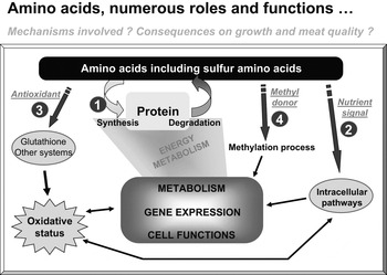
Regulation of metabolism, numerous roles played by amino acids: 1) precursors of protein synthesis, 2) nutrient signal, 3) antioxidant function and 4) methyl group donor. The figure represents a summary of findings discussed in the present review.

Conclusions
Amino acids are recognised to be essential in controlling metabolism (Figure 2). Despite the increased understanding of the role of amino acids, many questions still remain unanswered. Identifying the mechanisms by which amino acids regulate metabolism, signalling, gene expression and cell functioning is thus essential to improve the control of nutrient utilisation and to optimise dietary amino acid provision for animal production purposes.
Acknowledgements
The authors thank I. Seiliez (INRA, St Pée-sur-Nivelle, France) for valuable discussions, and D. Raine (Surrey, UK) for editing the English language.