Introduction
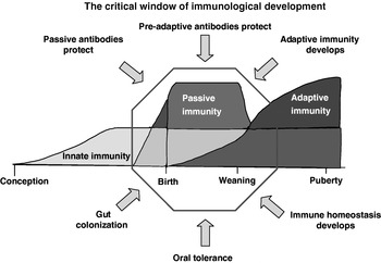
Continuation of a species or population depends on the survival of the offspring. Invertebrates and lower vertebrates (fish and amphibians) accomplish this by producing thousands of offspring with the goal that the species will continue even if only a very small number survive. Since birds, mammals and most reptiles conceive only 1–30 offspring per gestation, their survival depends more on parental care than sheer numbers. Parental care includes a direct physical bond between parent and offspring as well as passive immune protection. These protective factors have their major impact during the ‘critical window of immunological development’ (Fig. 1; Butler et al., Reference Butler, Sinkora, Wertz, Holtmeier and Lemke2006a; Butler and Sinkora, Reference Butler, Lemke, Weber, Sinkora and Lager2007). This period extends from late gestation to at least weaning. During this window, adaptive immunity develops and is superimposed on the intrinsically programmed innate immune system.
The critical window of immunological development extends from late gestation to weaning. Arrows depict critical factors or events within the ‘window’. The adaptive immune system that develops postnatally complements and is superimposed upon the intrinsic innate immune system that had developed during fetal life. Development of adaptive immunity including oral tolerance is dependent on gut colonization (see Fig. 4). Colonization and passive immunity are believed to be important for the development of immune homeostasis by tolerating commensal gut flora, dietary antigens and dampening responses to self-antigens. This developmental process proceeds during the window when offspring are protected from pathogens by passive antibody, natural antibodies and innate immunity. From Butler et al. (Reference Butler, Sinkora, Wertz, Holtmeier and Lemke2006a) and Butler and Sinkora (Reference Butler, Lemke, Weber, Sinkora and Lager2007).

Development of adaptive immunity not only involves gaining the ability to respond to non-self antigens in an adaptive manner, but also to down-regulate responses to those antigens which are not virulence factors. This has lead to the ‘no harm, no foul’ concept or the ‘danger hypothesis’ (Matzinger, Reference Matzinger2002). The development of adaptive immunity also includes the development of tolerance to dietary antigens and the establishment of ‘peaceful co-existence’ with normal gut flora. Various factors, including certain neonatal pathogens, failure to encounter certain other pathogens and commensals, nutritional factors, genetic factors and a failure of passive immunity, can interfere with the development of this ‘immune homeostasis’ during the critical window (Fig. 1). For example, the failure of neonates to become infested with certain helminth parasites predisposes them to the development of inflammatory bowel disease (IBD; Summers et al., Reference Summers, Elliott, Qadir, Urban, Thompson and Weinstock2003; Elliott et al., Reference Elliott, Setiawan, Metwali, Blum, Urban and Weinstock2004) and this disease can be exacerbated by certain commensal gut bacteria but not others (Heimesaat et al., Reference Heimesaat, Bereswill, Fischer, Fuchs, Struck, Niebergall, Jahn, Dunay, Moter, Gescher, Schumann, Goebel and Liesnfeld2006). Dietary deficiency of selenium and vitamin E (Smith et al., Reference Smith, Madden, Au Yeung, Zhao, Elfrey, Finkelman, Levander, Shea-Donohue and Urban2005) can reduce the timely expulsion of these helminths but ‘overstimulation’ of Th2 responses as a result of helminth infestation can have negative consequences for protective Th1 mediated immunity to Mycoplasma (Urban et al., Reference Urban, Steenhard, Solano-Aquilar, Dawson, Iweala, Nagler, Noland, Kumar, Anthony, Shea-Donohue, Weinstock and Gause2007). These studies indicate the importance of the delicate balance among the effects of colonization, helminth infection and nutrition. The increase in IBD and atopic allergy in highly developed countries have spawned various theories that are embodied in what has collectively become known as the ‘hygiene hypothesis’ (Rook and Stanford, Reference Rook and Stanford1998; Yazdanbaksh et al., Reference Yazdanbaksh, Kremsner and van Ree2002). This theory is regularly cited by the probiotic industry (Guarner and Malagelada, Reference Guarner and Malagelada2003; Walker and Buckley, Reference Walker and Buckley2006). The neonatal period is a time when the constituency of the normal gut flora is not yet established as it is in adults (Ducluzeau, Reference Ducluzeau1993; Zoentendal et al., Reference Zoentendal, Akkermans and De Vos1998; Tapiainen et al., Reference Tapiainen, Ylitalo, Eorola and Uhari2006; Walker and Buckley, Reference Walker and Buckley2006). During that period polyreactive B cells that recognize anti-nuclear antigens are numerous and persist in germfree mice (Maldonado et al., Reference Maldonado, Kakkanaiah, MacDonald, Chen, Reap, Balish, Jennette, Madalo, Kotzin and Eisenberg1999). The pathway to normal development of tolerance during this time is susceptible to dysregulation by agents like B cell superantigens (BSAgs) (Zouali, Reference Zouali1995; Silverman et al., Reference Silverman, Nayak and La Cava1997; Viau et al., Reference Viau, Cholly, Bjorck and Zouali2004). Among these is tolerance to dietary antigen, which depends on gut colonization for development (Sudo et al., Reference Sudo, Sawamura, Tanaka, Aiba, Kudo and Koga1997). Therefore many believe that development of immune homeostasis during this period depends on the response of innate immune receptors to pathogen-associated molecular patterns (PAMPs).
Herdsmen long before the growth of science recognized that if the offspring of domesticated animals like sheep, cattle, swine and horses failed to suckle during the critical window, they quickly died. Veterinary science would eventually show this to be not mere starvation, but death from infectious disease due to the lack of passive maternal antibodies (Fey and Margadant, Reference Fey and Margadant1961; Gay et al., Reference Gay, Anderson, Fisher and McEwan1965). In the future, we are likely to learn that other factors passively acquired during the critical window are also necessary for the neonate to develop a healthy adaptive immune system at homeostasis with its environment (Fig. 1).
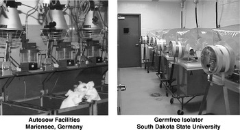
The reproductive and developmental nature of swine makes the piglet a convenient model to study events during the critical window. While the pathway for passive immunity differs between swine, rodents and humans, the end result is the same (Butler, Reference Butler, Larson and Smith1974; Butler and Kehrle, Reference Butler, Kehrle, Mestecky, Lamm, Strober, McGhee, Mayer and Bienenstock2005). While humans use IgG transport receptors in the placenta to actively transport protective antibodies to their fetus, the transport receptors for swine (and other artiodactyls) are on the acinar epithelial cells of the mammary gland. Circa 3 days after birth IgG transport in the mammary gland ceases and the Ig constituency, i.e. 80% IgA, becomes similar to that in humans. This makes piglets excellent models for fetal and newborn studies because piglets receive no maternal antibodies and probably no other maternal proteins in utero. Thus, the immunological development that occurs in fetal life is intrinsically driven, which removes the ambiguity encountered when studying immune development in fetal rodents or infants. Furthermore swine, like other large farm animals, give rise to precosial offspring, allowing them to be reared separate from their birth mothers in specific pathogen free (SPF) autosows or germfree isolators (Fig. 2) so the immediate postnatal environmental influences are under the control of the experimenter. These include control of gut flora, pathogens, diet and passive maternal immunity. Neonatal piglet models differ in the degree to which these external factors are experimentally controlled; increasing stringency in control of these factors is proportional to the cost of the experimental system, e.g. closed isolator systems are very expensive (see below).
Experimental rearing systems for neonatal piglets. Left: SPF autosow facilities for rearing of newborn or surgically derived piglets. Right: germfree isolators for Caesarian-derived piglets.

Fortunately for medical science, swine share various physiological features with humans. These include a similar respiratory tract (Pabst and Binns, Reference Pabst and Binns1994; Maina and Gils, Reference Maina and van Gils2001), an omnivorous diet with similar commensal flora and nutritional needs (Reeds and Odle, Reference Reeds, Odle, Tumbleson and Schook1996; Lesser et al., Reference Lesser, Amenuvor, Jensen, Linecrona, Boye and Moeller2002) and many shared contagion (Thacker, Reference Thacker2006). Pigs also have related lipoproteins and a propensity for atherosclerosis (Rapacz et al., Reference Rapacz, Hasler-Rapacz, Taylor, Checovich and Attie1986), a mucosal immune system that in many ways is more similar to that of humans than rodents and other artiodactyls (Butler, Reference Butler, Larson and Smith1974; Liebler-Tenorio and Pabst, Reference Liebler-Tenorio and Pabst2006) and susceptibility to dietary allergens (Barratt et al., Reference Barratt, Strachan and Porter1978; Bailey et al., Reference Bailey, Miller, Telemo, Stokes and Bourne1994; Helm et al., Reference Helm, Furuta, Stanley, Ye, Cockrell, Connaughton, Simpson, Bannon and Burks2002; Fu et al., Reference Fu, Jez, Kerley, Allee and Krishnan2007; Rupa et al., Reference Rupa, Hamilton, Cirinna and Wilkie2007). They also have organs of similar size to humans making them popular in transplantation research (Sachs et al., Reference Sachs, Sykes, Robson and Cooper2001). Recently they have become the model of choice for cystic fibrosis (Rogers et al., Reference Rogers, Stoltz, Meyerholz, Ostedgaard, Rokhlina, Taft, Rogan, Pezzulo, Karp, Itani, Kabel, Wohlford-Lenane, Davis, Hanfland, Smityh, Samuel, Wax, Murphy, Rieke, Whitworht, Uc, Starner, Brogden, Shilyansky, MvCray, Zabner, Prather and Welsh2008).
Despite the obvious advantages of piglets for addressing developmental issues during the critical window, adapting the piglet model for immunology has faced several obstacles; included are the paucity of reagents, cost and acceptance by veterinary science. When its use was originally considered in the mid- to late-1960s, the porcine immune system had not been characterized and progress was slow in the decades that followed. Major funding agencies in America (NIH and NSF) considered swine the responsibility of agriculture but the priorities of agriculture rarely include basic immunology. The delay in characterization of the swine immune system would result in delayed development of immunological reagents such as monoclonal antibodies (mAbs) to T- and B-cell subsets, immunoglobulin (Ig) and the many cluster of differentiation (CD) markers that are important in characterizing immunological responses. This ‘immunology deficiency’ in agricultural research has recently been recognized by the ARS (Gay and Richie, Reference Gay and Richie2007). Efforts to improve this situation are also ongoing through the establishment of the USDA-NRI Toolkit Project (http://www.medicine.uiowa.edu/cigw/news.htm) and through support from the National Porkboard. Significant advances in characterizing the porcine immune system have been made in the last 15 years through molecular genetic methods, results of which are later described. Furthermore, as certain mouse models of human diseases fail, alternatives are now being considered as exemplified by the CFTR knockout pig as a model for cystic fibrosis (Rogers et al., Reference Rogers, Stoltz, Meyerholz, Ostedgaard, Rokhlina, Taft, Rogan, Pezzulo, Karp, Itani, Kabel, Wohlford-Lenane, Davis, Hanfland, Smityh, Samuel, Wax, Murphy, Rieke, Whitworht, Uc, Starner, Brogden, Shilyansky, MvCray, Zabner, Prather and Welsh2008) and the development of B cell knockout pigs that can later be used to produce human therapeutic antibodies (M. Mendicino personal communication). These models have the potential for generating a level of financial support not previously available for basic research in an animal species. Further characterization of the porcine immune system will benefit both medical and veterinary science.
Reagent availability affects all studies in porcine immunology but studies using piglet models face additional hurdles. A variety of systems have been developed for using neonatal piglets especially as models for developmental immunology. These include Caesarian-derived colostrum-deprived (CDCD) piglets, autosow reared piglets and germfree isolators (Fig. 2). Piglets used in all systems may originate from SPF herds. The technical details of these systems have been recently reviewed (Butler et al., Reference Butler, Lager, Splichal, Francis, Kacskovics, Sinkora, Wertz, Sun, Zhao, Brown, DeWald, Dierks, Muyldermanns, Lunney, McCray, Rogers, Welsh, Navarro, Klobasa, Habe and Ramsoondar2008a). Whether one chooses to use isolator or SPF autosow facilities, both are very expensive, labor intensive and therefore a hard sell to funding agencies. In addition, some agricultural groups have been ambivalent in their embrace of such research, often considering the results to be artifacts and not models for what occurs in conventionally reared piglets. This appears to be largely a misunderstanding over the purpose of such studies. Using these types of neonatal piglet models is not a substitute for studies on conventionally reared animals but rather a way to reduce the number of experimental variables and put them under the control of the experimenter. This improves the chances of identifying virulence factors and the mechanisms involved in the innate and adaptive responses to pathogens.
Appropriately, a great deal of veterinary research is focused on vaccine development. However, the scope of such research contrasts with vaccine development studies that use mouse models. In the former, resources and energy are focused on the pathogens rather than the host. This is in part due to the lack of necessary reagents and animals of defined genetic background that can be reared in controlled environments. The veterinary approach is largely devoted to developing vaccines for combating especially current and emerging viral pandemics. Since these diseases impact conventional animals, there exists a bias against the use of isolator-based models as contrived and of limited relevance. This review will describe results obtained using neonatal piglet models that hopefully will reduce this bias.
By-products of neonatal piglet studies
Neonatal piglet models have a greater than 50-year history that has been accompanied by developments in technology and a broader understanding of nutrition, passive immunity and neonatal competence. This may be analogous to ‘climbing to the summit of a mountain’ in which discoveries made along the way are equally important as reaching the summit. Characterization of the porcine immune system has been an important by-product of neonatal piglet studies. Reviewed below are some of the characteristic if not unique features of porcine B and T cell systems that have been elucidated during ‘the climb’.
Igs: conservation and divergence
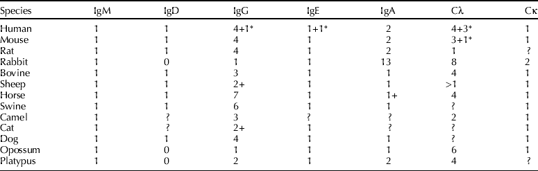
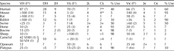
Table 1 compares the isotype diversity of the porcine Igs. Swine, like almost every other eutherian mammal, has genes expressing IgM, IgD, IgG, IgA, IgE, kappa light chains and lambda light chains. Among these, IgM, IgE, kappa and lambda are highly conserved, being present even in protherian mammals. Three features of the porcine Igs will be highlighted: IgD, IgG and light chain usage.
The C-region repertoire of common mammals
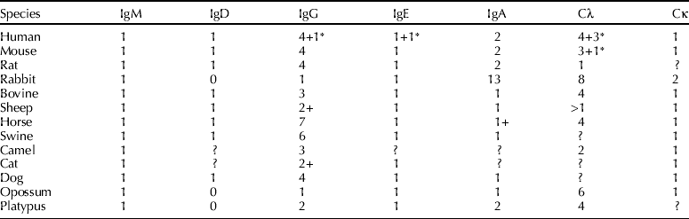
* =pseudogene.
+ =probably more than the number.
? =number not confirmed.
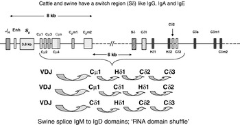
IgD is the least homologous Ig among mammals, including its absence in rabbits and protherian mammals (Table 1). This is particularly interesting since IgD is phylogenetically ‘old’ Ig like IgM. However, in the catfish and Xenopus, it is a nine-domain molecule (Wilson et al., Reference Wilson, Bengten, Miller, Clem, Du Pasquier and Warr1997; Zhao et al., Reference Zhao, Pan-Hammarstrom, Yu, Wertz, Zhang, Li, Butler and Hammarstrom2006) not the four-domain molecule of placental mammals (Fig. 3). The expression of IgD in cattle and swine is different than in mice. Instead of RNA splicing, both cattle and swine have a small switch region and this is functional at least in cattle (Fig. 3; Zhao et al., Reference Zhao, Pan-Hammarstrom, Kacskovics and Hammarstrom2002; Reference Zhao, Pan-Hammarstrom, Kacskovics and Hammarstrom2003). In addition, porcine IgD transcripts can include domain-spliced chimeras that also involve IgM domains (Fig. 3; Zhao et al., 2004). The significance of this alternative transcription is unknown. It is also unknown whether transcripts are translated and if IgD has any role in mammals or represents only an evolutionary vestige.
Transcriptional diversity in IgD expression in swine. Unlike mice that use RNA splicing to express IgD, the Cδ gene exons in the locus of swine, cattle and humans are preceded by a small switch region (Sδ) that may be functional in cattle (Zhao et al., Reference Zhao, Pan-Hammarstrom, Kacskovics and Hammarstrom2003). RNA splicing in swine can produce various chimeric transcripts comprised of both IgM and IgD domain exons. The exons (C or H for hinge) and the switch region (S) are designated for IgM (μ) and IgD (δ). m1 and m2 are membrane-spanning exons. Enh is the heavy chain enhancer. Cμs and Cδs designate the small exons encoding the C-terminal secretory tailpieces. The concave arrows depict the patterns of domain splicing.

The second isotype of special interest is IgG. IgG is the ‘flagship antibody’ of mammals. No other vertebrate class has IgG. Apart from IgD, it is the only antibody with a hinge domain and may have evolved from a common ancestor with IgF of Xenopus (Zhao et al., Reference Zhao, Pan-Hammarstrom, Yu, Wertz, Zhang, Li, Butler and Hammarstrom2006). As shown in Table 1, IgG has diversified into an array of subclasses in mammals ranging from one in rabbits to seven in the horse. This diversification came after speciation so that regardless of the nomenclature, each species has its own unique set of IgG subclasses (Butler, Reference Butler2006; Butler et al., Reference Butler, Wertz, Deschacht and Kacskovics2008b). This means that an IgG called IgG1 in swine, cattle, horse, mouse, human, is not homologous at the subclass level. Human and bovine IgG1 have well-described effector functions but these are different for each species, emphasizing that function cannot be extrapolated from a subclass of the same name in one species to another unless closely related species are involved, e.g. cattle and sheep.
Currently, 11 IgGs are transcribed in swine and these belong to six putative subclasses (Butler et al., Reference Butler, Wertz, Deschacht and Kacskovics2008b). Of particular interest is the number of in-frame splice acceptor sites in the CH2 domain. Also present are transcripts in which the rearranged VDJ can be spliced to the hinge domain which creates an antibody lacking CH1 and therefore potentially also a light chain. Assuming that all porcine IgG transcripts are expressed as antibodies, pigs can make more than 20 different IgGs. The significance of these alternative transcripts is unknown but if translated, they would create an IgG reagent nightmare. Currently, Toolkit grants from the National Porkboard and USDA-NRI are supporting efforts to ‘change this potential nightmare into sweet dreams’.
The third noteworthy feature of porcine Igs concerns light chain usage. Table 2 shows that swine are unusual among hoofed mammals; they equally express kappa and lambda light chains like humans rather than >90% lambda-like domesticated ruminants and the horse. Could this be important when using swine as models in medical science?
Variable region diversity and light chain usage among mammals

(F*) =number of families.
? =number not confirmed.
** =occurs as JλCλ duplicons.
+ =probably more than the number indicated.
Diversity in antibody repertoire development: different strokes for different folks
Antibody specificity is determined by the juxtapositioning of combinatorial determining regions (CDRs) of the heavy (VH) and light (VL) chain variable regions. Genes encoding VH and VL each contain CDR1 and CDR2 regions. CDR3 is the product of gene segment recombination among VH-DH-JH and VL-JL. The CDR3 of the heavy chain (HCDR3) occupies the center of the binding site of the antibody and is thought to account for most of the antibody specificity (Padlan, Reference Padlan1994). B cells diversify their repertoire by joining different V-D-J segments (Table 2; ‘combinatorial diversity’) and by the manner in which V-D-J are joined (‘junctional diversity’). Thereafter, activation-induced cytidine deaminase (AID)-mediated somatic hypermutation (SHM) or gene conversion acts to allow mutations to accumulate in CDR regions and create additional diversity. The degree of combinatorial diversity depends in part on the number of V-gene segments available in the genome.
Table 2 shows that the large numbers of VH genes available for human and mice contrast with that in swine. Furthermore, swine use primarily seven of their <30 VH genes, have only two functional DH segments and a single JH to generate >95% of their combinatorial pre-immune repertoire (Table 2; Sun et al., Reference Sun, Kacskovics, Brown and Butler1994; Butler et al., Reference Butler, Sun and Navarro1996, Reference Butler, Weber and Wertz2006b). This limited combinatorial diversity means that most of the pre-immune repertoire in swine can be attributed to HCDR3 diversity (Butler et al., Reference Butler, Weber, Sinkora, Sun, Ford and Christenson2000a). While combinatorial diversity is also limited in rabbits (Knight and Becker, Reference Knight and Becker1990) this species, like the chicken, but unlike swine, also uses gene conversion (Becker and Knight, Reference Becker and Knight1990). Authentic gene conversion has never been confirmed to occur in swine (Sinkora et al., Reference Sinkora, Sun and Butler2000). The limited combinatorial diversity in swine has simplified quantification of repertoire diversification in comparison to mice and humans (Butler et al., Reference Butler, Weber and Wertz2006b).
Conservation of the T cell and light chains systems
Tables 1 and 2 both show that the swine Ig gene system differs from that in other species. These tables also show that there has been considerable evolutionary divergence in the antibody gene system. This evolutionary divergence has been discussed by Marchalonis et al. (Reference Marchalonis, Schluter, Bernstein, Shanxiang and Edmundson1996). These authors note that while the B cell system has diversified, the T cell system has remained highly conserved. Swine exemplify this pattern based on the occurrence and distribution of α/β and γ/δ T cells, their surface CD markers and the conservation of the porcine TCR genes. Our study identified 19 families of TCRVβ genes, 17 of which have >70% sequence homology to human families and were therefore given the same family designation (Butler et al., Reference Butler, Wertz, Sun and Sacco2005a). Five families of TCRVδ genes have been identified that have sequence homology and therefore names corresponding to those in humans (Uenishi et al., Reference Uenishi, Hiraiwa, Yamamoto, Yasue, Takagaki, Shiina, Kikkawa, Inoko and Awata2003). Furthermore, there is also an invariant TCRVδ rearrangement similar to the one described in mice and its expression is also pronounced in fetal piglets (Holtmeier et al., Reference Holtmeier, Geisel, Bernert, Butler, Sinkora, Rehakova, Sinkora and Caspary2004). The so-called double-positive (CD4/CD8) peripheral T cells, first described in swine (Saalmueller et al., Reference Saalmueller, Reddehase, Buehriing, Jonjic and Koszinowski1987) have now been found in many species. It is now regarded that secondary expression of the CD8αα homodimers on CD4 T cells is a marker of activation (Zuckermann et al., Reference Zuckermann and Husmann1996).
Both the kappa and lambda loci in swine and the genes they encode also appear highly conserved. Like the porcine VH pre-immune repertoire, that of Vκ is also restricted (Butler et al., Reference Butler, Wertz, Wang, Sun, Chardon, Piumi and Wells2004, Reference Butler, Wertz, Sun, Wang, Lemke, Chardon, Puimi and Wells2005b).
Extrapolation without investigation
The previous section indicates that swine are not merely large mice with regard to their immune system. This is the reason to use caution in extrapolating immunological data from one species to another as discussed with the example of IgG subclasses. Unfortunately, this has already been done, which creates confusion in the literature. In addition to differences in antibody repertoire, the porcine system does not fit certain other paradigms established from studies of the murine immune system. Examples include: (1) the activity of terminal deoxynucleotide transferase (TdT), (2) class switch recombination (CSR) in fetal life, (3) the uncoupling of CSR from SHM, and (4) the anatomical site of B cell repertoire diversification. In swine, TdT is active at the time of the first VDJ rearrangements in yolk sac and its activity does not change during fetal life, resulting in HCDR3s of a constant length throughout fetal and neonatal development (Butler et al., Reference Butler, Weber, Sinkora, Sun, Ford and Christenson2000a; Sinkora et al., Reference Sinkora, Sun, Sinkorova, Christenson, Ford and Butler2003). In mice and humans, there is little TdT activity until late gestation and the onset of TdT activity seems correlated with an increase in HCDR3 length (Tonnelle et al., Reference Tonnelle, Cuisinier, Gauthhier, Guelpa-Fonlupt, Milili, Schiff and Fougereau1995). CSR in fetal piglets is another example. In mice, this is known to require AID, helper T cells responding to thymus-dependent environmental antigens and is associated with the formation of germinal centers (GC; Honjo et al., Reference Honjo, Kinoshitoa and Muramatsu2002; Durandy, Reference Durandy2003). Since healthy fetal piglets are not believed to encounter environmental antigen, the early switch to IgG and CSR to all isotypes including in fetal thymus is surprising (Cukrowska et al., Reference Cukrowska, Sinkora, Mandel, Splichal, Bianchi, Kovaru and Tlaskalova-Hogenova1996; Butler et al., Reference Butler, Sun, Weber, Ford, Rehakova, Sinkora and Lager2001, Reference Butler, Lager, Splichal, Francis, Kacskovics, Sinkora, Wertz, Sun, Zhao, Brown, DeWald, Dierks, Muyldermanns, Lunney, McCray, Rogers, Welsh, Navarro, Klobasa, Habe and Ramsoondar2008a; McAleer et al., Reference McAleer, Weber, Sun and Butler2005; Butler and Wertz, Reference Butler and Wertz2006). However, this CSR goes on in the absence of SHM (Butler et al., Reference Butler, Sun, Weber, Ford, Rehakova, Sinkora and Lager2001), which also requires AID. Many porcine viruses can cross the placenta, which may include a number of common poorly virulent pathogens. If harmless viral antigens are responsible for fetal CSR, they apparently do not trigger much SHM so the two splice/repair events described (CSR/SHM) appear uncoupled. Alternatively, not all CSR or SHM may require AID or even GC formation (Deenick et al., Reference Deenick, Hasbold and Hodgkins1999; Weller et al., Reference Weller, Faili, Garcia, Braun, Le Deist, de Saint Basile, Hermine, Fischer, Reynaud and Weill2001; Shen et al., Reference Shen, Tanaka, Bozek, Nicolae and Storb2006) so the swine system may not be unique but could be a valuable in vivo model in understanding both CSR and SHM.
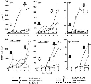
The effect of colonization of isolator piglets versus the administration of PAMPs on total serum Ig levels in μg/ml (top panel) and antibody responses in ELISA units/ml (lower panels). Vertical arrows indicate the time of immunization and boost with TNP-Ficoll and FLU-KLH. In top panels, †=IgM levels significantly greater than group C; *=IgM level significantly greater than group F. In bottom panels, ** and *=ELISA activity statistically greater on week 5 than CpG alone (group C); †=statistically greater than CpG + LPS (group F) or CpG alone (group C) on week 5. Group legend is given in the figure. See Butler et al. (Reference Butler, Francis, Freeling, Weber, Sun and Krieg2005c) for experimental details.

A major unresolved issue in swine B cell development concerns the role of hindgut lymphoid tissue. The ileal Peyers patches (IPP) extend like ‘rows of buttons’ along the terminal ileum and are distinct from the isolated jejunal Peyers patches that develop primarily after colonization or exposure to gut antigens (Pabst et al., Reference Pabst, Geist, Rothkotter and Fritz1988; Uhr, Reference Uhr1993; Liebler-Tenorio and Pabst, Reference Liebler-Tenorio and Pabst2006). The precise role of the IPP in diversification of the antibody repertoire in the manner of the chicken bursa or rabbit appendix remains undefined. Of interest is the preferential transcription of IgG3 in the IPP during the period when it is believed that the IPP may act as a primary lymphoid tissue (Butler and Wertz, Reference Butler and Wertz2006). Interestingly, IgG3 is the primordial IgG for swine and is best equipped to activate complement and bind Fcg receptors (Butler et al., Reference Butler, Wertz, Deschacht and Kacskovics2008b). Furthermore, the gene for IgG3 is located immediately downstream of IgD suggesting it is the first major secreted antibody class that results from CSR (Eguchi-Ogawa et al., Reference Eguchi-Ogawa, Wertz, Sun, Uenishi, Piumi, Chardon and Butler2009).
The fact that numerous paradigms established from studies in rodents and humans do not fit the swine system suggests that: (1) rodent/human immunologists have simply accepted these paradigms without deeper investigation or (2) that the immune system of each major mammalian species group has diverged. If the latter is true, it emphasizes the need to characterize the immune system of each economically important veterinary species. Thus reaching the ‘summit’ only allows a second higher summit to appear, i.e. applying this information to piglet models.
Application of neonatal piglet models
Total germfree systems for neonatal mammals were developed in the 1950s (Levenson et al., Reference Levenson, Mason, Huber, Malm, Horowitz and Einheber1959) and later extended to piglets (Meyer et al., Reference Meyer, Bohl and Kohler1964; Kim et al., Reference Kim, Bradley and Watson1966; Travnicek et al., Reference Travnicek, Mandel, Lanc and Ruzicka1966). In parallel, CDCD piglets begin to be used in research and Lecce (1969) developed a feeding system in which colostrum-deprived piglets were placed into the so-called ‘autosows’ in which their diet and exposure to environmental factors were limited, although colonization of the gut was not prevented. While rearing and microbial features were refined with time, their full value for immunology awaited characterization of the porcine immune system and the tools needed to study swine immunology.
Applying the autosow model to studies on passive immunity
Piglets reared in autosows have been most useful in studies on passive immunity (Klobasa et al., Reference Klobasa, Werhahn and Butler1981, Reference Klobasa, Habe, Werhahn and Butler1985a, Reference Klobasa, Habe, Werhahn and Butlerb, Reference Klobasa, Butler, Werhahn and Habe1986, Reference Klobasa, Butler and Habe1990; Werhahn et al., Reference Werhahn, Klobasa and Butler1981). These studies showed that: (a) absorption of maternal Igs from colostrum was restricted to ~12 h, i.e. ‘gut closure’, (b) maternal Ig can regulate de novo Ig synthesis in piglets, (c) gut closure is initiated by dietary carbohydrates and protein, (d) the intestinal absorptive capacity decays with time after birth, independent of feeding, and (e) delayed suckling is further exacerbated by changes in the quality and constituency of colostrum/milk. A full discussion of the subject is reviewed elsewhere (Butler et al., Reference Butler, Lager, Splichal, Francis, Kacskovics, Sinkora, Wertz, Sun, Zhao, Brown, DeWald, Dierks, Muyldermanns, Lunney, McCray, Rogers, Welsh, Navarro, Klobasa, Habe and Ramsoondar2008a).
Applying the isolator piglet model in colonization studies
Characterization of the porcine immune system was done so the neonatal piglet could be used to study immunological events that occur during the critical window (Fig. 1). The piglet model could then be applied to examine relevant issues in veterinary and medical science. One such issue is gut colonization. It had been suggested for some time that colonization with commensal flora was necessary for development of adaptive immunity (Thorbecke, Reference Thorbecke1959). This was tested in isolator piglets using a porcine exclusion culture provided by David Nisbet (Nisbet et al., Reference Nisbet, Corrier and Stanker1999; Harvey et al., Reference Harvey, Anderson, Genovese, Callaway and Nisbet2005). Colonization produced a 10–30-fold increase in serum Igs (Butler et al., Reference Butler, Sun, Weber and Francis2000b), which was consistent with early studies in rodents (Gustafsson and Laurell, Reference Gustafsson and Laurell1959; Thorbecke, Reference Thorbecke1959; Ohwaki et al., Reference Ohwaki, Yasutake, Yasui and Ogura1976). Serum IgA was preferentially elevated in piglets perhaps because bacterial antigens accessed the gut mucosa. However, we found no antibody activity to phosphoryl choline that might be expected, since it is expressed by some members of the colonizing flora. However, significant IgG responses to LPS were seen and these preferentially recognized the LPS of the colonizing Escherichia coli (A. Conklin and J. E. Butler, unpublished data).
When we immunized isolator piglets intraperitoneally with naked T-dependent (FLU-KLH) and type 2 T-independent (TNP-Ficoll) antigens (antigens without adjuvant) only those monoassociated with a benign E. coli and not those maintained germfree responded (Butler et al., Reference Butler, Weber, Sinkora, Baker, Schoenherr, Mayer and Francis2002). We then asked whether exposure to purified PAMPs (e.g. LPS, CpG-ODN and MDP) would have the same effect. Figure 4 shows that while colonization greatly elevated total IgG and IgA, only CpG-ODN and CpG-ODN plus MDP had a significant effect on serum Ig levels and this was on IgM levels only. When immunized with naked, defined antigens (TNP-Ficoll and FLU-KLH) there was no significant increase in serum Ig levels yet piglets responded with antibodies to TNP and FLU. While CpG-ODN given to germfree isolator piglets was the only TLR ligand tested that evoked a response, CpG-ODN+MDP+MDP (a NOD ligand) strongly influenced IgM and IgG responsiveness to both FLU and TNP. Surprisingly LPS alone had no effect but greatly augmented CpG-ODN induced responsiveness. Perhaps activation through TLR9 results in expression of TLR4, which then allows LPS to augment the response. IgA responses to TNP or FLU were not detected or were extremely weak, perhaps because of the site of administration of the antigen and TLR ligands (Butler et al., Reference Butler, Francis, Freeling, Weber, Sun and Krieg2005c).
Especially noteworthy was the very modest effect that TLR ligands especially had on total IgG and IgA levels in relationship to the response to TNP and FLU (Fig. 4). This was reflected in the fact that the specific activity of the response (antibody responsiveness compared to total Ig isotype levels) was four-fold greater for IgM anti-TNP in piglets receiving CpG-ODN+LPS than in colonized piglets, 60-fold higher for IgG anti-TNP responses and 80-fold higher for IgG anti-FLU responses (Butler et al., Reference Butler, Francis, Freeling, Weber, Sun and Krieg2005c). This could mean that the greatly elevated IgG and IgA levels in colonized piglets are the result of anti-bacterial immune responses or polyclonal B cell activation (Butler et al., Reference Butler, Francis, Freeling, Weber, Sun and Krieg2005c).
These results obtained using neonatal piglets reared in controlled environments have implication for vaccine development and vaccination of conventional piglets. First, adjuvant-mediated responses appear to preferentially affect specific antibody responses when given at the same site. Secondly, vaccine developers may consider that while bacterial DNA was the key ingredient in activating naïve B and probably dendritic cells, responses were significantly augmented through synergy (augmentation) with other PAMPs like MDP and LPS.
Applying piglet models to studies on viral pathogenesis
There has been growing concern for emerging diseases in veterinary species during the last 20 years. These include prion diseases, E. coli variants and neonatal viruses. One such disease is porcine reproductive and respiratory syndrome (PRRS), caused by a porcine arterivirus (PRRSV; Benfield et al., Reference Benfield, Nelson, Collins, Harris, Goyal, Robison, Christianson, Morrison, Gorcyca and Chladek1992; Conzelmann et al., Reference Conzelmann, Visser, Van Woensel and Thiel1993). This has become an infamous worldwide pandemic (Neumann et al., Reference Neumann, Kliebenstein, Johnson, Mabry, Bush, Seitzinger, Green and Zimmerman2005). In addition, a virulent form of circovirus (PCV-2) was also found to play a role in what became known as porcine multisystemic wasting syndrome (PMWS; Allan and Ellis, Reference Allan and Ellis2000; Chianini et al., Reference Chianini, Majo, Segales, Dominguez and Domingo2003). Subsequently, two genetic groups of PCV-2 were recognized with the Canadian form (PCV-2b) being somewhat more virulent (Cheung et al., Reference Cheung, Lager, Kohutyuk, Vincent, Henry, Baker, Rowland and Dunham2007; Gagnon et al., Reference Gagnon, Trembling, Tijssen, Venne, Houde and Elahi2007). Some reports from Asia suggest that the disease may have an even more complex etiology; perhaps those also involve African swine fever, other unidentified viruses, as well as bacterial infection (Tong et al., Reference Tong, Tian, Zhou, Hao, An, Wei, Qui and Cai2007; Yuan et al., Reference Yuan, Lu, Zhang, Li, Sun and Liu2007).
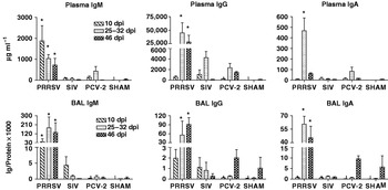
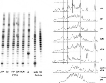
In recent years, we have used the isolator piglet model to address the issue of emerging neonatal viral diseases. Comparing PRRSV, PCV-2 and swine influenza virus (SIV) in isolator littermates revealed that PRRSV produced an extreme form of immune dysregulation (Lemke et al., Reference Lemke, Haynes, Spaete, Adolphson, Vorwald, Lager and Butler2004). This was characterized by hypergammaglobulinemia of all isotypes (Fig. 5), lymphoid adenopathy, and glomerular deposition of immune complexes, aggregated Igs and autoimmunity (Fig. 6). However, this was surprisingly associated with very little antibody repertoire diversification or selective expansion of B cells that used a certain VH gene (Lemke, Reference Lemcke2006; Butler et al., Reference Butler and Sinkora2007). When PRRSV-infected piglets were clonotyped, most lymphoid tissues from the same animal gave the same spectratype, indicating dissemination of the same selected clones throughout the animals (Fig. 7).
Ig concentrations in serum and bronchio-alveolar lavage (BAL) in isolator piglets infected with PRRS virus (PRRSV), SIV, porcine circovirus (PCV-2) and sham controls. Legend is given in the figure. Note the split Y-axis. Only PRRSV causes hypergammaglobulinemia both locally and in plasma. *Ig levels in PRRSV-infected piglets that are significantly higher than in all other treatment groups (P>0.05–0.001 depending on the groups compared). From Butler et al. (Reference Butler, Weber, Wertz and Lager2008c).

PRRSV-infected piglets suffer from lymph node adenopathy (TOP) and deposition of immune complexes in the glomerulus (dark stain; lower left) while possessing serum antibodies to autoantigens (perinuclear staining of HEP2 cells; lower right). From Lemke et al. (Reference Lemke, Haynes, Spaete, Adolphson, Vorwald, Lager and Butler2004).

Clonal distribution of B cells by spectratypic analysis. Left: autoradiograph of B cell spectratypes from various tissues of PRRSV-infected piglets. Right: scanogram of spectratype shown on the left. Vertical lines connect same length HCDR3s. Noteworthy is the very similar but selected spectratype of B cells in nearly all tissues of PRRSV-infected piglets whereas control spectratypes appear largely Gaussian. From Butler et al. (Reference Butler, Weber, Wertz and Lager2008c).

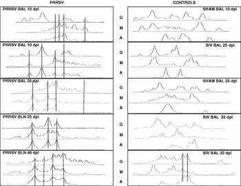
Clonotyping (or spectratyping) is a HCDR3 length analysis assay. Because of allelic exclusion, each B cell clone expresses only one length HCDR3 although two or more B cell clones could share a common length but would have unique sequences. Thus, both length and sequence must be examined. When the HCDR3 regions of the expanded clones were sequenced, a surprisingly high proportion shared the same sequence and nearly one-third were hydrophobic (Butler et al., Reference Butler and Sinkora2007). This was not seen in SIV-infected littermates. Furthermore, B cells expressing IgA, IgG and IgM from the same tissue of the same animal, shared the same spectratype and HCDR3 sequence whereas this was not seen with SIV (Fig. 8; Butler et al., Reference Butler, Weber, Wertz and Lager2008c). Random sampling of HCDR3 from the lung of PRRSV-infected piglets showed that one-third of all B cells had hydrophobic HCDR3s and furthermore, their VDJs were in germline configuration (Butler et al., Reference Butler, Weber, Wertz and Lager2008c). Interpretation of the result obtained with PRRSV-infected isolator piglets first requires a brief review of the pre-immune repertoire of mammals and the changes that occur as the adaptive response develops in neonates.
The pre-immune B cell repertoire in all mammals studied, including swine (Butler et al., Reference Butler, Weber, Sinkora, Sun, Ford and Christenson2000a) is comprised of B cell receptors (BCRs) in germline configuration, i.e. no SHM. The pre-immune repertoire harbors the so-called natural antibodies that have broad specificity so that a relatively small B cell population can engage a very broad range of antigens, including self-antigens (Dighiero et al., Reference Dighiero, Lymberi, Holmberg, Lundquist, Coutinho and Avrameas1985; Ochsenbein and Zinkernagel, Reference Ochsenbein and Zinkernagel2000; Marchalonis et al., Reference Marchalonis, Adelman, Schluter and Ramsland2006). Exposure to environmental antigens and the adjuvant effect of colonization (see above) stimulate development of a more specific adaptive B cell response by somatic modification of the pre-immune repertoire. This involves antigen-driven SHM, often CSR and antigen selection of B cells. It also involves deletion or suppression of auto-reactive B cells and death by neglect for those that fail to be antigen-selected. The latter group includes a high proportion of those with strongly hydrophobic BCRs. It is believed few antigenic epitopes are recognized by hydrophobic paratopes (Raaphorst et al., Reference Raaphorst, Raman, Nall and Teale1997).
A hydropathicity profile is a plot of the frequency distribution of HCDR3s according to their overall charge properties, which is calculated from the deduced amino acid sequence (Kyte and Doolittle, Reference Kyte and Doolittle1982). When this was done for PRRSV-infected piglets, aged matched piglets infected with SIV, newborn piglets (expressing the pre-immune repertoire) and heavily antigenized piglets (infected with parasites) the hydropathicity profile in PRRSV-infected animals resembled that of newborns with about 40% expressing hydrophobic HCDR3s. In parasite-infected and SIV-infected animals, the profile was shifted to favor B cells with hydrophilic, neutral or amphoteric-binding sites (Butler et al., Reference Butler, Weber, Wertz and Lager2008c). When this observation is combined with our results showing that the VDJ expressing hydrophobic HCDR3s were non-mutated and in germline configuration in B cells from different tissues (Fig. 7) and expressing different isotypes (Fig. 8), it suggested that the same B cell clones in PRRSV-infected piglets had been selectively proliferated without repertoire diversification. When B cell hydropathicity profiles are extrapolated to serum Igs, those with hydrophobic binding site should be uncommon in SIV and parasite-infected pigs but could comprise about 40% of the Igs in newborns and PRRSV-infected piglets. Considering the hypergammaglobulinemia associated with PRRS (Fig. 5; Albina et al., Reference Albina, Piriou, Hutet, Cariolet and Hospitalier1998; Lemke et al., Reference Lemke, Haynes, Spaete, Adolphson, Vorwald, Lager and Butler2004; Butler et al., Reference Butler and Sinkora2007, Reference Butler, Weber, Wertz and Lager2008c) and the weak anti-viral response in comparison to Ig levels (Lemke et al., Reference Lemke, Haynes, Spaete, Adolphson, Vorwald, Lager and Butler2004) it means that B cells in PRRSV-infected piglets have not only proliferated without repertoire development but have also differentiated to Ig-secreting plasma cells. Since the pre-immune repertoire harbors many self-reactive B cells, the presence of autoantibodies in PRRSV-infected piglets is not surprising (Fig. 6; Table 3).
Spectratypic analysis of local B cells in two viral respiratory disease. B cells from the BAL that express the major isotypes in PRRSV-infected piglets share a common spectratype (left) whereas those from SIV-infected littermates (right) do not. Vertical lines connect same length HCDR3s in different samples. Data for samples collected at various times after infection (dpi) are shown. From Butler et al. (Reference Butler, Weber, Wertz and Lager2008c).

Immune dysregulation in conventional (conv) versus isolator piglets

1 Data are mean±SD. Differences are obvious by inspection.
2 Fold increase compared to corresponding controls.
3 dpi=days post inoculation.
4 Conventional piglets have ingested maternal colostrum so one-fifth of the IgM, one-third of the IgG and one-half of the IgA is of maternal origin.
ND=not detectable; NA=not applicable.
Our studies on PRRSV-infected isolator piglets raise several questions: (1) what drives the generalized (polyclonal) activation of B cells? (2) Why do those with hydrophobic HCDR3s appear to be selected? (3) Why has the immune dysregulation we observed (Lemke et al., Reference Lemke, Haynes, Spaete, Adolphson, Vorwald, Lager and Butler2004) not been regularly observed by other PRRS researchers? (4) Does the phenomenon we describe suppress development of adaptive responses to the virus? These questions are discussed below.
First, many viral infections cause some level of polyclonal B cell activation although the reasons are mostly unknown (Goodman-Snitkoff et al., Reference Goodman-Snitkoff, Mannano and McSharry1981; Scalzo and Anders, Reference Scalzo and Anders1985; Coutelier et al., Reference Coutelier, Coulie, Wauters, Heremans and van der Logt1990; Hu et al., Reference Hu, Even and Plagemann1992; Rott et al., Reference Rott, Charreire, Mignon-Godefray and Cash1995; Stevenson and Doherty, Reference Stevenson and Doherty1999; Blutt et al., Reference Blutt, Crawford, Warfield, Lewis, Estes and Conner2004; Hunziker et al., Reference Hunziker, Recher, Macpherson, Ciurea, Freigang, Hengartner and Zinkernagel2003; Karupiah et al., Reference Karupiah, Sacks, Klinman, Frederickson, Hartley, Chen and Morse1998). The most likely candidate is a BSAg although none have been described that might selectively target B cells with hydrophobic binding sites. Most BSAgs and T cell superantigens target the framework region of the BCR or TCR (Silverman et al., Reference Silverman, Nayak and La Cava1997). It may be that BCRs with hydrophobic HCDR3s undergo a conformational change that provides greater access to a BSAg binding site within a framework region (Butler et al., Reference Butler, Weber, Wertz and Lager2008c). This is relevant to the second question since all porcine VH genes are VH3, and VH3-bearing B cells have been shown to be the target of BSAgs (Zouali, Reference Zouali1995; Silverman et al., Reference Silverman, Nayak and La Cava1997). Thus, the entire pre-immune repertoire should be proliferated, not only those B cells with hydrophobic binding sites. Although there are some subtle framework differences among the porcine VH genes (Butler et al., Reference Butler, Weber and Wertz2006b) but up to this point no selection for any VH gene has been seen (Butler et al., Reference Butler and Sinkora2007). Perhaps the apparent selection is merely statistical bias based on having analyzed too few sequences, given that so much of the pre-immune repertoire is hydrophobic.
The third question is more troubling since another arterivirus, lactate dehydrogenase elevating virus (LDV) produces a similar phenomenon (Cafruny and Hovinen, Reference Cafruny and Hovinen1988; Li et al., Reference Li, Hu, Harty, Even and Plagemann1990; Bradley et al., Reference Bradley, Broen and Cafruny1991; Hu et al., Reference Hu, Even and Plagemann1992; Plagemann and Moening, Reference Plagemann and Moenning1992; Rowland et al., Reference Rowland, Even, Anderson, Chen, Hu and Plagemann1994; Zitterkopf et al., Reference Zitterkopf, Jones, Bradley, Durick, Rowland, Plagemann and Cafruny2003). A BSAg has not yet been identified in LDV despite a considerable amount of work done on this viral infection. This suggests either that the subject has not been adequately pursued or that the BSAg is endogenous. If a BSAg is responsible, it need not be encoded by PRRSV or LDV but rather the infection could trigger expression of an endogenous BSAg, perhaps encoded by an activated retrovirus (Hsiao et al., Reference Hsiao, Lin, Tai, Chen and Huber2006).
The reason why the phenomenon we have described (Lemke et al., Reference Lemke, Haynes, Spaete, Adolphson, Vorwald, Lager and Butler2004; Butler et al., Reference Butler and Sinkora2007, Reference Butler, Weber, Wertz and Lager2008c) has not been reported by others, seems to be: (1) due to the type of animals studied and (2) the parameters measured. Data reported by Albina et al., (Reference Albina, Piriou, Hutet, Cariolet and Hospitalier1998) (one of the few to measure Ig levels) showed the same hypergammaglobulinemia that we observed in conventional piglets (Table 3). While investigators consider lymphoid adenopathy to be a part of the syndrome in PRRSV infections, even the most recent reports do not report Ig levels (Mulupuri et al., Reference Mulupuri, Zimmerman, Hermans, Johson, Cano, Yu, Dee and Murtaugh2008). However, most report anti-viral responses (Loemba et al., Reference Loemba, Mounir, Mardassi, Archambault and Dea1996; Mulupuri et al., Reference Mulupuri, Zimmerman, Hermans, Johson, Cano, Yu, Dee and Murtaugh2008), albeit delayed to all or certain viral epitopes (Batista and Molitor, Reference Bautista and Molitor1997; Lopez et al., Reference Lopez, Fuertes, Domenech, Alvarez, Ezquerra, Pominquea, Castro and Alovso1999; Mulupuri et al., Reference Mulupuri, Zimmerman, Hermans, Johson, Cano, Yu, Dee and Murtaugh2008). A delayed response to LDV was also reported (Cafruny and Plagemann, Reference Cafruny and Plagemann1982). Reviewing these reports and data given in Table 3 indicates the phenomenon we have reported in PRRSV-infected isolator piglets is not unique, but rather exaggerated. It is the exaggeration that allows this virulence effect of PRRSV to be pinpointed. Isolator piglets have been regularly used by others to take advantage of their sensitivity in identifying virulence factors (Francis et al., Reference Francis, Collins and Duimstra1986; Collins et al., Reference Collins, Bergeland, Bouley, Ducommun, Francis and Yeske1989; McKee et al., Reference McKee, Melton-Celsa, Moxley, Francis and O'Brien1995).
The third question and the contrasting observations made by us in conventional versus isolator-reared animals are important for another reason. Table 3 compares the degree of immune dysregulation in isolator versus conventional newborn piglets infected with PRRSV; these data show a striking reduction in the degree of PRRSV-induced immune dysregulation in conventional newborn piglets as indicated by the reduced hypergammaglobulinemia (5-fold versus 30–40-fold), absence of autoantibodies to dsDNA and reduced kidney pathology. The only differences between these conventional piglets and isolator piglets are that: (1) conventional piglets were colonized with a natural gut flora and (2) they received maternal colostrum and milk. By reference to Fig. 1, this suggests that passive maternal factors or intrinsic factors stimulated by either colonizing bacteria or factors in colostrum/milk can reduce immune dysregulation. There is reason to consider gut flora as a ‘forgotten organ’ (O'Hara and Shanahan, Reference O'Hara and Shanahan2006) and to regard the mammary gland merely as an organ for transferring passive antibody (Butler and Kehrle, Reference Butler, Kehrle, Mestecky, Lamm, Strober, McGhee, Mayer and Bienenstock2005; Nguyen et al., Reference Nguyen, Yuan, Azevedo, Jeong, Gonzalez and Saif2007). Regardless of the source of these factor(s) it strongly argues for a better understanding of the events that occur during the ‘critical window of immunological development’ (Fig. 1). The mechanism for suppressing immune dysregulation is speculative but perhaps involves inducing anergy in rapidly proliferating B cells, through FcγRIIB-BCR cross-linking by immune complexes in colostrum/milk or indirectly through the action of Tregs induced by colonization.
The final question of whether PRRSV-infection results in immunosuppression does not appear to be supported by studies on the response to specific glycoproteins (Mulupuri et al., Reference Mulupuri, Zimmerman, Hermans, Johson, Cano, Yu, Dee and Murtaugh2008). However, when considering the polyclonal hypergammaglobulinemia induced by PRRSV in isolator piglets (see above) the anti-viral response relative to the total Ig levels, might be considered subnormal while in absolute terms it is normal and similar to SIV (J. E. Butler, X-Z Sun and K. M. Lager, unpublished data). Nevertheless, the high level of non-virus-specific Igs resulting from polyclonal B cell activation could interfere with the proper effector activity of anti-viral antibodies. While direct suppression does not appear to occur, the observation that protective responses to both PRRSV and LDV are delayed could still imply immune dysregulation of anti-viral adaptive immunity.
The future for piglet models in studies on the development of adaptive immunity and biomedical research
The future of any model depends on convincing agencies that fund basic research that unique information can be obtained in a cost-effective manner. While isolator piglet research is expensive, the cost of using large numbers of conventional animals for which many variables cannot be controlled is also high. Moreover, the ambiguity resulting from poorly controlled studies in conventional animals may render the results to be of comparatively low value. Veterinary immunology continues to suffer unfavorable critiques from mainstream immunologists as compared to studies in rodents. This is in part because most veterinary studies are done using outbred populations for which many experimental variables cannot be controlled. This problem is likely to persist regardless of how many new anti-porcine reagents become available. In other words, the problem will persist mainly because of experimental design not reagent availability. It is the responsibility of all biological scientists to conceive of ways to control experimental variables and thereby expedite the identification of key host and pathogen mechanisms and their interactions and thereby move more rapidly to the control of animal disease. Presented in this review is an alternative that allows greater control of external factors. Provided that isolator piglet studies will continue to receive support, they can be valuable for addressing a number of issues. The first issue addresses hypotheses derived from Table 3, namely the role of colonizing microbes such as ‘exclusion flora’ in understanding gut microbe ecology and proper gut development. For example can colonization with specific microbes or exposure to certain viruses, provide insight into the mechanism of oral tolerance, tolerance to gut commensals and the development of immune homeostasis? Secondly, isolator piglets can continue to provide a more sensitive means of identifying virulence factors of bacteria and viruses, in the manner demonstrated for a virus like PRRSV (Lemke et al., Reference Lemke, Haynes, Spaete, Adolphson, Vorwald, Lager and Butler2004; Butler et al., Reference Butler and Sinkora2007, Reference Butler, Weber, Wertz and Lager2008c) and for enteric bacteria described by others (Francis et al., Reference Francis, Collins and Duimstra1986; Collins et al., Reference Collins, Bergeland, Bouley, Ducommun, Francis and Yeske1989; McKee et al., Reference McKee, Melton-Celsa, Moxley, Francis and O'Brien1995). If virulence in PRRS results from B cell immune dysregulation, it can theoretically be tested in B cell knockout animals reared in isolation. Thirdly, isolator piglets can also be valuable in resolving the role of the IPP in B cell diversification and whether the IPP serves only the gut or the entire B cell system. This is controversial in swine and other artiodactyls of economic importance. Piglets with resected IPP and reared in isolators and even as CDCD animals, can be used to resolve this controversy. Fourthly, controlled administration of colostrum/milk and its components, can shed light on the possible role of ‘maternal imprinting’, i.e. the ability of the mother to imprint the development of adaptive immunity. Fifthly, probiotics should have their greatest effect on neonates; isolator piglets provide a controlled setting for testing their effect. Finally, rearing CFTR knockout piglets (Rogers et al., Reference Rogers, Stoltz, Meyerholz, Ostedgaard, Rokhlina, Taft, Rogan, Pezzulo, Karp, Itani, Kabel, Wohlford-Lenane, Davis, Hanfland, Smityh, Samuel, Wax, Murphy, Rieke, Whitworht, Uc, Starner, Brogden, Shilyansky, MvCray, Zabner, Prather and Welsh2008) as germfree isolator piglets can establish whether cystic fibrosis is intrinsic, dependent on environmental pathogens or both. Because of their shared physiology, pathogens and diseases, results obtained with swine models could be more valuable to human medical science than studies in rodents.
Acknowledgments
The author acknowledges the many students, co-workers and collaborators who have contributed to the data reviewed. Special acknowledgement goes to Dr David Francis, South Dakota State University; Dr Kelly, M. Lager, National Animal Disease Center; Dr Marek Sinkora, Czech Academy of Science; Nancy Wertz, University of Iowa; Jishan Sun, Wake Forest University; and Dr Caitlin Lemke, Robarts Institute, London, Ontario, Canada. The research was supported by NSF-MCB 0077237; USDA-NRI 03–35204-13836; Carver Trust of the University of Iowa; National Porkboard 05-174 and 05-143, and USDA-ARS Cooperative Agreement 58-3625-4-155.