Obsessive–compulsive disorder (OCD) is a debilitating illness (Koran, Reference Koran2000; Koran, Thienemann, & Davenport, Reference Koran, Thienemann and Davenport1996) that has a lifetime prevalence of 2–3% in nearly every country for which epidemiological data are available (Bland, Newman, & Orn, Reference Bland, Newman and Orn1988; Horwath & Weissman, Reference Horwath and Weissman2000; Karno, Golding, Sorenson, & Burnam, Reference Karno, Golding, Sorenson and Burnam1988; Robins et al., Reference Robins, Helzer, Weissman, Orvaschel, Gruenberg and Burke1984; Sasson et al., Reference Sasson, Zohar, Chopra, Lustig, Iancu and Hendler1997; Weissman et al., Reference Weissman, Bland, Canino, Greenwald, Hwu and Lee1994). It affects an estimated 50 million people worldwide (Sasson et al., Reference Sasson, Zohar, Chopra, Lustig, Iancu and Hendler1997). OCD is typically characterized by the presence of both obsessions and compulsions, although the presence of either obsessions or compulsions alone is sufficient to make the diagnosis of OCD (American Psychiatric Association, 2000). Obsessions are recurrent, persistent, and intrusive ego-dystonic thoughts, impulses, or images; compulsions are repetitive behaviors or mental acts that are executed with the goal of preventing or reducing distress or preventing some dreaded event or situation (American Psychiatric Association, 2000).
Childhood- Versus Adult-Onset OCD
The age of onset of OCD seems to have a bimodal distribution, with a peak in childhood (at approximately 10 years of age) and another in early adulthood (Geller, Reference Geller2006; Geller, Biederman, Jones, Shapiro, et al., Reference Geller, Biederman, Jones, Shapiro, Schwartz and Park1998). Estimates of the prevalence of OCD among adolescents are of approximately 2–3.5% (Flament et al., Reference Flament, Whitaker, Rapoport, Davies, Berg and Kalikow1988; Valleni-Basile et al., Reference Valleni-Basile, Garrison, Jackson, Waller, McKeown and Addy1994; Zohar et al., Reference Zohar, Ratzoni, Pauls, Apter, Bleich and Kron1992). Approximately 40% of cases of childhood-onset OCD continue into adulthood, and that number increases to 60% when subthreshold presentations of OCD are also considered (Stewart et al., Reference Stewart, Geller, Jenike, Pauls, Shaw and Mullin2004). Conversely, one-third to one-half of adults with OCD have the onset of their symptoms in childhood or adolescence (Eichstedt & Arnold, Reference Eichstedt and Arnold2001).
Childhood- and adult-onset OCD differ in several ways. Compared to adult-onset OCD, childhood-onset OCD is more prevalent among males (Eichstedt & Arnold, Reference Eichstedt and Arnold2001; Geller, Biederman, Jones, Shapiro, et al., Reference Geller, Biederman, Jones, Shapiro, Schwartz and Park1998); is more familial (Bellodi, Sciuto, Diaferia, Ronchi, & Smeraldi, Reference Bellodi, Sciuto, Diaferia, Ronchi and Smeraldi1992; do Rosario-Campos et al., Reference do Rosario-Campos, Leckman, Curi, Quatrano, Katsovitch and Miguel2005; Nestadt et al., Reference Nestadt, Lan, Samuels, Riddle, Bienvenu and Liang2000; Pauls, Alsobrook, Goodman, Rasmussen, & Leckman, Reference Pauls, Alsobrook, Goodman, Rasmussen and Leckman1995); and is associated with a higher prevalence of tic disorders, both in the patients with OCD themselves and in their first-degree relatives (Chabane et al., Reference Chabane, Delorme, Millet, Mouren, Leboyer and Pauls2005; Grados et al., Reference Grados, Riddle, Samuels, Liang, Hoehn-Saric and Bienvenu2001; Pauls et al., Reference Pauls, Alsobrook, Goodman, Rasmussen and Leckman1995). These findings have led some authors to suggest that childhood-onset OCD is a distinct, “tic-related” subtype of OCD (Eichstedt & Arnold, Reference Eichstedt and Arnold2001; Taylor, Reference Taylor, Abramowitz and Houts2005). The distinction between childhood- and adult-onset OCD may also have treatment implications: adults with childhood-onset OCD respond less to treatment with clomipramine than do adults with adult-onset OCD, independent of illness duration or severity (Ackerman, Greenland, Bystritsky, Morgenstern, & Katz, Reference Ackerman, Greenland, Bystritsky, Morgenstern and Katz1994).
Most brain imaging studies that include adults with OCD use mixed samples that include adults with childhood- and adult-onset OCD. Given the evidence that brain activity differs in adults with childhood- versus adult-onset OCD (Busatto et al., Reference Busatto, Buchpiguel, Zamignani, Garrido, Glabus and Rosario-Campos2001), such heterogeneous samples may compromise statistical power and confound findings. In addition, many studies report only the mean and standard deviation of age of illness onset, which are inadequate to characterize what are likely to be bimodal distributions. Consequently, knowing whether findings apply to all adults with OCD or only to those with childhood- or adult-onset OCD is impossible for most existing studies that include adults with OCD.
In addition, few studies have compared the neural correlates of OCD across children and adults, severely limiting our knowledge of their similarities and differences. One of the aims of this review is to begin to redress this gap by systematically evaluating the similarities and differences in findings from studies of the neural bases of pediatric and adult OCD. We should note, however, that differences in findings across these age groups may be due to several factors. We list four, nonexhaustive possibilities. First, any such differences may be a consequence of comparing children with OCD with mixed samples that include adults with childhood-onset OCD and adults with adult-onset OCD. If childhood-onset OCD is a distinct biological subtype, this comparison confounds age or developmental effects with the effects of illness subtype. Second, such differences may be a consequence of compensatory behavioral, cognitive, or affective responses, which may be more developed, or simply different, in adults. Third, such differences may reflect the differences in phenomenology between pediatric and adult OCD (Geller, Biederman, Jones, Park, et al., Reference Geller, Biederman, Jones, Park, Schwartz and Shapiro1998). Fourth, such differences may be a consequence of brain development, affecting either the neural systems directly involved in the symptoms of OCD or closely related systems.
Functional Neuroimaging of OCD
Much of our understanding of the pathophysiology of OCD has been derived from functional neuroimaging studies. Broadly speaking, these can be divided into four categories, according to the experimental paradigm used (Saxena & Rauch, Reference Saxena and Rauch2000): (a) resting studies, which compare some measure of brain activity at rest in patients with OCD and controls; (b) symptom provocation studies, which compare brain activity before and after the provocation of symptoms (e.g., through contact with something dirty in patients with obsessions about germs and cleanliness); (c) treatment studies, which compare brain activity at rest before and after treatment with medication or psychotherapy; and (d) cognitive activation studies, which compare brain activity in patients with OCD and controls as they perform a cognitive task. Combinations of these categories are also possible: one can, for example, image cognitive activation or symptom provocation before and after treatment (Lázaro et al., Reference Lázaro, Caldú, Junqué, Bargalló, Andrés and Morer2008; Nakao et al., Reference Nakao, Nakagawa, Yoshiura, Nakatani, Nabeyama and Yoshizato2005b), or image cognitive activation before and after symptom provocation.
Three brain areas—the orbitofrontal cortex (OFC), the anterior cingulate cortex (ACC), and the head of the caudate nucleus—have been consistently implicated in a large number of resting, symptom provocation, and treatment studies of adults with OCD. These areas (a) are hyperactive at rest in adults with OCD relative to healthy controls, (b) become more active with symptom provocation, and (c) no longer show hyperactivity at rest following successful treatment with either medication or cognitive–behavioral therapy (Baxter, Clark, Iqbal, & Ackermann, Reference Baxter, Clark, Iqbal, Ackermann, Lichter and Cummings2001; Saxena, Bota, & Brody, Reference Saxena, Bota and Brody2001; Saxena, Brody, Schwartz, & Baxter, Reference Saxena, Brody, Schwartz and Baxter1998; Saxena & Rauch, Reference Saxena and Rauch2000; Schwartz, Reference Schwartz1998; Whiteside, Port, & Abramowitz, Reference Whiteside, Port and Abramowitz2004). These findings have generally been interpreted as evidence that abnormalities in these or closely related areas cause OCD (e.g., Baxter et al., Reference Baxter, Clark, Iqbal, Ackermann, Lichter and Cummings2001; Saxena et al., Reference Saxena, Brody, Schwartz and Baxter1998, Reference Saxena, Bota and Brody2001; Saxena & Rauch, Reference Saxena and Rauch2000).
Two studies have compared resting blood flow before and after pharmacological treatment in children with OCD (Castillo et al., Reference Castillo, Buchpiguel, de Araujo, Castillo, Asbahr and Maia2005; Diler, Kibar, & Avci, Reference Diler, Kibar and Avci2004). One of these studies (Diler et al., Reference Diler, Kibar and Avci2004) also compared resting blood flow in the children with OCD before treatment with resting blood flow in a group of healthy controls. The findings of this study were largely consistent with the findings in adults, including hyperactivity at rest in the caudate and ACC of treatment-naive children, which declined following treatment. The other study (Castillo et al., Reference Castillo, Buchpiguel, de Araujo, Castillo, Asbahr and Maia2005) failed to detect any differences between pre- and posttreatment scans. We are not aware of any symptom provocation studies in children with OCD.
The vast majority of functional imaging studies of OCD through the end of the 1990s used resting, symptom provocation, or pre- versus posttreatment designs. A comprehensive review of imaging studies of OCD that was published in 2001 listed 17 resting studies, 7 symptom provocation studies, 10 pre- versus posttreatment studies, and only 4 cognitive activation studies (Saxena et al., Reference Saxena, Bota and Brody2001). The situation has shifted in recent years, with the publication of a large number of cognitive activation studies in adults with OCD, using a wide variety of cognitive tasks (with an emphasis on executive function tasks). The tasks used have included the go/no-go (Maltby, Tolin, Worhunsky, O'Keefe, & Kiehl, Reference Maltby, Tolin, Worhunsky, O'Keefe and Kiehl2005; Roth et al., Reference Roth, Saykin, Flashman, Pixley, West and Mamourian2007), Stroop (Harrison et al., Reference Harrison, Yucel, Shaw, Kyrios, Maruff and Brewer2006; Nakao et al., Reference Nakao, Nakagawa, Yoshiura, Nakatani, Nabeyama and Yoshizato2005a; van den Heuvel, Veltman, Groenewegen, Witter, et al., Reference van den Heuvel, Veltman, Groenewegen, Witter, Merkelbach and Cath2005), Eriksen flanker (Fitzgerald et al., Reference Fitzgerald, Welsh, Gehring, Abelson, Himle and Liberzon2005), multisource interference (Yucel et al., Reference Yucel, Harrison, Wood, Fornito, Wellard and Pujol2007), stop signal (Woolley et al., Reference Woolley, Heyman, Brammer, Frampton, McGuire and Rubia2008), numeric conflict (Viard et al., Reference Viard, Flament, Artiges, Dehaene, Naccache and Cohen2005), reversal learning (Remijnse et al., Reference Remijnse, Nielen, van Balkom, Cath, van Oppen and Uylings2006), tower of London (van den Heuvel, Veltman, Groenewegen, Cath, et al., Reference van den Heuvel, Veltman, Groenewegen, Cath, van Balkom and van Hartskamp2005), spatial N-back (van der Wee et al., Reference van der Wee, Ramsey, Jansma, Denys, van Megen and Westenberg2003), continuous performance (Ursu, Stenger, Shear, Jones, & Carter, Reference Ursu, Stenger, Shear, Jones and Carter2003), task switching (Gu et al., Reference Gu, Park, Kang, Lee, Yoo and Jo2008), word generation (Pujol et al., Reference Pujol, Torres, Deus, Cardoner, Pifarre and Capdevila1999), and serial reaction time (Rauch et al., Reference Rauch, Savage, Alpert, Dougherty, Kendrick and Curran1997, Reference Rauch, Whalen, Curran, Shin, Coffey and Savage2001, Reference Rauch, Wedig, Wright, Martis, McMullin and Shin2007) tasks. The findings of these studies have generally been consistent with the findings of resting, symptom provocation, and treatment studies, highlighting functional disturbances in the OFC, ACC, basal ganglia, and related areas (Menzies et al., Reference Menzies, Chamberlain, Laird, Thelen, Sahakian and Bullmore2008). Rather than attempting to survey this large and varied literature, though, we wish to highlight a general difficulty with interpreting cognitive activation studies that applies across tasks.
Typically, a given cognitive task only activates those areas involved in the cognitive processes required to perform the task. Whether and how the cognitive processes involved in the tasks that have been used to study OCD may relate to the symptoms of OCD is not always obvious. Cognitive activation studies therefore often have less face validity than resting, symptom provocation, or treatment studies for addressing the neural bases of obsessive–compulsive symptoms. For example, most cognitive activation studies of OCD have used inhibitory control tasks. Patients with OCD exhibit behavioral deficits in at least some inhibitory control tasks (Chamberlain, Blackwell, Fineberg, Robbins, & Sahakian, Reference Chamberlain, Blackwell, Fineberg, Robbins and Sahakian2005), and imaging studies that employ those tasks are useful to determine the neural correlates of those deficits. However, the relevance of those studies to understanding the symptoms of OCD depends on the unproven assumption that those symptoms relate directly to a deficit in inhibitory control. The symptoms of OCD have indeed been suggested to be a consequence of deficient inhibitory control, with obsessions arising from a failure to inhibit intrusive thoughts, and compulsions arising from a failure to inhibit certain behaviors (Chamberlain et al., Reference Chamberlain, Blackwell, Fineberg, Robbins and Sahakian2005). Deficits in inhibitory control have, however, been reported for several psychiatric disorders, including attention-deficit/hyperactivity disorder (Barkley, Reference Barkley1997; Nigg, Reference Nigg2001), bipolar disorder (Larson, Shear, Krikorian, Welge, & Strakowski, Reference Larson, Shear, Krikorian, Welge and Strakowski2005; Quraishi & Frangou, Reference Quraishi and Frangou2002; Robinson et al., Reference Robinson, Thompson, Gallagher, Goswami, Young and Ferrier2006), schizophrenia (Crawford, Bennett, Lekwuwa, Shaunak, & Deakin, Reference Crawford, Bennett, Lekwuwa, Shaunak and Deakin2002; Weisbrod, Kiefer, Marzinzik, & Spitzer, Reference Weisbrod, Kiefer, Marzinzik and Spitzer2000), and addiction (Li & Sinha, Reference Li and Sinha2008; Yucel & Lubman, Reference Yucel and Lubman2007). The presence of deficits in inhibitory control in so many disorders with such widely varying phenotypes suggests caution in interpreting such deficits as the root cause of OCD.
Despite these limitations, cognitive activation studies can certainly be informative. Early cognitive activation studies, for example, found that healthy controls recruited the striatum during performance of an implicit (habit) learning task, whereas patients with OCD recruited the hippocampus, despite similar behavioral performances across the groups (Rauch et al., Reference Rauch, Savage, Alpert, Dougherty, Kendrick and Curran1997, Reference Rauch, Wedig, Wright, Martis, McMullin and Shin2001).Footnote 1 These findings suggested that patients with OCD might use hippocampus-dependent (declarative) learning to overcome deficits in striatum-dependent (implicit) learning. If so, the performance of patients with OCD should suffer more than that of healthy controls when a secondary task that taxes working memory is introduced, and this was confirmed experimentally (Deckersbach et al., Reference Deckersbach, Savage, Curran, Bohne, Wilhelm and Baer2002). These studies show that even in the absence of obvious differences in behavioral performance, cognitive activation studies may highlight differences in neural activity that reflect compensatory strategies. The pathogenic relation, if any, of deficits in implicit (habit) learning and the symptoms of OCD remains unclear however. Even if compulsions are maladaptive, exaggerated habits (Graybiel & Rauch, Reference Graybiel and Rauch2000), why patients with OCD would have difficulty using the striatal habit-learning system (Packard & Knowlton, Reference Packard and Knowlton2002) to learn other habits remains unclear. Post hoc, one can speculate that in patients with OCD the striatal habit-learning system is “overloaded” by compulsions, making it impossible for them to learn other habits. However, from the idea that OCD prominently involves exaggerated habits (the compulsions), one could equally plausibly have predicted the opposite pattern of findings: that the habit learning system in OCD should be especially powerful and effective. This highlights again the difficulties with interpreting the relevance of cognitive tasks and cognitive activation studies for the pathogenesis of OCD.
Cortico–Basal Ganglia–Thalamo–Cortical (CBGTC) Loops and the Pathophysiology of OCD
The OFC and ACC are intimately connected to the basal ganglia via CBGTC loops (Alexander, DeLong, & Strick, Reference Alexander, DeLong and Strick1986; Mega & Cummings, Reference Mega, Cummings, Salloway, Malloy and Duffy2001; Middleton & Strick, Reference Middleton, Strick, Lichter and Cummings2001b). Several CBGTC loops exist, and they seem to run largely parallel courses through the basal ganglia (Alexander et al., Reference Alexander, DeLong and Strick1986; Mega & Cummings, Reference Mega, Cummings, Salloway, Malloy and Duffy2001; Middleton & Strick, Reference Middleton, Strick, Lichter and Cummings2001b). Each of these loops receives inputs from multiple cortical areas and then projects back to one of its cortical areas of origin, thereby partly closing the loop (Alexander et al., Reference Alexander, DeLong and Strick1986). No consensus exists in the literature about exactly how many such loops there are, but a particularly influential formulation proposes the existence of five loops, whose target areas are (a) the supplementary motor area, (b) the frontal eye fields, (c) the dorsolateral prefrontal cortex (DLPFC), (d) the lateral OFC, and (e) the ACC (Alexander et al., Reference Alexander, DeLong and Strick1986). A more recent formulation adds two additional loops with target areas in the medial OFC and in the inferotemporal/posterior parietal cortex, and suggests that each of the loops may actually be further subdivided into multiple parallel loops (Middleton & Strick, Reference Middleton, Strick, Lichter and Cummings2001b). The main site in the striatum through which the CBGTC loops involving the OFC and ACC run is the head of the caudate nucleus (Alexander et al., Reference Alexander, DeLong and Strick1986; Mega & Cummings, Reference Mega, Cummings, Salloway, Malloy and Duffy2001; Middleton & Strick, Reference Middleton, Strick, Lichter and Cummings2001b). The findings from the resting, symptom provocation, and treatment studies described above may therefore be interpreted as implicating the CBGTC loops involving the OFC and ACC (henceforward referred to as OFC/ACC CBGTC loops) in OCD.
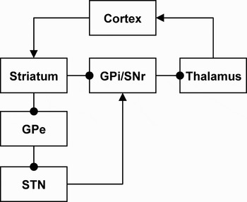
An influential idea based on these observations is that OCD results from an imbalance between the so-called “direct” and “indirect” pathways through the basal ganglia (Figure 1; Baxter et al., Reference Baxter, Clark, Iqbal, Ackermann, Lichter and Cummings2001; Saxena et al., Reference Saxena, Brody, Schwartz and Baxter1998, Reference Saxena, Bota and Brody2001; Saxena & Rauch, Reference Saxena and Rauch2000). The net effect of the direct pathway is excitatory, and the net effect of the indirect pathway is inhibitory (Figure 1), so these pathways are sometimes said to form positive and negative feedback loops, respectively. This simple model of opposing direct and indirect pathways has proven extremely useful in understanding hyperkinetic and hypokinetic movement disorders, such as Huntington and Parkinson diseases, respectively (Wichmann & DeLong, Reference Wichmann and DeLong1996). The idea is that excessive relative activity in the direct pathway disinhibits cortical motor programs, producing hyperkinetic symptoms, such as the chorea in Huntington disease. Conversely, excessive relative activity in the indirect pathway inhibits cortical motor programs, producing hypokinetic symptoms, such as the difficulty initiating movement in Parkinson disease.
The classical conceptualization of the anatomy of CBGTC loops in terms of direct and indirect pathways (Albin, Young, & Penney, Reference Albin, Young and Penney1989; DeLong, Reference DeLong1990). The direct pathway runs from the cortex to the striatum, then directly to the globus pallidus internal segment (GPi) and substantia nigra pars reticulata (SNr), then to the thalamus, and finally back to the cortex. The indirect pathway runs from the cortex to the striatum, then to the globus pallidus external segment (GPe), the subthalamic nucleus (STN), the GPi/SNr, the thalamus, and finally back to the cortex. Arrows represent excitatory (glutamatergic) connections and filled circles represent inhibitory (GABAergic) connections. The direct pathway contains an even number of inhibitory connections (2), so its net effect from the cortex back to the cortex is excitatory. The indirect pathway contains an odd number of inhibitory connections (3), so its net effect from the cortex back to the cortex is inhibitory.

These symptoms are in the motor domain, but different symptoms may occur if the balance of the direct and indirect pathways is compromised in nonmotor CBGTC loops. In particular, excessive relative activity in the direct pathway in OFC/ACC CBGTC loops has been suggested to result in a positive feedback loop in which obsessive thoughts become “trapped” (Baxter et al., Reference Baxter, Clark, Iqbal, Ackermann, Lichter and Cummings2001; Saxena et al., Reference Saxena, Brody, Schwartz and Baxter1998, Reference Saxena, Bota and Brody2001; Saxena & Rauch, Reference Saxena and Rauch2000). Consistent with this idea, the prevalence of obsessive–compulsive symptoms is significantly greater in Huntington disease than in the general population (Anderson, Louis, Stern, & Marder, Reference Anderson, Louis, Stern and Marder2001; Beglinger et al., Reference Beglinger, Langbehn, Duff, Stierman, Black and Nehl2007; De Marchi & Mennella, Reference De Marchi and Mennella2000), and some case reports describe onset of OCD following the onset of Huntington disease (Cummings & Cunningham, Reference Cummings and Cunningham1992; Scicutella, Reference Scicutella2000).
This theory of OCD is not without difficulties. In particular, it does not explain why patients with OCD should have specific obsessions, as opposed to obsessing about everything. This can be unpacked into two, related questions. The first is why one finds great similarity in the contents of obsessions cross-culturally (Sasson et al., Reference Sasson, Zohar, Chopra, Lustig, Iancu and Hendler1997); the second is why each individual patient tends to obsess only about a small subset of the larger set of common obsessions. One suggestion has been that “in persons with OCD, a response bias exists toward stimuli relating to socioterritorial concerns about danger, violence, hygiene, order, and sex—the themes of most obsessions in patients with OCD—mediated by orbitofrontal circuits” (Saxena & Rauch, Reference Saxena and Rauch2000). However, the OFC responds to a variety of both positive and negative emotional stimuli, as well as to neutral stimuli that have been previously paired with positive or negative outcomes (Elliott & Deakin, Reference Elliott and Deakin2005; Elliott, Dolan, & Frith, Reference Elliott, Dolan and Frith2000; Kringelbach & Rolls, Reference Kringelbach and Rolls2004; O'Doherty, Reference O'Doherty2007; Rolls, Reference Rolls1996, Reference Rolls1999, Reference Rolls2004; Schultz, Tremblay, & Hollerman, Reference Schultz, Tremblay and Hollerman2000; Zald & Kim, Reference Zald, Kim, Salloway, Malloy and Duffy2001). Why, then, “socioterritorial concerns” in particular should become locked in the OCD positive feedback loop is unclear. This suggestion also fails to explain why some patients with OCD will obsess about germs and wash compulsively, whereas others may obsess about whether they have locked the doors to their house and check the locks compulsively.
Searching for the Pathogenesis of OCD
As noted above, the findings that in patients with OCD the OFC, ACC, and caudate nucleus are hyperactive at rest, become more active under symptom provocation, and show less activity following treatment have generally been interpreted as evidence that hyperactivity in these areas generates the symptoms of OCD (e.g., Baxter et al., Reference Baxter, Clark, Iqbal, Ackermann, Lichter and Cummings2001; Saxena et al., Reference Saxena, Brody, Schwartz and Baxter1998, Reference Saxena, Bota and Brody2001; Saxena & Rauch, Reference Saxena and Rauch2000). However, several alternative interpretations of these findings are equally plausible. We know, for example, that when healthy controls are exposed to pictures depicting OCD-relevant scenes (specifically, pictures that are washing relevant, checking relevant, and hoarding relevant) and are asked to imagine related scenarios (e.g., “imagine that you must come into contact with what's shown in the following pictures without washing yourself afterward”), they activate regions similar to those that are hyperactive in OCD (Mataix-Cols et al., Reference Mataix-Cols, Cullen, Lange, Zelaya, Andrew and Amaro2003). One possible interpretation of these findings is that rather than being the origin of obsessional content, these regions become hyperactive as a normal consequence of the obsessional content (Shafran & Speckens, Reference Shafran, Speckens, Abramowitz and Houts2005). We also know that the OFC and ACC are involved in emotion regulation (Ochsner, Bunge, Gross, & Gabrieli, Reference Ochsner, Bunge, Gross and Gabrieli2002; Ochsner & Gross, Reference Ochsner and Gross2005). Thus, again hyperactivity in these areas may be a consequence of an attempt by patients with OCD to regulate their anxiety, rather than being the cause of their symptoms.
A related, although slightly different idea is that hyperactivation of the OFC, ACC, and caudate nucleus simply reflects the need to inhibit compulsive behavior during the scan (Peterson, Reference Peterson2003). This idea is supported by the finding that the ACC and the head of the caudate nucleus are activated when patients with Tourette syndrome (TS) voluntarily inhibit their tics (Peterson et al., Reference Peterson, Skudlarski, Anderson, Zhang, Gatenby and Lacadie1998). The genetic, phenomenological, and pathophysiological similarities between TS and OCD (Fineberg, Saxena, Zohar, & Craig, Reference Fineberg, Saxena, Zohar and Craig2007; Marsh, Leckman, Bloch, Yazgan, & Peterson, Reference Marsh, Leckman, Bloch, Yazgan, Peterson, Nelson and Luciana2008) suggest that excessive activity in these areas in patients with OCD might reflect the inhibition of compulsions. Further support for this idea comes from the findings that the OFC, ACC, and caudate are activated in a variety of tasks that require the suppression of a prepotent response (Peterson, Reference Peterson2003). Weighing against this thesis are human lesion studies suggesting that response inhibition may be localized to the right inferior frontal cortex, rather than in the OFC, ACC, or caudate (Aron, Fletcher, Bullmore, Sahakian, & Robbins, Reference Aron, Fletcher, Bullmore, Sahakian and Robbins2003; Aron, Robbins, & Poldrack, Reference Aron, Robbins and Poldrack2004). The evidence from functional imaging for an involvement of the OFC in response inhibition, as studied using the go/no-go task, also remains inconclusive, with block designs typically showing OFC activation, but event-related designs failing to show such activation (Elliott & Deakin, Reference Elliott and Deakin2005).
Other authors have similarly suggested that OFC hyperactivation may reflect patients' efforts to inhibit their symptoms during the scanning procedure (Adler et al., Reference Adler, McDonough-Ryan, Sax, Holland, Arndt and Strakowski2000; Roth et al., Reference Roth, Saykin, Flashman, Pixley, West and Mamourian2007), remaining agnostic as to whether that inhibition is targeted specifically at obsessions, compulsions, anxiety, or possibly all of them. Consistent with the idea that OFC activity plays an inhibitory role, keeping the obsessions, compulsions, or anxiety in check, greater activation of the OFC during symptom provocation is associated with a smaller increase in reported symptoms with the provocation (Adler et al., Reference Adler, McDonough-Ryan, Sax, Holland, Arndt and Strakowski2000; Rauch et al., Reference Rauch, Jenike, Alpert, Baer, Breiter and Savage1994). If OFC hyperactivity played a role in causing, rather than in inhibiting symptoms, then greater activation of the OFC during symptom provocation should be associated with a greater, not a lesser, increase in symptoms.
Our goal is not to suggest that one of these various interpretations is the correct one. Instead, we wish to emphasize that all are consistent with the findings that the OFC, ACC, and caudate nucleus are hyperactive at rest, become more active under symptom provocation, and show less activity following treatment. For example, if the activation of these areas reflects an attempt to inhibit symptoms, such activation could be expected to be higher at rest in patients with OCD than in healthy controls, to increase with symptom provocation, when such inhibition becomes more necessary, and to decrease after treatment, when such inhibition becomes less necessary.
The problem with interpreting these activations as the cause of the symptoms is that these functional imaging findings are inherently correlational: they demonstrate only that activity in these areas correlates with a symptomatic state in OCD. One might hope that anatomical imaging studies would resolve the difficulties in establishing a causal role for these areas in OCD. However, much evidence suggests that repeatedly engaging in a class of behaviors or cognitive processes can change brain structure (Lazar et al., Reference Lazar, Kerr, Wasserman, Gray, Greve and Treadway2005; Mechelli et al., Reference Mechelli, Crinion, Noppeney, O'Doherty, Ashburner and Frackowiak2004; Pascual-Leone, Reference Pascual-Leone2001; Schlaug, Reference Schlaug2001; Schlaug, Norton, Overy, & Winner, Reference Schlaug, Norton, Overy and Winner2005). Thus, even anatomical differences in the brains of patients with OCD may be a consequence rather than a cause of the disorder. This highlights the usefulness of studying patients with OCD as close to symptom onset as possible, when such epiphenomenal changes may be less prominent. OCD is, however, often diagnosed long after the onset of symptoms, making this strategy relatively impractical. Furthermore, subclinical patterns of thinking and behavior that may long precede clinically significant symptoms (Rasmussen & Eisen, Reference Rasmussen, Eisen, Davis, Charney, Coyle and Nemeroff2002) may also conceivably produce epiphenomenal changes in brain structure that could be apparent by the time of symptom onset.
The problem is that, like functional imaging studies, anatomical studies only provide information about the correlates of the disorder. Several valuable animal models of OCD have been developed in efforts to study causal mechanisms in OCD more directly (Joel, Reference Joel2006; Korff & Harvey, Reference Korff and Harvey2006). These have, however, been criticized because they only model repetitive behaviors; compulsive behaviors in OCD are intimately tied to obsessions, and we have no way of assessing whether animals have obsessions (Shafran & Speckens, Reference Shafran, Speckens, Abramowitz and Houts2005).
Other lines of evidence may, however, help establish causality in humans. We will explore three such lines of evidence. First, certain brain lesions due to accidents, stroke, and other naturalistic causes seem to cause OCD. Second, some cases of OCD and of related psychiatric disorders, such as TS, seem to develop as a consequence of an autoimmune reaction in which antibodies to Group A beta-hemolytic streptococcus attack and damage the basal ganglia (Hoekstra & Minderaa, Reference Hoekstra and Minderaa2005; Leonard & Swedo, Reference Leonard and Swedo2001; Murphy, Husted, & Edge, Reference Murphy, Husted and Edge2006; Snider & Swedo, Reference Snider and Swedo2004); these are usually termed pediatric autoimmune neuropsychiatric disorders associated with streptococcal infections (PANDAS). Third, several neurosurgical procedures make localized lesions in attempts to provide symptom relief in severe, treatment-refractory cases of OCD.
Each of these lines of evidence has important limitations in establishing a causal role for abnormalities in specific brain regions in producing OCD. First, studies of OCD as a consequence of brain injury typically involve a small number of subjects who have diffuse lesions that vary greatly across subjects. Furthermore, demonstrating that lesions that affect certain brain circuits result in OCD does not prove a pathogenic role for those circuits in most cases of OCD. Second, the validity of the PANDAS construct remains controversial (Harris & Singer, Reference Harris and Singer2006; Kurlan, Reference Kurlan2004; Kurlan & Kaplan, Reference Kurlan and Kaplan2004; Singer & Loiselle, Reference Singer and Loiselle2003), with substantial difficulties involved in establishing a causal relation between infection with Group A beta-hemolytic streptococcus and the development of neuropsychiatric disorders (and additional difficulties in proving that the possible autoimmune reaction affects the basal ganglia). Third, neurosurgical studies lack appropriate controls, and the mechanisms by which they attenuate OCD symptoms are not fully understood. Moreover, an improvement in symptoms following certain neurosurgical lesions does not prove that the circuits that were lesioned caused the symptoms (just as having gastric bypass surgery and losing weight as a result does not prove that the cause of excess weight was an enlarged stomach).
Although each of the aforementioned lines of evidence (anatomical imaging, OCD as a consequence of brain injury, PANDAS, and neurosurgery) in isolation is insufficient to establish causality, their convergence with functional imaging studies in implicating OFC/ACC CBGTC loops in OCD would provide some reassurance that those loops may indeed play a causal role in OCD. We will therefore review each of these lines of evidence in turn.
Anatomical and Spectroscopy Studies in Adults With OCD
Studies based on regions of interest (ROIs)
Volumetric studies measure the volumes of specific ROIs in the brain. Proton magnetic resonance spectroscopy (1H-MRS) studies measure the concentrations of certain metabolites in ROIs. N-Acetyl-aspartate (NAA), one of those metabolites, has been of particular interest in studies of disease processes because decreases in NAA levels may indicate neuronal loss or dysfunction (Barker, Reference Barker2001; Dager & Steen, Reference Dager and Steen1992; Maier, Reference Maier1995). NAA levels may be more sensitive than volumetric measures to detect neuronal abnormalities (Bartha et al., Reference Bartha, Stein, Williamson, Drost, Neufeld and Carr1998; Cendes, Andermann, Preul, & Arnold, Reference Cendes, Andermann, Preul and Arnold1994; Ebisu, Rooney, Graham, Weiner, & Maudsley, Reference Ebisu, Rooney, Graham, Weiner and Maudsley1994).
The vast majority of anatomical imaging studies of OCD have been conducted after the early functional imaging findings implicated the OFC, ACC, and caudate nucleus in the pathophysiology of OCD. Those studies were therefore strongly influenced by the theory that OFC/ACC CBGTC loops are centrally involved in the pathogenesis of OCD, and have therefore tended to look specifically at brain regions that are part of those loops: the OFC, ACC, caudate nucleus or striatum, and thalamus. Fewer studies have focused specifically on the globus pallidus, another key component of those loops.
OFC
Several studies have reported bilateral reductions in OFC volumes in adults with OCD (Atmaca et al., Reference Atmaca, Yildirim, Ozdemir, Aydin, Tezcan and Ozler2006; Atmaca, Yildirim, Ozdemir, Tezcan, & Poyraz, Reference Atmaca, Yildirim, Ozdemir, Tezcan and Poyraz2007; Szeszko et al., Reference Szeszko, Robinson, Alvir, Bilder, Lencz and Ashtari1999); one study reported a volume reduction only in the left OFC (Kang et al., Reference Kang, Kim, Choi, Kim, Kim and Youn2004). A study that measured specifically the volume of the medial OFC also found it to be reduced bilaterally in adults with OCD (Cardoner et al., Reference Cardoner, Soriano-Mas, Pujol, Alonso, Harrison and Deus2007). Another study that divided the OFC into anterior and posterior regions reported a volume reduction in the left anterior OFC, but not in the left posterior or right anterior or posterior OFC (Choi et al., Reference Choi, Kang, Kim, Ha, Lee and Youn2004). Both left and right OFC volumes have been found to correlate inversely with the severity of OCD symptoms (Atmaca et al., Reference Atmaca, Yildirim, Ozdemir, Tezcan and Poyraz2007; Kang et al., Reference Kang, Kim, Choi, Kim, Kim and Youn2004) and to be larger posttreatment in patients who responded to a trial of selective serotonin reuptake inhibitors than in treatment-naive or treatment-refractory patients (Atmaca et al., Reference Atmaca, Yildirim, Ozdemir, Aydin, Tezcan and Ozler2006). In summary, considerable evidence suggests that OFC volumes are significantly reduced in adults with OCD, with greater reductions accompanying more severe symptoms.
ACC
Several 1H-MRS studies have reported decreased levels of NAA in the ACC of adults with OCD (Ebert et al., Reference Ebert, Speck, Konig, Berger, Hennig and Hohagen1997; Jang et al., Reference Jang, Kwon, Jang, Moon, Lee and Ha2006; Sumitani, Harada, Kubo, & Ohmori, Reference Sumitani, Harada, Kubo and Ohmori2007; Yucel et al., Reference Yucel, Harrison, Wood, Fornito, Wellard and Pujol2007). Furthermore, treatment with the selective serotonin reuptake inhibitor citalopram increases levels of NAA in the ACC (and other regions of the prefrontal cortex [PFC]) in adults with OCD (Jang et al., Reference Jang, Kwon, Jang, Moon, Lee and Ha2006), although no significant correlation was detected between increases in NAA and decreases in symptom severity. Several studies have compared ACC volumes in adults with OCD and normal controls, but have generally failed to detect significant differences (Atmaca et al., Reference Atmaca, Yildirim, Ozdemir, Aydin, Tezcan and Ozler2006, Reference Atmaca, Yildirim, Ozdemir, Tezcan and Poyraz2007; Grachev et al., Reference Grachev, Breiter, Rauch, Savage, Baer and Shera1998; Kellner et al., Reference Kellner, Jolley, Holgate, Austin, Lydiard and Laraia1991; Riffkin et al., Reference Riffkin, Yucel, Maruff, Wood, Soulsby and Olver2005; Szeszko et al., Reference Szeszko, Robinson, Alvir, Bilder, Lencz and Ashtari1999), even when the ACC has been subdivided into anterior and posterior regions (Kang et al., Reference Kang, Kim, Choi, Kim, Kim and Youn2004).
To the extent that decreased levels of NAA are a more sensitive indicator of neuronal loss or dysfunction than traditional volumetric measurements (Bartha et al., Reference Bartha, Stein, Williamson, Drost, Neufeld and Carr1998; Cendes et al., Reference Cendes, Andermann, Preul and Arnold1994; Ebisu et al., Reference Ebisu, Rooney, Graham, Weiner and Maudsley1994), these findings may indicate the presence of subtle neuronal abnormalities in the ACC of adults with OCD. The fact that the reductions in NAA are reversible with administration of citalopram (Jang et al., Reference Jang, Kwon, Jang, Moon, Lee and Ha2006) suggests that those reductions may reflect reversible abnormalities either in metabolic processes or in neuropil (axonal or dendritic arbors and synapses), rather than reductions in the number of neurons (Barker, Reference Barker2001).
Striatum
Studies comparing caudate volumes in adults with OCD and healthy controls have yielded highly variable findings. Although most studies have not detected significant differences (Atmaca et al., Reference Atmaca, Yildirim, Ozdemir, Aydin, Tezcan and Ozler2006, Reference Atmaca, Yildirim, Ozdemir, Tezcan and Poyraz2007; Aylward et al., Reference Aylward, Harris, Hoehn-Saric, Barta, Machlin and Pearlson1996; Kang et al., Reference Kang, Kim, Choi, Kim, Kim and Youn2004; Kellner et al., Reference Kellner, Jolley, Holgate, Austin, Lydiard and Laraia1991; Riffkin et al., Reference Riffkin, Yucel, Maruff, Wood, Soulsby and Olver2005), two studies have reported decreased caudate volumes in OCD (Luxenberg et al., Reference Luxenberg, Swedo, Flament, Friedland, Rapoport and Rapoport1988; Robinson et al., Reference Robinson, Wu, Munne, Ashtari, Alvir and Lerner1995). One study reported increases in the volume of the head of the right caudate in adults with OCD (Scarone et al., Reference Scarone, Colombo, Livian, Abbruzzese, Ronchi and Locatelli1992), but two studies failed to detect significant differences in volumes of the head of the caudate between adults with OCD and healthy controls (Bartha et al., Reference Bartha, Stein, Williamson, Drost, Neufeld and Carr1998; Kellner et al., Reference Kellner, Jolley, Holgate, Austin, Lydiard and Laraia1991). One study reported increased ventral striatum volumes in adults with OCD (Cardoner et al., Reference Cardoner, Soriano-Mas, Pujol, Alonso, Harrison and Deus2007). Studies using 1H-MRS have been slightly more consistent, with two studies reporting decreased levels of NAA in the striatum of adults with OCD (Bartha et al., Reference Bartha, Stein, Williamson, Drost, Neufeld and Carr1998; Ebert et al., Reference Ebert, Speck, Konig, Berger, Hennig and Hohagen1997), but a third study failing to detect such differences (Sumitani et al., Reference Sumitani, Harada, Kubo and Ohmori2007).
The variability in findings for the caudate may be, in part, a consequence of the heterogeneous nature of the circuits that traverse it. Different portions of the caudate participate in different CBGTC loops, and disturbances in only one or two of those loops (e.g., the ones involving the OFC and ACC) may not be of sufficient magnitude to yield differences in overall caudate volume. In fact, different portions of the caudate could differ from healthy controls both in the direction and magnitude of the effect. Thus, the overall volume of the caudate could be normal (Atmaca et al., Reference Atmaca, Yildirim, Ozdemir, Aydin, Tezcan and Ozler2006, Reference Atmaca, Yildirim, Ozdemir, Tezcan and Poyraz2007; Aylward et al., Reference Aylward, Harris, Hoehn-Saric, Barta, Machlin and Pearlson1996; Kang et al., Reference Kang, Kim, Choi, Kim, Kim and Youn2004; Kellner et al., Reference Kellner, Jolley, Holgate, Austin, Lydiard and Laraia1991; Riffkin et al., Reference Riffkin, Yucel, Maruff, Wood, Soulsby and Olver2005) or even reduced (Luxenberg et al., Reference Luxenberg, Swedo, Flament, Friedland, Rapoport and Rapoport1988; Robinson et al., Reference Robinson, Wu, Munne, Ashtari, Alvir and Lerner1995) in adults with OCD, whereas the volumes of the head of the caudate (Scarone et al., Reference Scarone, Colombo, Livian, Abbruzzese, Ronchi and Locatelli1992) and ventral striatum (Cardoner et al., Reference Cardoner, Soriano-Mas, Pujol, Alonso, Harrison and Deus2007) could be increased. Volume increases in the head of the caudate and ventral striatum, if confirmed, would provide additional evidence for the involvement of OFC/ACC CBGTC loops in OCD, because these are the regions of the striatum through which such loops run.
Globus pallidus
Most components of the OFC/ACC CBGTC loops—the OFC, ACC, striatum, and thalamus—have been relatively well investigated in ROI studies. However, two other key components of these loops—the subthalamic nucleus and the globus pallidus—remain largely unstudied. Volumes of the subthalamic nucleus have not been assessed because this is a small, poorly demarcated structure that is exceedingly difficult to measure. The few studies that have measured volumes of the globus pallidus or overall volumes of the lenticular nuclei in adults with OCD generally failed to detect differences from healthy controls (Choi et al., Reference Choi, Kim, Yoo, Kang, Kim and Lee2007; Jenike et al., Reference Jenike, Breiter, Baer, Kennedy, Savage and Olivares1996; Luxenberg et al., Reference Luxenberg, Swedo, Flament, Friedland, Rapoport and Rapoport1988).
Thalamus
Volumes of the thalamus have been reported to be larger in treatment-naive adults with OCD than in healthy controls (Atmaca et al., Reference Atmaca, Yildirim, Ozdemir, Aydin, Tezcan and Ozler2006, Reference Atmaca, Yildirim, Ozdemir, Tezcan and Poyraz2007). Patients who responded to medication treatment had smaller thalami posttreatment than either treatment-naive or treatment-refractory patients, with no differences detected between the latter two groups (Atmaca et al., Reference Atmaca, Yildirim, Ozdemir, Aydin, Tezcan and Ozler2006). Patients were not scanned before and after treatment, so whether treatment responders had smaller thalami to begin with or as a consequence of treatment is unclear. Nevertheless, a study that scanned children with OCD before and after medication treatment did detect medication-induced reductions in thalamic volume that moreover correlated with symptom improvement (Gilbert et al., Reference Gilbert, Moore, Keshavan, Paulson, Narula and Mac Master2000), suggesting that the findings of smaller thalami in treatment-responding adults may also have been a consequence of treatment. Thalamic volumes were found to correlate positively with symptom severity in both treatment-naive and treatment-refractory patients (Atmaca et al., Reference Atmaca, Yildirim, Ozdemir, Aydin, Tezcan and Ozler2006, Reference Atmaca, Yildirim, Ozdemir, Tezcan and Poyraz2007).
Two studies failed to detect differences in volumes of the thalamus between adults with OCD and healthy controls (Kang et al., Reference Kang, Kim, Choi, Kim, Kim and Youn2004; Kwon et al., Reference Kwon, Shin, Kim, Kim, Youn and Han2003). However, approximately 70% of the patients in one of these studies (Kang et al., Reference Kang, Kim, Choi, Kim, Kim and Youn2004) had previously been treated with medication (although all were medication free for at least 4 weeks at the time of study), and the medication history and medication status of patients in the other study (Kwon et al., Reference Kwon, Shin, Kim, Kim, Youn and Han2003) were not reported. Given the evidence that medication treatment may reduce thalamic volumes in patients with OCD, the failure of these studies to detect differences between adults with OCD and controls could conceivably be due to the effects of medication.
Other areas
Other areas that strongly influence the OFC/ACC CBGTC loops, by virtue of their projections to the same regions of the striatum as the OFC or ACC, include the superior temporal gyrus and hippocampus (Alexander et al., Reference Alexander, DeLong and Strick1986; Middleton & Strick, Reference Middleton, Strick, Lichter and Cummings2001b). These areas have received less attention in studies of OCD. Nevertheless, volume reductions have been reported for both the superior temporal gyrus (Choi et al., Reference Choi, Kim, Yoo, Ha, Chang and Kim2006) and hippocampus (Kwon et al., Reference Kwon, Shin, Kim, Kim, Youn and Han2003) in adults with OCD. Two studies failed to detect volumetric abnormalities in the hippocampus of adults with OCD (Jenike et al., Reference Jenike, Breiter, Baer, Kennedy, Savage and Olivares1996; Szeszko et al., Reference Szeszko, Robinson, Alvir, Bilder, Lencz and Ashtari1999), but one of these (Jenike et al., Reference Jenike, Breiter, Baer, Kennedy, Savage and Olivares1996) had a small sample and failed to find differences in most areas, and the other (Szeszko et al., Reference Szeszko, Robinson, Alvir, Bilder, Lencz and Ashtari1999) found a trend for less asymmetry in hippocampus volumes in adults with OCD.
An area that is closely related, anatomically and functionally, to the OFC and ACC is the amygdala (Amaral, Price, Pitkanen, & Carmichael, Reference Amaral, Price, Pitkanen, Carmichael and Aggleton1992; Carmichael & Price, Reference Carmichael and Price1995; Cavada, Company, Tejedor, Cruz-Rizzolo, & Reinoso-Suarez, Reference Cavada, Company, Tejedor, Cruz-Rizzolo and Reinoso-Suarez2000; Rolls, Reference Rolls1999; Zald & Kim, Reference Zald, Kim, Salloway, Malloy and Duffy2001). The amygdala also projects strongly to the mediodorsal nucleus of the thalamus (Amaral et al., Reference Amaral, Price, Pitkanen, Carmichael and Aggleton1992), the final relay station before the OFC/ACC CBGTC loops project back to the cortex (Alexander et al., Reference Alexander, DeLong and Strick1986; Middleton & Strick, Reference Middleton, Strick, Lichter and Cummings2001b), and it is therefore critically positioned to influence the output of these loops. The amygdala's key role in mediating normal fear and anxiety (LeDoux, Reference LeDoux2000, Reference LeDoux2007; Phelps & LeDoux, Reference Phelps and LeDoux2005) and its prominent involvement in anxiety disorders (Bremner, Reference Bremner2004; Miller, Taber, Gabbard, & Hurley, Reference Miller, Taber, Gabbard and Hurley2005; Rauch, Shin, & Wright, Reference Rauch, Shin and Wright2003) highlight its potential relevance for understanding the pathophysiology of OCD, a disorder in which anxiety plays a crucial role (Foa, Steketee, & Ozarow, Reference Foa, Steketee, Ozarow, Mavissakalian, Turner and Michelson1985; Rachman & Hodgson, Reference Rachman and Hodgson1980). The few studies that have examined volumes of the amygdala in adults with OCD have, however, yielded inconsistent findings, with one study reporting bilateral reductions in amygdala volume (Szeszko et al., Reference Szeszko, Robinson, Alvir, Bilder, Lencz and Ashtari1999) and another reporting an increase in left amygdala volume (Kwon et al., Reference Kwon, Shin, Kim, Kim, Youn and Han2003).
Summary
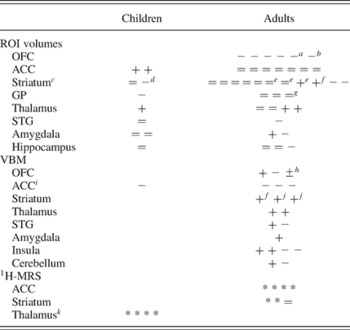
Volumetric studies in adults with OCD provide convincing evidence for reduced OFC volumes, suggestive evidence for increased thalamic and normal ACC volumes, and inconsistent evidence for caudate or striatum volumes. 1H-MRS studies in adults with OCD provide convincing evidence for decreased NAA levels in the ACC and suggestive evidence for decreased NAA levels in the striatum. Together, these findings point to abnormalities in volumes or NAA levels in all components of the OFC/ACC CBGTC loops that have been studied in detail. Abnormalities in closely related areas, such as the hippocampus, amygdala, and superior temporal gyrus, have also been reported. The findings from volumetric and 1H-MRS studies of adults with OCD are summarized in Table 1.
Summary of volumetric, VBM, and 1H-MRS findings in children and adults with OCD

Note: In a given cell, each symbol represents one study. Symbols in different cells may refer to the same study. ACC, anterior cingulate cortex; GP, globus pallidus;
1 H-MRS, proton magnetic resonance spectroscopy; OCD, obsessive–compulsive disorder; OFC, orbitofrontal cortex; ROI, region of interest; STG, superior temporal gyrus; VBM, voxel-based morphometry; (+) increased volumes or increased gray matter density in patients; (−) decreased volumes or decreased gray matter density in patients; (=) no significant differences between patients and healthy controls; (*) abnormal concentrations of neurometabolites (N-acetyl-aspartate, choline, or creatine/phosphocreatine) in patients.
a Medial OFC.
b Left anterior OFC.
c Studies refer to the caudate unless otherwise noted.
d Decreased putamen but not caudate volumes.
e Head of the caudate.
f Ventral striatum.
g Lenticular nuclei.
h Gray matter increases and decreases were reported for differing regions of OFC.
i All gray matter decreases were reported for both the ACC and the medial frontal gyrus.
j Putamen.
k All studies refer to the medial thalamus.
Limitations of ROI approaches
ROI approaches suffer from two serious limitations. First, because manual delimitation of ROIs is laborious, ROI studies often analyze only a small number of regions for which there are prior suspicions of abnormality, which limits the opportunity to find abnormalities in unanticipated regions. Automated methods to delimit ROIs partly alleviate this problem, but they are significantly less accurate than manual methods. Second, anatomical ROIs may not correspond to functionally meaningful units because gross anatomical structures or landmarks do not always reflect anatomical connectivity or cytoarchitecture. For example, several CBGTC loops run through the caudate nucleus (Alexander et al., Reference Alexander, DeLong and Strick1986; Middleton & Strick, Reference Middleton, Strick, Lichter and Cummings2001b) and each of those loops also partly seems to run through the putamen (Haber, Reference Haber2003). Considering each nucleus (caudate or putamen) as a whole misses both the distinctions between different loops within the nucleus and the fact that portions of one nucleus should be considered together with portions of the other. A meta-analysis of positron emission tomography (PET) and single-photon emission computed tomography studies of OCD that found reliable differences between patients with OCD and normal controls in the orbital gyrus and the head of the caudate nucleus, but not in the OFC or caudate nucleus as a whole (Whiteside et al., Reference Whiteside, Port and Abramowitz2004), illustrates the importance of moving toward finer anatomical distinctions in the study of OCD.
These limitations are addressed by two techniques: voxel-based morphometry (VBM) and tensor-based morphometry. VBM is a popular whole-brain approach that gives voxel-by-voxel measurements (Ashburner & Friston, Reference Ashburner and Friston2000). It remains controversial, however, because errors in spatial normalization may confound all subsequent computations, resulting in unreliable findings (Bookstein, Reference Bookstein2001). Tensor-based morphometry is used to analyze local morphological changes on the surface of cortical or subcortical structures (a procedure often termed “surface analysis”) or to measure cortical thickness. This approach gives highly localized, point-by-point measurements. Surface analyses, however, still require the prior delimitation of ROIs, as it is within a previously delimited ROI that surface analysis gives detailed, point-by-point measurements.
Cortical thickness
The findings from the only extant study of cortical thickness in OCD (Shin et al., Reference Shin, Yoo, Lee, Ha, Lee and Lee2007) are consistent with a pathophysiological model that emphasizes disturbances in the OFC, but also suggest abnormalities in a number of areas that are not traditionally considered involved in the pathophysiology of OCD. This study found cortical thinning in several areas of the left hemisphere of adults with OCD, including not only the OFC but also the ventrolateral prefrontal, middle frontal, precentral, superior temporal, parahippocampal, and lingual cortices. No abnormalities were detected in the ACC or anywhere in the right hemisphere. The findings from this study should be interpreted with care because most patients were medicated. Nevertheless, the finding of thinning in the superior temporal cortex is consistent with the volume reductions that have been reported for the superior temporal gyrus (Choi et al., Reference Choi, Kim, Yoo, Ha, Chang and Kim2006), and the finding of thinning in the parahippocampal cortex may point to broader abnormalities in the tissue surrounding the hippocampus that would be consistent with the volume reductions that have been reported for the hippocampus proper (Kwon et al., Reference Kwon, Shin, Kim, Kim, Youn and Han2003).
Surface analysis
Only two studies, both conducted by the same group and with adults, have used surface analysis to analyze subcortical structures in OCD. One of these studies focused on the hippocampus and reported shape changes concentrated on the hippocampus head (Hong et al., Reference Hong, Shin, Kim, Yoo, Lee and Kim2007). These results are consistent with the previous report by the same group of volumetric abnormalities in the hippocampus of adults with OCD (Kwon et al., Reference Kwon, Shin, Kim, Kim, Youn and Han2003). The other study focused on the basal ganglia and found outward deformities concentrated in the dorsal anterior caudate nucleus bilaterally and, to a lesser extent, in the ventral lateral part of the left putamen (Choi et al., Reference Choi, Kim, Yoo, Kang, Kim and Lee2007). These results are surprising, given that these are not the striatal regions through which the OFC/ACC CBGTC loops run (Alexander et al., Reference Alexander, DeLong and Strick1986; Haber, Reference Haber2003; Lehericy et al., Reference Lehericy, Ducros, Van de Moortele, Francois, Thivard and Poupon2004; Middleton & Strick, Reference Middleton, Strick, Lichter and Cummings2001b). The results of these surface analysis studies need to be interpreted with care because neither study corrected for multiple comparisons, and in both studies the majority of patients had a history of medication (although all were medication free for at least 4 weeks prior to scanning). Surface analysis techniques are also sufficiently new that replication by other laboratories would be important.
VBM
Several studies have used VBM to compare gray matter in adults with OCD and healthy controls (Kim et al., Reference Kim, Lee, Kim, Kim, Kim and Han2001; Pujol et al., Reference Pujol, Soriano-Mas, Alonso, Cardoner, Menchon and Deus2004; Valente et al., Reference Valente, Miguel, Castro, Amaro, Duran and Buchpiguel2005; Yoo et al., Reference Yoo, Roh, Choi, Kang do, Ha and Lee2008). Findings from these studies have been generally consistent with the pathophysiological model of OCD that emphasizes the OFC and ACC and their loops through the basal ganglia. Both increases (Kim et al., Reference Kim, Lee, Kim, Kim, Kim and Han2001; Valente et al., Reference Valente, Miguel, Castro, Amaro, Duran and Buchpiguel2005) and decreases (Pujol et al., Reference Pujol, Soriano-Mas, Alonso, Cardoner, Menchon and Deus2004; Valente et al., Reference Valente, Miguel, Castro, Amaro, Duran and Buchpiguel2005) in gray matter have been reported for differing subregions of the OFC, highlighting the potential value of a finer level of morphological analysis than is typically achieved in ROI-based studies. Decreases in gray matter have been consistently reported in the ACC and surrounding medial frontal gyrus (Pujol et al., Reference Pujol, Soriano-Mas, Alonso, Cardoner, Menchon and Deus2004; Valente et al., Reference Valente, Miguel, Castro, Amaro, Duran and Buchpiguel2005; Yoo et al., Reference Yoo, Roh, Choi, Kang do, Ha and Lee2008). Increases in gray matter have generally been reported in the striatum (Pujol et al., Reference Pujol, Soriano-Mas, Alonso, Cardoner, Menchon and Deus2004; Valente et al., Reference Valente, Miguel, Castro, Amaro, Duran and Buchpiguel2005; Yoo et al., Reference Yoo, Roh, Choi, Kang do, Ha and Lee2008). These increases have included the ventral striatum (Pujol et al., Reference Pujol, Soriano-Mas, Alonso, Cardoner, Menchon and Deus2004), which is consistent with the known neuroanatomy of OFC/ACC CBGTC loops, but also the putamen (Valente et al., Reference Valente, Miguel, Castro, Amaro, Duran and Buchpiguel2005; Yoo et al., Reference Yoo, Roh, Choi, Kang do, Ha and Lee2008), which is less clearly consistent with the standard pathophysiological model of OCD. Increases in gray matter have also been reported in the thalamus (Kim et al., Reference Kim, Lee, Kim, Kim, Kim and Han2001; Yoo et al., Reference Yoo, Roh, Choi, Kang do, Ha and Lee2008). VBM studies have also reported abnormalities in other areas that have been found abnormal in volumetric studies or in the only study of cortical thickness in OCD to date (Shin et al., Reference Shin, Yoo, Lee, Ha, Lee and Lee2007) and that relate closely to OFC/ACC CBGTC loops. For example, VBM studies have reported both increases (Kim et al., Reference Kim, Lee, Kim, Kim, Kim and Han2001) and decreases (Yoo et al., Reference Yoo, Roh, Choi, Kang do, Ha and Lee2008) in gray matter in the superior temporal gyrus, as well as increases in gray matter in the parahippocampal gyrus, extending to the amygdala (Valente et al., Reference Valente, Miguel, Castro, Amaro, Duran and Buchpiguel2005).
The only other areas for which abnormalities have been found in at least two VBM studies in adults with OCD are the insula (Kim et al., Reference Kim, Lee, Kim, Kim, Kim and Han2001; Pujol et al., Reference Pujol, Soriano-Mas, Alonso, Cardoner, Menchon and Deus2004; Valente et al., Reference Valente, Miguel, Castro, Amaro, Duran and Buchpiguel2005; Yoo et al., Reference Yoo, Roh, Choi, Kang do, Ha and Lee2008) and cerebellum (Kim et al., Reference Kim, Lee, Kim, Kim, Kim and Han2001; Pujol et al., Reference Pujol, Soriano-Mas, Alonso, Cardoner, Menchon and Deus2004). The insula is contiguous with the posterior OFC and is heavily interconnected with both the OFC and the ACC (Mesulam & Mufson, Reference Mesulam and Mufson1982; Mufson & Mesulam, Reference Mufson and Mesulam1982; Ongur & Price, Reference Ongur and Price2000). The cerebellum and frontal cortex are interconnected in parallel corticocerebellar loops (Kelly & Strick, Reference Kelly and Strick2003; Middleton & Strick, Reference Middleton and Strick1997, Reference Middleton and Strick2000, Reference Middleton and Strick2001a), and the cerebellum can also influence the striatum via a disynaptic connection through the thalamus (Hoshi, Tremblay, Feger, Carras, & Strick, Reference Hoshi, Tremblay, Feger, Carras and Strick2005). The potential involvement of the insula and cerebellum in OCD may therefore be consistent with the pathophysiological model that emphasizes disturbances in the OFC/ACC CBGTC loops and related areas in OCD.
Although abnormalities in several other areas have been reported in single VBM studies, they have not been replicated across studies. Although this does not exclude the possible involvement of other areas, it does provide some measure of reassurance that the main structural abnormalities in adults with OCD are concentrated along the OFC/ACC CBGTC loops and related areas. The findings from VBM studies of adults with OCD are summarized in Table 1.
Anatomical and Spectroscopy Studies in Children With OCD
Studies based on ROIs
OFC
We are not aware of any volumetric studies of the OFC in pediatric OCD. This is a major gap in the literature, given that reduced volumes in the OFC are the most consistent finding in the volumetric literature on adult OCD.
ACC
One study reported larger volumes of the ACC in treatment-naive children with OCD than in matched controls, with larger ACC volumes accompanying more severe symptoms (Rosenberg & Keshavan, Reference Rosenberg and Keshavan1998). Another study reported increased volumes of gray matter, but not of white matter, in the ACC of medication-naive children with OCD (Szeszko, MacMillan, McMeniman, Chen, et al., Reference Szeszko, MacMillan, McMeniman, Chen, Baribault and Lim2004). These findings stand in contrast to those in adults with OCD, which have generally failed to detect volumetric abnormalities in the ACC. A possible explanation for this difference in findings across age groups is that the ACC may develop differently in patients with OCD and healthy controls. One cross-sectional study reported a nearly significant correlation between age and ACC volumes in healthy children but not in children with OCD (Rosenberg & Keshavan, Reference Rosenberg and Keshavan1998). If this finding is confirmed, ideally in a longitudinal study, it could mean that ACC volumes are larger in children with OCD but then stay relatively constant with advancing age, whereas ACC volumes in healthy children might be smaller but then increase with age, resulting in similar ACC volumes in patients with OCD and healthy controls by adulthood.
Striatum
Two studies detected no significant differences in caudate volumes between children with OCD and matched controls (Rosenberg et al., Reference Rosenberg, Keshavan, O'Hearn, Dick, Bagwell and Seymour1997; Szeszko, MacMillan, McMeniman, Chen, et al., Reference Szeszko, MacMillan, McMeniman, Chen, Baribault and Lim2004), although one of these studies reported smaller putamen volumes in children with OCD (Rosenberg et al., Reference Rosenberg, Keshavan, O'Hearn, Dick, Bagwell and Seymour1997). The finding of a putamen abnormality is somewhat surprising from the perspective of the pathophysiological model of OCD that emphasizes the OFC/ACC CBGTC loops, given that those loops run mostly (although not exclusively) through the caudate; replication of this finding would therefore be important. If confirmed, this finding would also emphasize the need for volumetric studies of the putamen in adults with OCD, because virtually all volumetric studies of the striatum in adults with OCD have focused on the caudate.
Globus pallidus
One study reported smaller globus pallidus volumes in medication-naive children with OCD compared with matched healthy controls (Szeszko, MacMillan, McMeniman, Chen, et al., Reference Szeszko, MacMillan, McMeniman, Chen, Baribault and Lim2004). This finding stands in contrast to those in adults with OCD, which have generally failed to detect volumetric abnormalities in the globus pallidus.
Thalamus
One study reported increased thalamic volumes in medication-naive children with OCD that decreased to normal levels following treatment with the selective serotonin reuptake inhibitor paroxetine (Gilbert et al., Reference Gilbert, Moore, Keshavan, Paulson, Narula and Mac Master2000). Furthermore, the reduction in thalamic volumes with treatment correlated with the improvement in OCD symptoms. The finding of increased thalamic volumes in children with OCD is consistent with similar findings in adults. The finding that paroxetine treatment in children with OCD reduces thalamic volumes is also consistent with the indirect evidence that medication reduces thalamic volumes in adults with OCD. Another study found no differences in volumes of the thalamus measured before and after successful cognitive–behavioral therapy (Rosenberg, Benazon, Gilbert, Sullivan, & Moore, Reference Rosenberg, Benazon, Gilbert, Sullivan and Moore2000), suggesting that the reduction in volumes of the thalamus seen with paroxetine treatment is not necessary for symptom improvement. This study, however, did not include a control group, so the possibility that the children with OCD in this study might not have significantly enlarged thalami prior to treatment cannot be excluded.
Several studies by the same group using 1H-MRS have reported abnormalities in the medial thalamus of treatment-naive children with OCD. The first such study detected reduced ratios of NAA to choline and NAA to creatine/phosphocreatine + choline levels in the medial but not the lateral thalami of treatment-naive children with OCD (Fitzgerald, Moore, Paulson, Stewart, & Rosenberg, Reference Fitzgerald, Moore, Paulson, Stewart and Rosenberg2000). The findings were interpreted as indicative of reduced NAA levels in the medial thalami in the patient group, with the caveat that an increase in choline could produce the same results. Indeed, subsequent studies from the same group did report increases in choline (Rosenberg, Amponsah, Sullivan, MacMillan, & Moore, Reference Rosenberg, Amponsah, Sullivan, MacMillan and Moore2001; Smith et al., Reference Smith, Russell, Lorch, Banerjee, Rose and Ivey2003) and creatine/phosphocreatine levels (Mirza et al., Reference Mirza, O'Neill, Smith, Russell, Smith and Banerjee2006) in the medial thalami of children with OCD, suggesting that the earlier findings reflected elevated levels of choline and creatine/phosphocreatine rather than decreased levels of NAA. In fact, measurement of absolute NAA concentrations in the medial thalamus did not reveal differences between children with OCD and matched controls (Rosenberg et al., Reference Rosenberg, Amponsah, Sullivan, MacMillan and Moore2001). The significance of elevated choline and creatine/phosphocreatine levels in the medial thalamus remains to be fully elucidated. Nevertheless, it is noteworthy that these abnormalities occur in the medial thalamus, where the mediodorsal nucleus (the primary nucleus of the thalamus in OFC/ACC CBGTC loops) is located.
Other areas
The only study that measured volumes of the superior temporal gyrus and hippocampus in children with OCD did not detect significant differences from healthy controls (Rosenberg & Keshavan, Reference Rosenberg and Keshavan1998). This is in partial contrast to the findings in adults, which tentatively suggest that these structures may be abnormal in adults with OCD. Two studies failed to detect significant differences in amygdala volumes between children with OCD and healthy controls (Rosenberg & Keshavan, Reference Rosenberg and Keshavan1998; Szeszko, MacMillan, McMeniman, Lorch, et al., Reference Szeszko, MacMillan, McMeniman, Lorch, Madden and Ivey2004), although one of these studies found that children with OCD, unlike healthy controls, had significantly larger left than right amygdalae (Szeszko, MacMillan, McMeniman, Lorch, et al., Reference Szeszko, MacMillan, McMeniman, Lorch, Madden and Ivey2004). Abnormalities in other areas, for example, increased NAA levels in the left but not right DLPFC of children with OCD (Russell et al., Reference Russell, Cortese, Lorch, Ivey, Banerjee and Moore2003) and decreased pituitary volumes in boys but not girls with OCD (MacMaster et al., Reference MacMaster, Russell, Mirza, Keshavan, Banerjee and Bhandari2006), have been reported sporadically but need to be replicated.
Summary
Significantly fewer volumetric or 1H-MRS studies exist for children than for adults with OCD. Nevertheless, two findings receive convergent support from more than one study of children with OCD: increased volumes of the ACC and abnormal metabolite concentrations in the thalamus. Findings that were obtained in only one study and require replication include reduced putamen and globus pallidus volumes and increased thalamus volume. The findings from volumetric and 1H-MRS studies of children with OCD are summarized in Table 1.
The findings of increased thalamic volumes in children with OCD echo similar findings in adults. The findings of increased ACC volumes in children with OCD, however, are in contrast to the findings in adults with OCD, which have generally failed to detect volumetric abnormalities in the ACC, although they are consistent with several 1H-MRS studies that have reported decreased NAA levels in the ACC of adults with OCD. The finding of reduced globus pallidus volumes in children with OCD is also in contrast to the findings of the few existing volumetric studies of the globus pallidus in adults with OCD. Volumetric studies in adults with OCD have generally not measured the putamen, so whether reduced putamen volumes are found in adults with OCD remains unclear.
Most extant volumetric and 1H-MRS studies of OCD have focused on comparing volumes or metabolite levels between patients with OCD and healthy controls, paying little to no attention to how the volumes or metabolite levels change with age in persons with and without OCD. This information is, however, vitally important for a developmental perspective on the pathogenesis of OCD, as well as to understand how the abnormalities reported in children with OCD ultimately evolve to the abnormalities reported in adults with OCD, at least for adults with childhood-onset OCD. Similarly, studies of adults with OCD should distinguish better whether their findings relate to adult- or childhood-onset OCD, or to both.
VBM
No cortical thickness or surface analysis studies of children with OCD have been reported in the literature. However, two VBM studies have been recently published (Carmona et al., Reference Carmona, Bassas, Rovira, Gispert, Soliva and Prado2007; Gilbert et al., Reference Gilbert, Keshavan, Diwadkar, Nutche, Macmaster and Easter2008). One of these (Gilbert et al., Reference Gilbert, Keshavan, Diwadkar, Nutche, Macmaster and Easter2008) reported decreased gray matter in the left ACC and in the medial frontal gyrus bilaterally in children with OCD, consistent with similar findings in adults (Pujol et al., Reference Pujol, Soriano-Mas, Alonso, Cardoner, Menchon and Deus2004; Valente et al., Reference Valente, Miguel, Castro, Amaro, Duran and Buchpiguel2005; Yoo et al., Reference Yoo, Roh, Choi, Kang do, Ha and Lee2008) and with the hypothesized involvement of the ACC in OCD. The consistency of the findings of decreased gray matter in the medial frontal gyrus in children and adults underscores the need to consider not only the ACC proper, but also the surrounding medial frontal gyrus as a potential locus of abnormality in OCD.
The other VBM study in children with OCD (Carmona et al., Reference Carmona, Bassas, Rovira, Gispert, Soliva and Prado2007) also reported a large cluster of decreased gray matter in the cingulate cortex, although that finding did not remain significant after correcting for multiple comparisons. Other large clusters of decreased gray matter were found in the middle frontal gyrus, with the cluster in the right middle frontal gyrus remaining significant after correcting for multiple comparisons. Additional clusters were also reported, but they were substantially smaller and none remained significant after correcting for multiple comparisons. Additional studies are necessary to determine whether the finding in the middle frontal gyrus is reproducible, especially because of the 18 children with OCD, 11 had comorbid tic disorder and 10 were on medication.
The only consistent finding between the two existing VBM studies of children with OCD is reduced gray matter in the ACC. Although this is consistent with similar findings in VBM studies of adults with OCD, it contrasts with the findings of volumetric studies in children with OCD, which have generally found increased ACC volumes (Rosenberg & Keshavan, Reference Rosenberg and Keshavan1998; Szeszko, MacMillan, McMeniman, Chen, et al., Reference Szeszko, MacMillan, McMeniman, Chen, Baribault and Lim2004). A possible explanation for this discrepancy comes from the observation that, depending on the details of the VBM procedure used, VBM may characterize gray matter density rather than volume (Good et al., Reference Good, Johnsrude, Ashburner, Henson, Friston and Frackowiak2001). In fact, one of the studies (Gilbert et al., Reference Gilbert, Keshavan, Diwadkar, Nutche, Macmaster and Easter2008) states precisely that its findings concern gray matter densities, not volumes, and that this may explain their discrepancy with prior volumetric studies. The findings from VBM studies of children with OCD are summarized in Table 1.
PANDAS
Another potential line of evidence for the involvement of the basal ganglia in OCD comes from the study of PANDAS. This designation refers to childhood-onset OCD or tic disorders with abrupt onset or an episodic symptom course, in which symptom onset or exacerbation is temporally associated with infection with Group A beta-hemolytic streptococcus (GAS; Swedo et al., Reference Swedo, Leonard, Garvey, Mittleman, Allen and Perlmutter1998). Several authors have suggested that PANDAS are autoimmune disorders caused by antibodies to GAS that interact with the basal ganglia, in particular the striatum (Hoekstra & Minderaa, Reference Hoekstra and Minderaa2005; Leonard & Swedo, Reference Leonard and Swedo2001; Murphy, Husted, & Edge, Reference Murphy, Husted and Edge2006; Snider & Swedo, Reference Snider and Swedo2004). Controversy remains, however, regarding not only this causal hypothesis but the validity of the PANDAS construct itself (Harris & Singer, Reference Harris and Singer2006; Kurlan, Reference Kurlan2004; Kurlan & Kaplan, Reference Kurlan and Kaplan2004; Singer & Loiselle, Reference Singer and Loiselle2003).
GAS infection was first observed to produce Sydenham chorea, an illness associated with rheumatic fever and characterized primarily by involuntary rapid, jerky movements, which are often accompanied by other motor, behavioral, and emotional problems, including obsessive–compulsive symptoms and OCD (Swedo et al., Reference Swedo, Rapoport, Cheslow, Leonard, Ayoub and Hosier1989, Reference Swedo, Leonard, Schapiro, Casey, Mannheim and Lenane1993). Later observations identified putative cases of abrupt childhood-onset OCD or TS that followed GAS infections but did not meet criteria for Sydenham chorea (Allen, Leonard, & Swedo, Reference Allen, Leonard and Swedo1995; Swedo et al., Reference Swedo, Leonard, Garvey, Mittleman, Allen and Perlmutter1998). Establishing a clear and definitive causal role for GAS infections in these cases has proven difficult, however, in part because GAS infections are so common in childhood (Murphy, Sajid, & Goodman, Reference Murphy, Sajid and Goodman2006).
Experimental evidence suggests but does not prove a causal role for GAS in the onset or exacerbation of OCD or TS. Several studies have reported elevated anti basal ganglia antibodies in the sera of patients with TS or OCD (Dale, Heyman, Giovannoni, & Church, Reference Dale, Heyman, Giovannoni and Church2005; Kiessling, Marcotte, & Culpepper, Reference Kiessling, Marcotte and Culpepper1994; Rizzo, Gulisano, Pavone, Fogliani, & Robertson, Reference Rizzo, Gulisano, Pavone, Fogliani and Robertson2006; Singer et al., Reference Singer, Giuliano, Hansen, Hallett, Laurino and Benson1998; Wendlandt, Grus, Hansen, & Singer, Reference Wendlandt, Grus, Hansen and Singer2001), although at least one study failed to detect differences in anti basal ganglia antibodies between patients with PANDAS and healthy controls (Singer et al., Reference Singer, Loiselle, Lee, Minzer, Swedo and Grus2004). One study reported that immunomodulatory treatment of PANDAS led to improvements in both OCD and TS symptoms (Perlmutter et al., Reference Perlmutter, Leitman, Garvey, Hamburger, Feldman and Leonard1999), although this study has been criticized on multiple grounds (Singer, Reference Singer1999). Two studies have reported that antibiotic prophylaxis and treatment was effective in reducing symptoms in PANDAS (Murphy & Pichichero, Reference Murphy and Pichichero2002; Snider, Lougee, Slattery, Grant, & Swedo, Reference Snider, Lougee, Slattery, Grant and Swedo2005), although these studies should be considered preliminary because neither of them included a placebo group. Finally, infusion of sera from TS patients who have elevated antineuronal antibodies into the striatum of rats has been reported to produce an increase in stereotypies (Hallett, Harling-Berg, Knopf, Stopa, & Kiessling, Reference Hallett, Harling-Berg, Knopf, Stopa and Kiessling2000; Taylor et al., Reference Taylor, Morshed, Parveen, Mercadante, Scahill and Peterson2002), although these findings have not always been replicated (Loiselle, Lee, Moran, & Singer, Reference Loiselle, Lee, Moran and Singer2004; Singer et al., Reference Singer, Mink, Loiselle, Burke, Ruchkina and Morshed2005).
The caudate, putamen, and globus pallidus are enlarged in patients with PANDAS (Giedd, Rapoport, Garvey, Perlmutter, & Swedo, Reference Giedd, Rapoport, Garvey, Perlmutter and Swedo2000), just as they are in Sydenham chorea (Giedd et al., Reference Giedd, Rapoport, Kruesi, Parker, Schapiro and Allen1995). Changes in basal ganglia volumes are commonly found in OCD and TS, however, so the possibility that the volumetric changes found in patients with PANDAS simply reflect changes associated with TS or OCD, independently of an association with GAS infection, cannot be excluded. Stronger evidence for a causal link between an autoimmune reaction and enlarged basal ganglia volumes would be obtained if immunomodulatory treatment was shown to reduce the size of enlarged basal ganglia. A case study of an adolescent boy with exacerbation of OCD symptoms after a streptococcal pharyngitis showed precisely that, and the reduction in basal ganglia volumes was also accompanied by a reduction in symptom severity (Giedd, Rapoport, Leonard, Richter, & Swedo, Reference Giedd, Rapoport, Leonard, Richter and Swedo1996). However, larger controlled studies are needed to confirm these findings.
To summarize, the existing evidence suggests but does not prove that a GAS infection may, in some cases, trigger an autoimmune response that attacks the basal ganglia, leading to OCD, TS, Sydenham chorea, or a combination of these disorders. The specific CGBTC loops affected may determine the neuropsychiatric presentation, with disturbances in motor loops causing TS or Sydenham chorea and disturbances in OFC/ACC CBGTC loops causing OCD.
OCD as a Consequence of Brain Injury
The onset of OCD following focal brain lesions can provide valuable clues regarding the anatomical bases of OCD. In principle, simply because a lesion in a given brain region can produce OCD, that does not necessarily imply that all or even some cases of non-lesion-related OCD involve impairments in that region. Nevertheless, if lesion studies highlight the same general circuits that have been implicated in anatomical and functional imaging studies of OCD, the case for a causal role for those circuits in the pathogenesis of OCD becomes stronger.
Multiple reports describe cases of OCD following lesions confined to the basal ganglia (Carmin, Wiegartz, Yunus, & Gillock, Reference Carmin, Wiegartz, Yunus and Gillock2002; Chacko, Corbin, & Harper, Reference Chacko, Corbin and Harper2000; Laplane et al., Reference Laplane, Levasseur, Pillon, Dubois, Baulac and Mazoyer1989; Lopez-Rodriguez, Gunay, & Glaser, Reference Lopez-Rodriguez, Gunay and Glaser1997; Rodrigo Escalona, Adair, Roberts, & Graeber, Reference Rodrigo Escalona, Adair, Roberts and Graeber1997; Weilburg et al., Reference Weilburg, Mesulam, Weintraub, Buonanno, Jenike and Stakes1989; Weiss & Jenike, Reference Weiss and Jenike2000); two describe cases following lesions confined to the OFC (Kim & Lee, Reference Kim and Lee2002; Ogai, Iyo, Mori, & Takei, Reference Ogai, Iyo, Mori and Takei2005); a few describe cases following lesions involving broader expanses of frontal cortex (Swoboda & Jenike, Reference Swoboda and Jenike1995; Ward, Reference Ward1988; Weiss & Jenike, Reference Weiss and Jenike2000); and a few others describe cases following more widespread lesions, which nonetheless also involve the frontal lobes (typically including the OFC) or the basal ganglia (Berthier, Kulisevsky, Gironell, & Lopez, Reference Berthier, Kulisevsky, Gironell and Lopez2001; Gamazo-Garran, Soutullo, & Ortuno, Reference Gamazo-Garran, Soutullo and Ortuno2002; Max et al., Reference Max, Smith, Lindgren, Robin, Mattheis and Stierwalt1995). In addition, two case reports describe improvements in preexisting OCD after hemorrhage in the basal ganglia (Fujii, Otsuka, Suzuki, Endo, & Yamadori, Reference Fujii, Otsuka, Suzuki, Endo and Yamadori2005; Yaryura-Tobias & Neziroglu, Reference Yaryura-Tobias and Neziroglu2003).
The consistency of the brain areas involved in these case reports with those implicated in anatomical and functional imaging studies of patients with OCD suggests a causal role for the frontal cortices (in particular, the OFC) and the basal ganglia in the pathogenesis of OCD. Some caution is warranted when interpreting the findings of case reports, however, for three main reasons. First, the number of subjects involved is relatively small, even when considering the findings of all available case reports. Second, the stress associated with having a brain lesion could, by itself, aggravate or even precipitate some cases of OCD. In fact, greater psychosocial adversity is associated with the development of obsessive–compulsive symptoms following traumatic brain injury (Grados et al., Reference Grados, Vasa, Riddle, Slomine, Salorio and Christensen2008). Third, the possibility of reporting bias cannot be excluded, given that several of the cases mentioned above were reported after theories of the involvement of the OFC and respective loops through the basal ganglia had appeared in the literature, and clinicians may have been more willing to report cases consistent with that theoretical framework. To the extent that childhood- and adult-onset OCD may have differing etiologies, these cases may also bear mostly on adult-onset illness, as the vast majority of them involve adults (often older adults).
A large, prospective study assessed new-onset obsessive–compulsive symptoms in 80 children and adolescents (ages 6–18) following severe traumatic brain injury (Grados et al., Reference Grados, Vasa, Riddle, Slomine, Salorio and Christensen2008). This study reported a high prevalence of new-onset obsessions or compulsions 1 year after injury, with 21 out of the 80 subjects reporting obsessions or compulsions at that time but not before the lesion. Conversely, five subjects who had obsessions or compulsions before the lesion no longer had them 1 year after the lesion. Medial prefrontal and temporal lobe lesions were associated with new-onset obsessions, but this effect disappeared when obsessions and compulsions were considered conjointly. OFC lesions seemed to be associated with fewer symptoms, suggesting that anatomical integrity of the OFC is required for the onset of OCD. This is consistent with neurosurgical treatments for OCD that disrupt the OFC or its connections, but it contrasts with the case reports that have implicated lesions of the OFC in the onset of OCD (Kim & Lee, Reference Kim and Lee2002; Ogai et al., Reference Ogai, Iyo, Mori and Takei2005). A possible explanation for this discrepancy is that lesions to certain subregions of the OFC may cause OCD, whereas lesions to other subregions may prevent its expression.
This study suffered from three important limitations. First, it used an ROI approach that examined only the areas that have traditionally been implicated in OCD (OFC, medial PFC, basal ganglia, and thalamus), plus the temporal lobe. Second, symptoms were assessed 1 year after injury, and their onset or disappearance could, at least in some cases, be due simply to the passing of time. The absence of a group of matched noninjured controls in the study precludes any inferences regarding the extent to which the changes in symptoms were caused by the injury. Third, scanning and symptom assessment occurred 9 months apart. In addition, the focus of the study was on obsessive–compulsive symptoms, not full-blown OCD. In fact, only 2 out of the 80 children had new-onset OCD following the lesion.
In summary, several reports describe cases of OCD following lesions of the basal ganglia or frontal cortex (with an emphasis on the OFC); two reports describe cases of improvement in OCD after lesions of the basal ganglia. A larger, prospective study (Grados et al., Reference Grados, Vasa, Riddle, Slomine, Salorio and Christensen2008) also suggests a role for the OFC in obsessive–compulsive symptoms following brain injury, although in that study lesions to the OFC were inversely related to symptom severity. The same study also reported an association between lesions to the medial PFC (which included the ACC) and obsessions. All of these findings are consistent with the hypothesis that the pathogenesis of OCD involves CBGTC loops, in particular, the loops involving the OFC and possibly the ACC.
Despite the convergence of findings from lesion studies with those from anatomical and functional imaging studies, the correct way to conceptualize the relation of lesion findings with functional imaging findings remains elusive. For example, we have seen above that the OFC is often hyperactive in OCD. Do we interpret this as evidence that activity of the OFC is somehow causally related to the symptoms of OCD and therefore expect a lesion of the OFC to improve symptoms, given that a lesioned OFC would not be active? Or do we interpret the imaging data as evidence that the OFC is working in overdrive to try to inhibit or control the expression of OCD symptoms and therefore expect a lesion of the OFC to worsen the symptoms? Or do we interpret the hyperactivity in OFC not as a sign of heightened function but rather of dysfunction in that region and therefore expect that lesions of the OFC might produce OCD, by making the OFC dysfunctional? Different versions of all of these hypotheses can be found implicitly or explicitly in the literature. Existing lesion studies do not, however, help us discriminate between these alternatives, given that lesions of the OFC have been associated with both increases and decreases in OCD symptoms. More research is needed to address these questions.
Another question, raised specifically by findings of the prospective study described above (Grados et al., Reference Grados, Vasa, Riddle, Slomine, Salorio and Christensen2008), is whether the temporal lobe may also be involved in the pathogenesis of OCD. In support of this possibility, temporal lobe epilepsy has been associated with OCD and obsessive–compulsive symptoms (Kroll & Drummond, Reference Kroll and Drummond1993; Monaco et al., Reference Monaco, Cavanna, Magli, Barbagli, Collimedaglia and Cantello2005). In addition, several studies have reported anatomical abnormalities in the superior temporal gyrus in patients with OCD (Choi et al., Reference Choi, Kim, Yoo, Ha, Chang and Kim2006; Kim et al., Reference Kim, Lee, Kim, Kim, Kim and Han2001; Shin et al., Reference Shin, Yoo, Lee, Ha, Lee and Lee2007; Yoo et al., Reference Yoo, Roh, Choi, Kang do, Ha and Lee2008). One possibility, which we have already discussed, is that the temporal lobe, in particular the superior temporal gyrus, is involved in OCD via its connections with the regions of the striatum that are part of the OFC/ACC CBGTC loops (Alexander et al., Reference Alexander, DeLong and Strick1986). However, additional research is needed to test this hypothesis.
Neurosurgical Lesions
Neurosurgical lesions are sometimes made to attenuate symptoms in extremely severe cases of OCD that do not respond to psychotherapy and medication. Four types of neurosurgical lesions are used: anterior capsulotomy, subcaudate tractotomy, cingulotomy, and limbic leucotomy (for reviews, see, e.g., Greenberg, Murphy, & Rasmussen, Reference Greenberg, Murphy and Rasmussen2000; Greenberg et al., Reference Greenberg, Price, Rauch, Friehs, Noren and Malone2003; Mindus, Rasmussen, Lindquist, & Noren, Reference Mindus, Rasmussen, Lindquist, Noren, Salloway, Malloy and Duffy2001). All are small bilateral lesions that interrupt white matter tracts that connect the OFC or ACC with subcortical structures involved in CBGTC loops. Their success, although modest, provides additional converging evidence for the involvement of the OFC, ACC, and their associated CBGTC loops in the pathophysiology of OCD.
The lesions in anterior capsulotomy are placed in the anterior limb of the internal capsule. Such lesions are believed to interrupt the connections between the OFC and the mediodorsal nucleus of the thalamus (Greenberg et al., Reference Greenberg, Murphy and Rasmussen2000; Mindus et al., Reference Mindus, Rasmussen, Lindquist, Noren, Salloway, Malloy and Duffy2001), which is the thalamic nucleus with densest reciprocal connections with the OFC and the primary nucleus in the thalamus involved in CBGTC loops that connect to the OFC (Fuster, Reference Fuster1997; Ongur & Price, Reference Ongur and Price2000). The lesions in subcaudate tractotomy are placed below and immediately anterior to the head of the caudate nucleus. They seem to be restricted to white matter, as they are anterior to the substantia innominata and rarely include the cortex (Malhi & Bartlett, Reference Malhi and Bartlett1998; Newcombe, Reference Newcombe1975). These lesions are believed to disconnect the OFC from the thalamus, basal ganglia, and limbic system (Feldman, Alterman, & Goodrich, Reference Feldman, Alterman and Goodrich2001; Malhi & Bartlett, Reference Malhi and Bartlett1998). They have also been shown to produce degeneration in the anterior limb of the internal capsule, which can be traced back to the mediodorsal nucleus of the thalamus (Mindus et al., Reference Mindus, Rasmussen, Lindquist, Noren, Salloway, Malloy and Duffy2001). The lesions in cingulotomy are placed in the cingulate bundle (i.e., cingulum). The mechanisms by which cingulotomy reduces the severity of OCD symptoms are unknown, but may involve the fibers that connect the ACC and the caudate nucleus (Rauch et al., Reference Rauch, Kim, Makris, Cosgrove, Cassem and Savage2000). Limbic leucotomy refers simply to a combination of subcaudate tractotomy and cingulotomy.
Evaluating the success of these procedures is complicated by the small number of subjects in each study and the obvious ethical difficulties with conducting randomized double-blind studies involving neurosurgical lesions (Earp, Reference Earp1979). In addition, different studies use different criteria (e.g., remission of symptoms, percent improvement over preoperative symptom severity) to evaluate the success of these procedures. Nevertheless, estimated success rates are generally approximately 50% or more for all procedures (Jenike, Reference Jenike1998; Mindus et al., Reference Mindus, Rasmussen, Lindquist, Noren, Salloway, Malloy and Duffy2001). Similar rates are, however, often seen in open trials of medication or psychotherapy and typically drop substantially in blinded, controlled treatment trials.
The fact that these procedures primarily target fiber bundles that connect the OFC or ACC with related subcortical structures, notably the thalamus and basal ganglia, provides additional evidence for the involvement of these circuits in the pathogenesis of OCD. Such evidence remains circumstantial, however, for three reasons. First, all of these procedures were developed largely empirically between 30 and 60 years ago (Bingley, Leksell, Meyerson, & Rylander, Reference Bingley, Leksell, Meyerson, Rylander, Sweet, Obrador and Martín-Rodríguez1977; Kelly et al., Reference Kelly, Richardson, Mitchell-Heggs, Greenup, Chen and Hafner1973; Knight, Reference Knight1964; Richardson, Reference Richardson1973; Talairach, Hecaen, David, Monnier, & Ajuriaguerra, Reference Talairach, Hecaen, David, Monnier and Ajuriaguerra1949; Whitty, Duffield, Tov, & Cairns, Reference Whitty, Duffield, Tov and Cairns1952), often based on neuroanatomical theories that are now outdated. Consequently, in most cases we still do not fully know all of the neuroanatomical connections that they affect. Second, the improvement in symptoms is often gradual following surgery, occurring over a time span of weeks to months (Jenike, Reference Jenike1998). This suggests that the anatomical changes underlying such improvement may be a secondary consequence of the lesion, possibly involving retrograde degeneration or a neuroplastic response. Indeed, volume reductions in distant structures have been observed after localized neurosurgical lesions (Rauch et al., Reference Rauch, Kim, Makris, Cosgrove, Cassem and Savage2000; Taren, Curtis, & Gebarski, Reference Taren, Curtis and Gebarski1994). Third, the fact that a lesion in a given circuit improves symptoms does not necessarily imply that an abnormality at that lesion site or even in that circuit caused the symptoms. In fact, the same neurosurgical procedures used for the treatment of OCD are also used for a variety of other conditions, including other anxiety disorders (Cosgrove & Rauch, Reference Cosgrove and Rauch2003), depression (Cosgrove & Rauch, Reference Cosgrove and Rauch2003; Malhi & Bartlett, Reference Malhi and Bartlett2000), and intractable pain (Wilkinson, Davidson, & Davidson, Reference Wilkinson, Davidson and Davidson1999). For all of these reasons, whether these procedures target directly and specifically the circuits involved in the pathogenesis of OCD remains unclear.
Development of OFC/ACC CBGTC Circuits
Knowledge of the normal development of OFC/ACC CBGTC circuits may provide clues to the neurodevelopmental processes that go awry in patients with OCD. Although brain weight and volume nearly reach their adult values by 5–6 years of age (Courchesne et al., Reference Courchesne, Chisum, Townsend, Cowles, Covington and Egaas2000; Dekaban & Sadowsky, Reference Dekaban and Sadowsky1978; Giedd, Snell, et al., Reference Giedd, Snell, Lange, Rajapakse, Casey and Kozuch1996; Kretschmann, Kammradt, Krauthausen, Sauer, & Wingert, Reference Kretschmann, Kammradt, Krauthausen, Sauer, Wingert and Kretschmann1986; Reiss, Abrams, Singer, Ross, & Denckla, Reference Reiss, Abrams, Singer, Ross and Denckla1996; Sahni, Jit, & Sodhi, Reference Sahni, Jit and Sodhi1998), the brain continues to develop throughout childhood, adolescence, and early adulthood. The total volume of gray matter starts to decrease after around 5 years of age (Courchesne et al., Reference Courchesne, Chisum, Townsend, Cowles, Covington and Egaas2000; Pfefferbaum et al., Reference Pfefferbaum, Mathalon, Sullivan, Rawles, Zipursky and Lim1994; Reiss et al., Reference Reiss, Abrams, Singer, Ross and Denckla1996; Sowell et al., Reference Sowell, Peterson, Thompson, Welcome, Henkenius and Toga2003), with cortical thickness and gray matter density decreasing in much of the cortex (Gogtay et al., Reference Gogtay, Giedd, Lusk, Hayashi, Greenstein and Vaituzis2004; Sowell et al., Reference Sowell, Thompson, Leonard, Welcome, Kan and Toga2004). In contrast, white matter volume increases throughout childhood, adolescence, and early adulthood (Courchesne et al., Reference Courchesne, Chisum, Townsend, Cowles, Covington and Egaas2000; Giedd et al., Reference Giedd, Blumenthal, Jeffries, Castellanos, Liu and Zijdenbos1999; Matsuzawa et al., Reference Matsuzawa, Matsui, Konishi, Noguchi, Gur and Bilker2001; Pfefferbaum et al., Reference Pfefferbaum, Mathalon, Sullivan, Rawles, Zipursky and Lim1994; Reiss et al., Reference Reiss, Abrams, Singer, Ross and Denckla1996; Sowell et al., Reference Sowell, Peterson, Thompson, Welcome, Henkenius and Toga2003). In general, sensorimotor areas mature earlier and higher order areas, including the OFC, mature later (Benes, Reference Benes1989; Gogtay et al., Reference Gogtay, Giedd, Lusk, Hayashi, Greenstein and Vaituzis2004; Yakovlev & Lecours, Reference Yakovlev, Lecours and Minkowski1967). During the years in which childhood-onset OCD typically starts, the PFC, basal ganglia, and thalamus are undergoing major developmental changes. Furthermore, some of those changes seem to affect primarily boys, which may explain the higher prevalence of OCD among boys than among girls. This section reviews these developmental changes and their possible relation to the pathogenesis of OCD.
Development of the PFC
The PFC grows markedly between 5 and 14 years of age (Kanemura, Aihara, Aoki, Araki, & Nakazawa, Reference Kanemura, Aihara, Aoki, Araki and Nakazawa2003; Sowell et al., Reference Sowell, Thompson, Leonard, Welcome, Kan and Toga2004; Thompson et al., Reference Thompson, Giedd, Woods, MacDonald, Evans and Toga2000). Such growth seems to include the most ventral aspects of the PFC (Sowell et al., Reference Sowell, Thompson, Leonard, Welcome, Kan and Toga2004; Thompson et al., Reference Thompson, Giedd, Woods, MacDonald, Evans and Toga2000), possibly including the OFC. However, the studies that have mapped the growth of the PFC did not include the ventral surface of the brain, so the growth patterns of the OFC remain to be elucidated. Some evidence suggests that the fastest growth in the PFC occurs between 8 and 14 years of age (Kanemura et al., Reference Kanemura, Aihara, Aoki, Araki and Nakazawa2003), the period during which most cases of childhood-onset OCD begin, raising the possibility that abnormalities in that growth could cause childhood-onset OCD—a hypothesis that is consistent with the findings of reduced OFC volume in patients with OCD. The PFC, including the OFC, continues to mature through adulthood (Gogtay et al., Reference Gogtay, Giedd, Lusk, Hayashi, Greenstein and Vaituzis2004), however, so abnormalities in later stages of maturation of the OFC could conceivably cause adult-onset OCD.
The growth of the PFC during childhood and adolescence may be largely driven by the development of white matter (Barnea-Goraly et al., Reference Barnea-Goraly, Menon, Eckert, Tamm, Bammer and Karchemskiy2005; Reiss et al., Reference Reiss, Abrams, Singer, Ross and Denckla1996). In fact, gray matter density and cortical thickness in PFC seem to decrease during childhood, adolescence, and young adulthood (Gogtay et al., Reference Gogtay, Giedd, Lusk, Hayashi, Greenstein and Vaituzis2004; Sowell, Thompson, Holmes, Jernigan, & Toga, Reference Sowell, Thompson, Holmes, Jernigan and Toga1999; Sowell et al., Reference Sowell, Thompson, Leonard, Welcome, Kan and Toga2004). Furthermore, postmortem studies indicate that the density of synapses and the sizes of neuronal cell bodies and dendritic fields in PFC increase only until approximately 2 to 3 years of age (Huttenlocher, Reference Huttenlocher1979; Huttenlocher & Dabholkar, Reference Huttenlocher and Dabholkar1997; Koenderink, Uylings, & Mrzljak, Reference Koenderink, Uylings and Mrzljak1994; Petanjek, Judas, Kostovic, & Uylings, Reference Petanjek, Judas, Kostovic and Uylings2008). Whether this means that OCD is caused primarily by abnormalities in the development of white matter or in the regressive processes that fine-tune synaptic organization in OFC is unknown.
Future studies should address more directly the normal development of the OFC. Several of the magnetic resonance imaging (MRI) studies that have mapped cortical development did not map the inferior surface of the brain (Sowell et al., Reference Sowell, Peterson, Thompson, Welcome, Henkenius and Toga2003, Reference Sowell, Thompson, Leonard, Welcome, Kan and Toga2004; Thompson et al., Reference Thompson, Giedd, Woods, MacDonald, Evans and Toga2000) and postmortem developmental studies have generally focused on the lateral PFC (Huttenlocher, Reference Huttenlocher1979; Huttenlocher & Dabholkar, Reference Huttenlocher and Dabholkar1997; Koenderink et al., Reference Koenderink, Uylings and Mrzljak1994; Petanjek et al., Reference Petanjek, Judas, Kostovic and Uylings2008). Our knowledge of the development of the OFC is therefore indirect, and the extent to which the OFC and the lateral and dorsal PFC share similar developmental trajectories is unknown. One study reported that while the volume of white matter in dorsal PFC increased throughout childhood and adolescence, the volume of white matter in the OFC decreased (Reiss et al., Reference Reiss, Abrams, Singer, Ross and Denckla1996), raising the possibility that these structures may develop differently. Even within the OFC, decreases in gray matter density throughout childhood and adolescence seem to follow medial–lateral and anterior–posterior gradients (Gogtay et al., Reference Gogtay, Giedd, Lusk, Hayashi, Greenstein and Vaituzis2004). A better understanding of the development of the OFC in healthy children and in children with OCD could substantially advance our understanding of the pathogenesis of this disorder.
Current knowledge of the development of the ACC is similarly limited. Volumes of the cingulate seem to change less during childhood and adolescence than do volumes of other regions such as the frontal or medial temporal lobes (Sowell, Trauner, Gamst, & Jernigan, Reference Sowell, Trauner, Gamst and Jernigan2002). Gray matter density also seems to change less across the life span in the ACC than in many other cortical regions (Sowell et al., Reference Sowell, Peterson, Thompson, Welcome, Henkenius and Toga2003). Furthermore, cortical thickness and size of the dorsal ACC seem to change relatively little between 5 and 11 years of age, although cortical thickness and size of the ventral ACC may increase in that age range (Sowell et al., Reference Sowell, Thompson, Leonard, Welcome, Kan and Toga2004). Together, these findings may suggest that anatomical development is less prominent in the ACC than in other portions of the OFC/ACC CBGTC loops during the years in which OCD typically begins. The volume of the right ACC has, however, been reported to increase during childhood and adolescence (Casey et al., Reference Casey, Trainor, Giedd, Vauss, Vaituzis and Hamburger1997; Marquardt et al., Reference Marquardt, Levitt, Blanton, Caplan, Asarnow and Siddarth2005). Furthermore, larger volumes of the right ACC have been associated with better performance on an attentional task in children and adolescents (Casey et al., Reference Casey, Trainor, Giedd, Vauss, Vaituzis and Hamburger1997). Some authors have argued that development of the ACC during infancy, childhood, and adolescence may underlie the improvements in self-regulatory capacities that characterize these periods (Posner & Rothbart, Reference Posner and Rothbart1998; Posner, Rothbart, Sheese, & Tang, Reference Posner, Rothbart, Sheese and Tang2007). Whether abnormalities in the development of the ACC may lead to deficits in the self-regulation of intrusive thoughts, images, or feelings, or to the well-documented deficits in inhibitory control in OCD (Chamberlain et al., Reference Chamberlain, Blackwell, Fineberg, Robbins and Sahakian2005) is unknown.
Development of the basal ganglia and thalamus
The volume of the caudate nucleus has generally been reported to decrease during childhood and adolescence (Giedd, Castellanos, Rajapakse, Vaituzis, & Rapoport, Reference Giedd, Castellanos, Rajapakse, Vaituzis and Rapoport1997; Giedd, Snell, et al., Reference Giedd, Snell, Lange, Rajapakse, Casey and Kozuch1996; Jernigan, Trauner, Hesselink, & Tallal, Reference Jernigan, Trauner, Hesselink and Tallal1991). Some evidence suggests that such decreases may be most pronounced between 4 and 12 years of age (Giedd, Snell, et al., Reference Giedd, Snell, Lange, Rajapakse, Casey and Kozuch1996), and marked tissue loss has been reported in the head of the caudate between 7 and 13 years of age (Thompson et al., Reference Thompson, Giedd, Woods, MacDonald, Evans and Toga2000). These age ranges are consistent with the ages during which childhood-onset OCD typically begins, raising the possibility that abnormal development of the caudate could cause childhood-onset OCD. Volumes of the lenticular nuclei (Giedd et al., Reference Giedd, Castellanos, Rajapakse, Vaituzis and Rapoport1997; Giedd, Snell, et al., Reference Giedd, Snell, Lange, Rajapakse, Casey and Kozuch1996; Jernigan et al., Reference Jernigan, Trauner, Hesselink and Tallal1991; Reiss et al., Reference Reiss, Abrams, Singer, Ross and Denckla1996; Sowell et al., Reference Sowell, Trauner, Gamst and Jernigan2002) and thalamus (Jernigan et al., Reference Jernigan, Trauner, Hesselink and Tallal1991; Reiss et al., Reference Reiss, Abrams, Singer, Ross and Denckla1996; Sowell et al., Reference Sowell, Trauner, Gamst and Jernigan2002) also decrease throughout childhood and adolescence. In addition, white matter tracts within the basal ganglia and between the basal ganglia and thalamus also develop during childhood and adolescence (Barnea-Goraly et al., Reference Barnea-Goraly, Menon, Eckert, Tamm, Bammer and Karchemskiy2005). Together with the findings of maturation of frontal cortex during childhood and adolescence (Barnea-Goraly et al., Reference Barnea-Goraly, Menon, Eckert, Tamm, Bammer and Karchemskiy2005; Gogtay et al., Reference Gogtay, Giedd, Lusk, Hayashi, Greenstein and Vaituzis2004; Kanemura et al., Reference Kanemura, Aihara, Aoki, Araki and Nakazawa2003; Klingberg, Vaidya, Gabrieli, Moseley, & Hedehus, Reference Klingberg, Vaidya, Gabrieli, Moseley and Hedehus1999; Reiss et al., Reference Reiss, Abrams, Singer, Ross and Denckla1996; Sowell et al., Reference Sowell, Thompson, Holmes, Jernigan and Toga1999, Reference Sowell, Trauner, Gamst and Jernigan2002, Reference Sowell, Thompson, Leonard, Welcome, Kan and Toga2004; Thompson et al., Reference Thompson, Giedd, Woods, MacDonald, Evans and Toga2000), these results may reflect the integrated development of CBGTC loops. OCD may be caused by abnormalities in the development of one or more of the key structures or white matter tracts in these loops.
Gender differences in the development of CBGTC circuits
Many studies have reported sexual dimorphisms in the human brain (Gilmore et al., Reference Gilmore, Lin, Prastawa, Looney, Vetsa and Knickmeyer2007; Goldstein et al., Reference Goldstein, Seidman, Horton, Makris, Kennedy and Caviness2001; Luders et al., Reference Luders, Narr, Thompson, Woods, Rex and Jancke2005; Rabinowicz, Dean, Petetot, & de Courten-Myers, Reference Rabinowicz, Dean, Petetot and de Courten-Myers1999; Sowell et al., Reference Sowell, Peterson, Kan, Woods, Yoshii and Bansal2007). These tend to occur in regions that have high concentrations of sex steroid receptors during brain development (Goldstein et al., Reference Goldstein, Seidman, Horton, Makris, Kennedy and Caviness2001), suggesting a role for gonadal hormones in causing such dimorphisms. Gender differences in brain structure and development during childhood and adolescence may provide an explanation for the significantly higher prevalence of childhood-onset OCD in boys than in girls. In fact, existing evidence suggests that both the caudate and the OFC, two key components of the OFC/ACC CGBTC loops, differ markedly and develop differently in boys and girls.
On average, male brains are larger than female brains across all age groups (Caviness, Kennedy, Richelme, Rademacher, & Filipek, Reference Caviness, Kennedy, Richelme, Rademacher and Filipek1996; Courchesne et al., Reference Courchesne, Chisum, Townsend, Cowles, Covington and Egaas2000; Giedd et al., Reference Giedd, Castellanos, Rajapakse, Vaituzis and Rapoport1997; Giedd, Snell, et al., Reference Giedd, Snell, Lange, Rajapakse, Casey and Kozuch1996; Goldstein et al., Reference Goldstein, Seidman, Horton, Makris, Kennedy and Caviness2001; Lenroot et al., Reference Lenroot, Gogtay, Greenstein, Wells, Wallace and Clasen2007; Reiss et al., Reference Reiss, Abrams, Singer, Ross and Denckla1996; Sowell et al., Reference Sowell, Peterson, Kan, Woods, Yoshii and Bansal2007). Comparisons of volumes of brain structures between males and females are therefore usually performed after adjusting for overall brain volume. When this is done, girls have greater caudate volumes than boys (Caviness et al., Reference Caviness, Kennedy, Richelme, Rademacher and Filipek1996; Giedd et al., Reference Giedd, Castellanos, Rajapakse, Vaituzis and Rapoport1997; Giedd, Snell, et al., Reference Giedd, Snell, Lange, Rajapakse, Casey and Kozuch1996; Sowell et al., Reference Sowell, Trauner, Gamst and Jernigan2002). In addition, the decline in volumes of the caudate and lenticular nuclei during childhood and adolescence may be restricted to boys (Giedd et al., Reference Giedd, Castellanos, Rajapakse, Vaituzis and Rapoport1997; Giedd, Snell, et al., Reference Giedd, Snell, Lange, Rajapakse, Casey and Kozuch1996; Sowell et al., Reference Sowell, Trauner, Gamst and Jernigan2002). Childhood and adolescence may therefore be particularly critical for the development of the basal ganglia in boys, which may explain why boys are more susceptible than girls to OCD and other disorders of CBGTC circuitry such as TS and attention-deficit/hyperactivity disorder.
Potential sexual dimorphisms in the human OFC during childhood or adolescence remain to be studied. In adulthood, women have larger OFC volumes than men (Goldstein et al., Reference Goldstein, Seidman, Horton, Makris, Kennedy and Caviness2001), and in a sample spanning childhood to old age, females had thicker OFC cortices than males (Sowell et al., Reference Sowell, Peterson, Kan, Woods, Yoshii and Bansal2007). These anatomical differences have functional implications, as males perform better on average than females on tasks that rely on OFC function (Overman, Reference Overman2004). Such performance differences seem to apply across most age groups, and have been found even in children younger than 3 years of age (Overman, Reference Overman2004). Similar findings have been reported in monkeys, with male infant monkeys performing better than female infant monkeys on an object discrimination reversal task, which is known to depend on the OFC (Clark & Goldman-Rakic, Reference Clark and Goldman-Rakic1989). The OFC also appears to develop earlier in male monkeys than in female monkeys (Clark & Goldman-Rakic, Reference Clark and Goldman-Rakic1989; Goldman, Crawford, Stokes, Galkin, & Rosvold, Reference Goldman, Crawford, Stokes, Galkin and Rosvold1974). In short, the structure, function, and development of the OFC seem to differ between males and females. The contribution of these differences to the differing susceptibilities of boys and girls to OCD remains to be elucidated however.
Several lines of evidence suggest that these differences in OFC structure, function, and development in males and females are caused by the effects of gonadal hormones on the OFC during development. First, the OFC has high affinity for androgen binding (Clark, MacLusky, & Goldman-Rakic, Reference Clark, MacLusky and Goldman-Rakic1988) and high aromatase activity (MacLusky, Clark, Naftolin, & Goldman-Rakic, Reference MacLusky, Clark, Naftolin and Goldman-Rakic1987; MacLusky, Naftolin, & Goldman-Rakic, Reference MacLusky, Naftolin and Goldman-Rakic1986) in the developing monkey, suggesting that it may be particularly susceptible to the influence of gonadal hormones. Second, treating female infant monkeys with androgen early in postnatal life abolishes the performance differences between male and female monkeys in the object discrimination reversal task (Clark & Goldman-Rakic, Reference Clark and Goldman-Rakic1989). Third, female rats have more extensive dendritic fields in the OFC than do male rats, but those differences are abolished by neonatal gonadectomy (Kolb, Pellis, & Robinson, Reference Kolb, Pellis and Robinson2004). The finding of more extensive dendritic fields in the OFC of female rats may also help to explain the findings of larger volume (Goldstein et al., Reference Goldstein, Seidman, Horton, Makris, Kennedy and Caviness2001) and thicker cortex (Sowell et al., Reference Sowell, Peterson, Kan, Woods, Yoshii and Bansal2007) in the OFC of women.
Conclusions
All of the lines of research reviewed herein—anatomical imaging, PANDAS, OCD as a consequence of brain injury, and neurosurgery for treatment-refractory OCD—suggest a role for OFC/ACC CBGTC loops in the pathogenesis of OCD, but each suffers from important limitations. Anatomical imaging findings point to abnormalities in the OFC/ACC CBGTC loops in both children and adults with OCD. However, these findings are inherently correlational and therefore do not license any conclusions regarding a causal role for those abnormalities in OCD. Studies of PANDAS and of OCD as a consequence of brain injury suggest that insults to OFC/ACC CBGTC loops may indeed cause OCD. Specifically, studies of PANDAS suggest that an autoimmune insult to the basal ganglia may cause OCD; however, controversy remains regarding this putative pathogenic mechanism and even the very existence of PANDAS. Studies of OCD as a consequence of brain injury suggest that lesions in key components of these loops may also cause OCD; however, much of this evidence is based on case reports. Finally, neurosurgical lesions used for treatment-refractory OCD suggest that lesions placed in key components of the OFC/ACC CBGTC loops can be effective in attenuating the symptoms of OCD; however, the improvement in symptoms is often gradual, raising the possibility that it may be due to degeneration or plasticity elsewhere in the brain, rather than an effect of the primary lesion itself. Despite the limitations when each of these lines of evidence is considered in isolation, the remarkable convergence of findings across all of them does suggest a causal role for the OFC/ACC CBGTC loops in OCD, in both children and adults.
Assuming that OCD is indeed caused by abnormalities in OFC/ACC CBGTC loops, do the findings reviewed above suggest a particular locus within those loops that may be responsible for OCD? In adults with OCD, anatomical imaging studies using ROI approaches point to volumetric or NAA abnormalities in all components of the OFC/ACC CBGTC loops that have been studied in detail: the OFC, ACC, striatum, and thalamus. Whole-brain approaches such as VBM have also found gray matter abnormalities in all of these areas. Abnormalities in other areas closely related to the OFC/ACC CBGTC loops, including the superior temporal gyrus, hippocampus, and amygdala, have also been found in both ROI and whole-brain anatomical studies. Several VBM studies have also highlighted abnormalities in the insula, another area closely related to the OFC/ACC CBGTC loops (Mesulam & Mufson, Reference Mesulam and Mufson1982; Mufson & Mesulam, Reference Mufson and Mesulam1982; Ongur & Price, Reference Ongur and Price2000).
The implications for the pathogenesis of OCD of such widespread abnormalities within OFC/ACC CBGTC loops and potentially also in related areas in adults with OCD can be interpreted in at least four ways. First, the etiology of OCD may involve some mechanism that causes widespread abnormalities in all of these structures. Second, the etiology of OCD may involve some mechanism that causes an abnormality in just one or a small number of these structures, with the abnormalities in the remaining structures being a consequence of dynamic changes prompted by that primary etiologic abnormality. As we have seen, anatomical imaging studies after neurosurgery have shown that localized lesions in one structure or fiber bundle of the OFC/ACC CBGTC loops can cause volume changes in remote, albeit related structures (Rauch et al., Reference Rauch, Kim, Makris, Cosgrove, Cassem and Savage2000; Taren et al., Reference Taren, Curtis and Gebarski1994). Thus, abnormalities in one or a few structures in these loops could translate into structural abnormalities in all of the structures within the loops and related structures elsewhere. Third, the etiology of OCD may, as in the second hypothesis, consist of an initial abnormality in just one or a few structures, with the abnormalities in the remaining structures following from that, but with different cases of OCD possibly arising from initial abnormalities in different structures. Lesion studies suggest that damage to any one of several components of the OFC/ACC CBGTC loops is sufficient to cause OCD, raising the possibility that even non-lesion-related cases of OCD may be a consequence of different initial abnormalities. Fourth, the widespread abnormalities may actually be an artifact of averaging across subjects, with individual subjects having more localized abnormalities that vary across subjects.
One way of trying to disentangle these possibilities would be to conduct longitudinal anatomical imaging studies that followed patients with OCD over extended period of time, starting as close to their diagnosis as possible. Such studies could determine whether the abnormalities start in only one or a few structures (with the initial site of the abnormality possibly varying by case) and then spread to other structures. A cruder and less conclusive alternative is to compare the findings from anatomical imaging studies across children and adults with OCD to determine whether the findings in children suggest abnormalities in a smaller subset of regions than do the findings in adults. If so, this could indicate that abnormalities in OCD start in a specific subset of the regions that have been found abnormal in adults with OCD. Differences between findings in children and adults with OCD could, however, be due to multiple factors, including a comparison across childhood- and adult-onset subtypes, differing types and rates of comorbidity, differing ascertainment biases, and differing durations of illness.
Unfortunately, the extant literature does not allow us to conclude with any certainty which areas are and are not affected in children with OCD. Existing studies clearly point to anatomical abnormalities in the ACC and metabolite abnormalities in the thalamus. No anatomical abnormalities have yet been reported in the other structures that are affected in adults with OCD, including the OFC and caudate, but that may be attributable either in whole or in part to the fact that those structures have not yet been properly studied in children with OCD. For example, even though the most consistently replicated volumetric finding in adults with OCD is a reduction in volume of the OFC, no ROI studies have analyzed the volume of the OFC in children with OCD. Similarly, even though the most consistent finding for the striatum of adults with OCD is a reduction in NAA levels, no studies have analyzed NAA levels in the striatum of children with OCD. In addition, of the two VBM studies of pediatric OCD, one had fairly small samples (Gilbert et al., Reference Gilbert, Keshavan, Diwadkar, Nutche, Macmaster and Easter2008), and in the other the majority of children were on medication (Carmona et al., Reference Carmona, Bassas, Rovira, Gispert, Soliva and Prado2007).
A large-scale longitudinal study that followed children or adults with high familial risk for OCD from before their diagnosis through several years later would clearly provide a much richer and more dynamic view of the unfolding of pathology in the brain affected by OCD. Such a study should ideally be multimodal, including, for example, anatomical MRI, functional MRI, 1H-MRS, investigation of white matter fiber tracts using diffusion tensor imaging, and investigation of neurotransmitter and receptor levels using PET. The results of such a study could then inform a more detailed search for the ultimate cause or causes of OCD: clearly, the focus of such a search would be vastly different if OCD were found to be a consequence of, say, an abnormality localized initially in the striatum, than if it were found to be a consequence of abnormalities affecting, from the very beginning, several structures of the OFC/ACC CBGTC loops and related structures elsewhere.