Implications
The present study demonstrates that pre-pubertal active immunization against gonadotropin-releasing hormone (GnRH) generates testicular physiological impairment in three different mammalian species (lagomorphs, hystricomorphs and ovine species). Anti-GnRH products are usually targeted to bovine and swine species. Our findings suggest that the use of anti-GnRH vaccines, once developed, can be extended to a broad spectrum of mammalian species but the efficacy may vary as we found that germ cell arrest was detected in all the species studied, but some individuals within hystricomorphs and ovine species had normal sexual hormones levels and spermatogenesis. This is the first multispecies report of variable anti-GnRH efficacy in mammals.
Introduction
Conventional invasive surgical castration, also known as gonadectomy, has been commonly conducted to provide different advantages to animal producers, consumers and owners (Miesner and Anderson, Reference Marti, Devant, Amatayakul-Chantler, Jackson, Lopez, Janzen and Schwartzkopf-Genswein2015). Advantages include fertility control (prevention of undesired pregnancies), meat taint reduction, improvement of carcass quality and better husbandry by controlling unwanted aggressive and sexual behaviors (Marti et al., Reference Martí, Aparicio and García-Herreros2015). However, surgical castration usually comes with complications, such as surgical trauma, infections, hemorrhage, stress, pain, discomfort, potential death and other inconveniences such as high economical expenses, lower feed efficiency and growth rate, prolonged setbacks on productivity and excessive deposition of fat (Ison et al., Reference Howard and Reed2016). Gonadotropin-releasing hormone is a decapeptide expressed in selected hypothalamic nuclei that controls the release of the gonadotropins LH and FSH from the adenohypophysis and therefore orchestrates the reproductive development and functioning in mammals (Simoni et al., Reference Rodríguez and Wettstein1999; Clarke and Pompolo, Reference Clayton2005). Immunization against GnRH has proved a promising option for immunocastration of farm and companion animal species (Jago et al., Reference Ison, Clutton, Di Giminiani and Rutherford1997; Janett et al., Reference Jago, Bass and Matthews2009 and Reference Janett, Gerig, Tschuor, Amatayakul-Chantler, Walker, Howard, Piechotta, Bollwein, Hartnack and Thun2012). Reducing LH (and as a consequence reducing testosterone (T) secretion) through the use of GnRH antagonists may be an alternative for inhibiting male reproductive function (Simoni et al., Reference Rodríguez and Wettstein1999). Another alternative using long-term doses of GnRH agonists has been carried out (Kauffold et al., Reference Jimenez-Liñan, Rubin and King2010). This can effectively desensitize the hypothalamic–pituitary–gonadal axis to GnRH by a down-regulation physiological mechanism which reduces the concentrations of testicular hormones, testes size and weight as well as the diameter of seminiferous tubules through the inhibition of the function of Leydig and Sertoli cells and sperm production (arrest of spermatogenesis) (Kauffold et al., Reference Jimenez-Liñan, Rubin and King2010). Thus, active immunization against GnRH is a very promising alternative to surgical castration. Active immunization against GnRH is commonly called immunocastration. This procedure involves autoimmune responses that directly or indirectly affect the structure and function of the gonads, reducing sex hormone levels and interfering with the capacity to produce gametes (Occhio et al., Reference Niewiesk2001). Although several GnRH-based vaccines exist, contraceptive efficacy is still low, maybe due to low immune response against a self-antigen, low-affinity antibodies or different adjuvants and carrier molecules (Goubau et al., Reference Goubau, Silversides, Gonzalez, Laarveld, Mapletoft and Murphy1989). An immunocontraceptive vaccine based on GnRH should be highly immunogenic in order to induce a sufficient titre of neutralizing anti-GnRH antibodies, with only few injections required and it must minimize the risk of immunological cross-reactions while avoiding unintended interference with other physiological processes (Kutzler and Wood, Reference Kauffold, Rohrmann, Boehm and Wehrend2006). Moreover, the immunological response may differ among species, among breeds and among individuals within the same breed. Therefore, it cannot be assumed a priori that vaccination effectiveness can be extrapollable among species.
Growth and maturation of the testes in pre-pubertal and early puberal individuals are acutely dependent on gonadotrophic stimulation by increasing serum T concentrations (Hernandez-Medrano et al., Reference Hernandez-Medrano, Williams, Peters, Hannant, Campbell and Webb2012; Janett et al., Reference Janett, Gerig, Tschuor, Amatayakul-Chantler, Walker, Howard, Piechotta, Bollwein, Hartnack and Thun2012; Han et al., Reference Han, Cheng, Chen, Du, Cao and Zeng2014). Accordingly, there may be specific windows during development (probably short-lived) when GnRH antibodies could successfully affect the hypothalamic–pituitary–gonadal axis by impairing reproductive function in a species-specific fashion (Hernandez-Medrano et al., Reference Hernandez-Medrano, Williams, Peters, Hannant, Campbell and Webb2012; Janett et al., Reference Janett, Gerig, Tschuor, Amatayakul-Chantler, Walker, Howard, Piechotta, Bollwein, Hartnack and Thun2012; Han et al., Reference Han, Cheng, Chen, Du, Cao and Zeng2014). It has been suggested that the age at primary immunization is an important determinant of the magnitude of the immunization response and that the response seems to be optimal when immunization occurs before puberty (Janett et al., Reference Janett, Gerig, Tschuor, Amatayakul-Chantler, Walker, Howard, Piechotta, Bollwein, Hartnack and Thun2012; Han et al., Reference Han, Cheng, Chen, Du, Cao and Zeng2014). However, to the best of our knowledge, there are limited studies detailing variations in the inter-specific effect of early (pre-pubertal) vaccination with GnRH based vaccines in different domestic animal species. As comprehensive studies in the efficacy of the inter-species active immunization against GnRH in pre-pubertal individuals are lacking, the aims of the present study were as follows: (i) to design an effective vaccine using the rabbit (Oryctolagus cuniculus) as a model for active immunization against GnRH, (ii) to determine the effectiveness of the anti-GnRH vaccine on testicular morphometry, histopathological alterations and endocrine suppression in pre-pubertal individuals after its homologous administration to rabbits and (iii) to evaluate the effects of the anti-GnRH vaccine optimized in rabbits for active immunization in pre-pubertal individuals of guinea pig (Cavia porcellus) and sheep (Ovis aries).
Material and methods
Ethical statement
The current study was carried out under a Project License (PROM-CEB-013-2014) from the National Secretariat of Higher Education, Science, Technology, and Innovation (SENESCYT). We confirm that this study was carried out in strict accordance with the recommendations in the legal framework (Animal Welfare Law) currently in place in AGROCALIDAD Ethics Committee (MAGAP-AGROCALIDAD), for all Ecuadorian Public and Private Laboratories and Higher Education Institutions. All procedures were performed in accordance to the Ecuadorian Animal Protection Law (COIP Art. 249-2014).
Animal husbandry
A total of 40 New Zealand breed rabbits (entire males with a starting age of 2 months) were used for the different experiments (20 individuals for anti-GnRH vaccine optimization and 20 individuals for vaccination with the anti-GnRH vaccine). All animals were kept in indoor individual mesh-cages (0.90×0.60×0.45 m). A total of 20 entire male guinea pigs (Peruvian genetic-line, 1 month of age) were used for the experiments. All animals were housed by experimental groups in indoor individual boxes (1.5×1.5 m). A total of 20 entire ram lambs (Dorper breed, entire males with a starting age of 2 months) were maintained in indoor individual pens (5×5 m). In all cases, the buildings housing the different species were disinfected with quaternary ammonium (day 7). Afterwards, cleaning and disinfection was performed every 7 days. Upon arriving to the housing facilities, animals were given vitamins and dextrose per os to attenuate transportation and environmental adaptation stress. Before the experiments began, all the animals were placed in quarantine for 15 days to ensure they were disease-free and totally adapted to the new environment. During that time (day 8) all the animals were dewormed and kept in an enriched environment. All animals were fed alfalfa forage and standard formulated concentrate-food and water ad libitum during the experimental period.
Vaccine production and vaccination scheme in rabbits
All reagents and media used in the present study were purchased from Sigma (Sigma-Aldrich, St Louis, MO, USA) unless otherwise specified. The anti-GnRH vaccine was intended to be a slow delivery compound. Every 2 ml contained 570 μg of GnRH conjugated with Clostridium septicum (4×10³ ppm) and Clostridium chauvoei toxoid (16×107 ppm). Aluminum hydroxide was added as adjuvant (15 mg). The vehicle for the injectable suspension was mQ water with 2% formaldehyde. Dosis optimization was done in rabbits. Four applications spaced 15 days showed the best dosis to (1) reduce LH, FSH and T to non-detectable levels, (2) reduce testis size and (3) impair spermatogenesis (with total sperm absence). Best dosis was 500 μg in 0.3 ml.
An experimental group of rabbits was administered 500 μg/0.3 ml of anti-GnRH vaccine subcutaneously and a booster 15 days afterwards. A control group was administered saline solution instead (NaCl 0.9%/0.3 ml, two administrations spaced 15 days); five additional animals were castrated at the start of the vaccinations to assess initial reproductive status. Animals were randomly allocated to the treatment groups. The same scheme was used with the other two species used. In order to evaluate the gross vaccination effect on the size of the testes of the individuals from different species, testicular length and width of animals were measured early in the morning with a measuring tape, every 15 days from the day of arrival to the end of the experiments.
Blood samples and hormone analyses
Blood samples (1 ml) from rabbits were aseptically taken by cardiac puncture. Samples were used for hormone analysis (LH, FSH and T in rabbits and additionally cortisol in guinea pigs and ram lambs). In guinea pigs, 1 ml blood samples were aseptically taken by making a small incision at the interdigital space. In ram lambs, 5 ml blood samples were obtained from the external jugular vein. The first sample was taken a day before the start of the experiments to have a reference profile for comparisons with the vaccination group; 1 day after the first anti-GnRH dose, two blood samples per animal/treatment were taken every 15 days (total blood samples=3). Finally, in the group of animals that were castrated at the beginning of the experiments, one blood sample was taken before surgery. Hormonal assays were performed at a private accredited bioanalysis laboratory. Hormonal assays were performed using ELISA kits (Abnova, Taipei City, Taiwan). Immediately, after whole blood sample collection (EDTA-treated tubes), the cells were removed from plasma by centrifugation at 2000 r.p.m. for 5 min with a refrigerated centrifuge (4°C). The resulting supernatant fraction (plasma) was transferred into clean polypropylene tubes. If the ELISA analysis was postponed, the plasma samples were divided into aliquots and stored at −20°C for further analysis. For the direct quantitative determination of FSH, LH, T and cortisol, specific monoclonal antibodies (anti-FSH-radish peroxidase (RP), anti-LH-RP, anti-testosterone-RP and anti-cortisol-RP, respectively) were used. A two-step assay was carried out for each hormone quantification. The fist monoclonal specific antibodies were used as first immobilized factors onto the microwell plate and the second specific antibodies, conjugated with RP, were designed for a different specific region of the FSH, LH, T or cortisol hormones, respectively. Plasma samples (10 µl per duplicate) were allowed to bind to the plate for 30 min at room temperature (22°C to 23°C) in a plate shaker (250 r.p.m.). After a gently wash (300 µl washing buffer per well×3 times) they were incubated with the second monoclonal antibody (1 : 500 v/v) conjugated with RP. After a second wash, a stop-reaction buffer (50 µl) was added and the absorbance was assessed by using a microtiter plate reader (450 nm) within 20 min after stop-reaction buffer addition. Sensitivity was set at 0.50 m IU/ml for FSH (CV<6%), 0.52 m IU/ml for LH (CV<10%), 0.05 ng/ml for T (CV<11%) and 0.003 ng/ml for cortisol (CV<10%). Sensitivity was calculated at the lower detection limit from the standard curve by determining the resulting concentration of the mean OD of the first calibrator (10 replicate analyses). Cortisol assays were carried on to monitor stress in experimental animals.
Testicular tissue sampling and processing
After 15 days the last anti-GnRH vaccine application all animals were castrated (control and vaccinated groups). In total, three animals per species/experimental group were castrated to evaluate the overall developmental stage of the testis at the start of the experiments. Testes were obtained early in the morning and were kept on ice (at ~4°C) before histological fixation. The tunica vaginalis and epididymis were excised and testis weight recorded. Volume was estimated by the water displacement method using a graduated cylinder. Testes did not show any apparent macroscopical lesions. Testes were transversally excised and fixed in neutral buffered formalin (4% formaldehyde, 48 h) and stored in 70% ethanol (4°C).
Testicular tissue pieces (~0.5 cm3) were transferred into biopsy cassettes for further histological processing. Samples were dehydrated in alcohol series: ethanol 70% (30 min twice), ethanol 96% (30 min twice), ethanol 100% (30 min twice), xylene (45 min twice) and paraffin infiltrated in a histoprocessor (Slee MTM, Mainz, Germany). Samples were embedded in paraffin blocks and cut 5 μm thick with a microtome (Cut 6062, Slee). Slides were deparaffinized, stained with Harris hematoxylin and mounted with synthetic Canada balsam (Biopack, Buenos Aires, Argentina).
Testicular morphometrical and histological analyses
Testicular tissues were analyzed for general morphology and stereologic and morphometric parameters were estimated. Images were captured with a digital video camera (Tucsen Imaging Technology Co., Ltd, Beijing, China). Volume density of testicular compartments (seminiferous tubules and interstitial or intertubular space) were estimated with a 30-point 195-μm-spaced graticule, digitally superimposed at random positions over testicular tissue images corresponding to the treatments used. A total of 20 images per treatment were used and 600 points per treatment were counted. Tubule diameters were measured on the digital images. Tubular diameters were calculated as the average of the major and minor axes of randomly chosen near-round cross-section profiles of seminiferous tubules. Diameters were measured with image-J software (downloaded from http://imagej.nih.gov/ij/) and ~55 near-round cross-section tubule profiles/treatment were evaluated. Determinations of the stages of seminiferous tubules showing spermatozoa were according to the literature: Swierstra and Foote (Reference Simoni, Weinbauer, Gromoll and Nieschlag1963), Morton (Reference Miller, Johns and Killian1988) (Rabbits); Clermont (Reference Ciaramella, Chianese, Pariante, Fasano, Pierantoni and Meccariello1960), Rodríguez and Wettstein (Reference Occhio, Aspden and Trigg2004) (Guinea pigs); Bilaspuri and Guraya (Reference Bilaspuri and Guraya1986) (Rams). Germ cell identification was done on high resolution digital images based on criteria such as close contact with the basal membrane and ovoid shape. Absolute numbers of spermatogonia were estimated through unbiased stereological methods. For this purpose, a consecutive pair of every tenth 5-mm thick sections (with a random microtome sectioning start) were used. Spermatogonial density (spermatogonia per volume unit of parenchyma) was estimated through the physical disector. Images from paired sections were digitalized. The image pairs were exposed simultaneously side by side on a wide computer screen and square-shaped counting frames were digitally and randomly superimposed. Type A spermatogonia that appeared in the first image (reference section) but did not in the same area in the second section (look-up section) were counted (whenever cells appeared in both sections, they were not counted) (Howard and Reed, Reference Tanaka, Kanatsu-Shinohara, Lei, Rao and Shinohara1998). The area of the counting frames was recorded and used to calculate the sample volume. This volume was equal to the counting frame area times the length of the space between the consecutive pair of sections (5 μm). The division of the reference volume by the total cells counted yielded sures of density, that is number of type A spermatogonia per volume unit (spermatogonia/mm3). Absolute numbers of type A spermatogonia were estimated by the product of the volume of the testis times the spermatogonial density. A total of five pairs of sections per testis were used.
Statistical analysis
All statistical analyses were performed using SPSS for Windows® software, v17. General statistical analysis of stereologic and morphometric variables was performed using one-way ANOVA. The level of significance was set at P<0.05 and when the analyses were statistically significant, multiple comparisons were performed by means of Bonferroni post hoc test.
Results
Effects of anti-gonadotropin-releasing hormone vaccine in rabbits
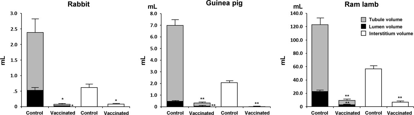
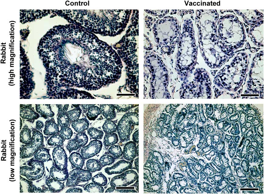
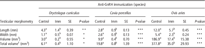
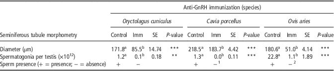
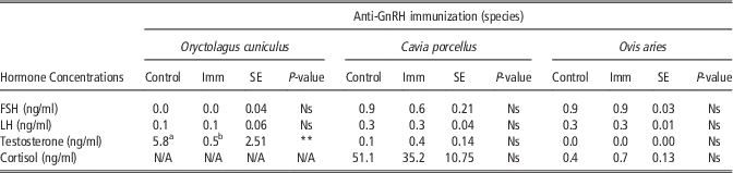
All the parameters studied (testicular length, width, volume and total volume) were significantly reduced in vaccinated rabbits with respect to the control group (Table 1). The anti-GnRH vaccine generated a ~1500% reduction in testicular volume in the experimental group (P<0.05). Testis weight was also significantly reduced in the experimental group (~375%) (control, 3.54±0.32 g v. vaccinated, 0.94±0.10 g; P<0.05). Tubule diameter decreased approximately twofold in the vaccinated group with respect to the control, that is about 200% (P<0.001) (Table 2). Both tubule (parenchyma) and intertubular volumes significantly decreased in vaccinated animals (P<0.05) (Figure 1). There was also a significant reduction of the lumen volume between control and vaccinated animals (P<0.05). Spermatogonial populations decreased in the vaccinated group of rabbits. They went from more than a million spermatogonia per testis to <100 000 in the vaccinated group (P<0.01) (Table 2). In contrast with the control group, no sperm was found in any of the seminiferous tubule cross-sections in the vaccinated group (Figure 2). Follicle-stimulating hormone and LH levels had considerable fluctuations during the experimental period in both control and vaccinated animals. However no significant differences were observed between the two groups regarding these gonadotropins at any time during the experiments (Table 3). At the end of the period, testosterone levels dropped by about 22-fold in the vaccinated animals with respect the control group (P<0.01).
Absolute volumes of testicular compartments of control (non-vaccinated) and anti-gonadotropin-releasing factor vaccinated rabbits, guinea pigs and ram lambs (*P<0.05, **P<0.01).

Testicular morphology of control (non-vaccinated) and anti-gonadotropin-releasing factor vaccinated rabbits. Hematoxylin staining. High magnification, bar=60 μm; low magnification, bar=240 μm.

Testis morphometric parameters in control (non-immunized) and anti-gonadotropin-releasing hormone (GnRH) immunized rabbits, guinea pigs and ram lambs at the end of the experimental period (in vivo measurements)

Control=control group; Imm=immunized group.
All individuals (rabbits: n=40; guinea pigs: n=20; and ram lambs: n=20) were pre-pubertal and clinically healthy. Volume was estimated from length and width data using the prolate spheroid formula.
1 Volumes of left and right testes.
a,bValues within a row/treatment with different superscripts differ significantly at P<0.05.
*P<0.05, **P<0.01, ***P<0.001
Testis micromorphometric parameters in control (non-immunized) and anti-gonadotropin-releasing hormone (GnRH) immunized rabbits, guinea pigs and ram lambs at the end of the experimental period

Control=control group; Imm=immunized group.
All histopathological analyses were carried out in individuals from the three different species (rabbits: n=40; guinea pigs: n=20; and ram lambs: n=20).
1 Negative presence of sperm (80% effectiveness).
2 Negative presence of sperm (60% effectiveness).
a,bValues within a row/treatment with different superscripts differ significantly at P<0.05 (**P<0.01, ***P<0.001).
Hormone concentrations in control (non-immunized) and anti-gonadotropin-releasing hormone (GnRH) immunized rabbits, guinea pigs and ram lambs at the end of the experimental period

Control=control group; Imm=immunized group; N/A=not available.
a,bValues within a row/treatment with different superscripts differ significantly at P<0.05 (**P<0.01).
Blood samples were collected from all individuals (rabbits: n=40; guinea pigs: n=20; and ram lambs: n=20) to carry out different hormonal analyses.
Effects of anti-gonadotropin-releasing hormone vaccine in guinea pigs
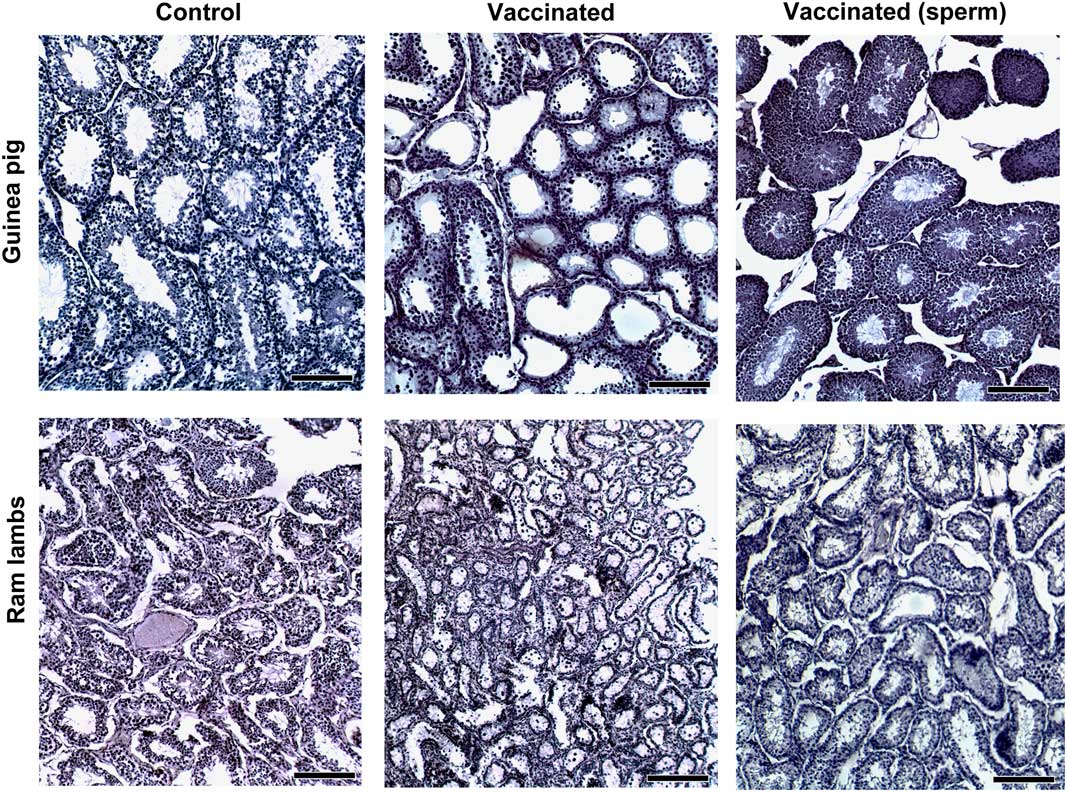
Just as in rabbits, all the parameters studied (testicular length, width, volume and total volume) were significantly reduced in vaccinated guinea pigs with respect to the control group (Table 1). The anti-GnRH vaccine generated an almost 2700% reduction in testicular volume in the experimental group (P<0.001). Tubule diameter decreased ~1.2-fold in the vaccinated group with respect to the control, that is about 120% (P<0.001) (Table 2). As in rabbits, both tubule (parenchyma) and intertubular volumes significantly decreased in vaccinated animals (P<0.01), (Figure 1). There was also a highly significant reduction of the lumen volume in vaccinated animals (P<0.01) (Figure 1). Spermatogonial populations also decreased in the vaccinated group of guinea pigs. They went from more than a million spermatogonia per testis to <30 000 in the vaccinated group (P<0.001) (Table 2). As expected, the control group showed sperm in the corresponding stage VII seminiferous tubules. In the vaccinated group, no spermatozoa were found in the seminiferous tubule cross-sections (80% of cases). However, 20% of animals showed adult spermatogenesis (Table 2, Figure 3). No significant differences were observed between control and vaccinated guinea pigs regarding LH, FSH or T levels at the end of the experimental period (Table 3). Cortisol levels were not statistically significant either (Table 3).
Testicular morphology of control (non-vaccinated) and anti-gonadotropin-releasing factor vaccinated guinea pigs and ram lambs. Hematoxylin staining. Some vaccinated animals showed sperm in the lumen of the seminiferous tubules (20% guinea pigs and 40% ram lambs, right panel). Bar=200 μm.

Effects of anti-gonadotropin-releasing hormone vaccine in ram lambs
Testis morphometry
Similarly as in rabbits and guinea pigs, all the parameters studied (testicular length, width, volume and total volume) were significantly reduced in vaccinated ram lambs with respect to the control group (Table 1). The anti-GnRH vaccine produced about a 110% reduction in testicular volume in the experimental group (P<0.001). Tubule diameter was reduced ~3.5-fold in the vaccinated group, that is about 350%, with respect to the control group (P<0.001) (Table 2). As in rabbits and guinea pigs, both tubule (parenchyma) and intertubular volumes significantly decreased in vaccinated ram lambs (P<0.05) (Figure 1). There was also a highly significant reduction of the lumen volume in vaccinated animals (P<0.05).
Spermatogonial populations also decreased in the vaccinated group of male sheep. They went from more than near 23 million spermatogonia per testis to around one million in the vaccinated group (P<0.001) (Table 2). As expected, the control group showed sperm in the corresponding stage VIII and I seminiferous tubules of the ram. No sperm was found in any of the seminiferous tubule cross-sections (60% of cases) in the vaccinated group. However, 40% of individuals showed normal adult spermatogenesis (Table 2, Figure 3). No significant differences were observed between control and vaccinated animals regarding LH, FSH or T levels at the end of the experimental period (Table 3). Cortisol levels were not statistically significant (Table 3).
Discussion
Active immunization against GnRH has been proposed as an alternative to surgical castration and represents an important advance in the control of fertility in domestic animals. The present study examines the effects of active immunization against GnRH on male individuals of three mammalian species (rabbit, guinea pig and ram lambs) on testicular morphometry, histopathology and endocrine responses during the pre-pubertal period. On present study, there were important variations in the immune responses among the species tested when an anti-GnRH vaccine was administered. In general, the regression in testis size observed was primarily a consequence of germ cell arrest in all the species studied, shown by a significant impairment of spermatogenesis, as previously found in immunoneutered ruminants (Adams et al., Reference Adams, Daley, Adams and Sakurai1996; Occhio et al., Reference Niewiesk2001). Nevertheless, of the three species studied, endocrine levels appeared to be altered only in rabbits, showing a significant decrease in T levels after two vaccinations (initial vaccination and booster). On the other hand, our results showed a different and unexpected trend in the other two species vaccinated (guinea pigs and ram lambs) which had an incomplete suppression of spermatogenesis and unchanged levels in serum concentrations of FSH, LH and T when compared with the control group. These contrasting results may be explained by possible differences on effects over intrinsic and extrinsic factors in spermatogenesis. First of all, species-specific factors probably determine different immune responses against GnRH. Interestingly, the amino acid sequence of GnRH is highly conserved in mammals, but in guinea pigs the decapeptide shows a variation that falls out of the rule, being that histidine is switched for tyrosine in the second position while leucine is substituted by valine in the seventh position (Jimenez-Liñan et al., Reference Janett, Stump, Burger and Thun1997). Although the substitutions involve chemically similar amino acids, this probably results on a different immune response on this species, being the vaccine based on GnRH with the general mammalian sequence. Alternatively, unaltered levels of endocrine profiles in vaccinated guinea pigs and ram lambs could be probably attributed to low blood sample frequencies implemented, especially during the 1st weeks of age, when pre-pubertal individuals show episodic pulses of hormonal release that occur randomly during time (Hernandez-Medrano et al., Reference Hernandez-Medrano, Williams, Peters, Hannant, Campbell and Webb2012) with variations upon the specific physiology of the species studied (Han et al., Reference Han, Cheng, Chen, Du, Cao and Zeng2014). Another significant factor involved on the variation of the immune responses among species is the individual genotype or interferences of species-specific maternal derived antibodies (Niewiesk, Reference Morton2014). Other hormonal interactions such as the negative feedback of T or inhibin on the release of FSH are unlikely because the effects of this gonadotropin tend to appear after individuals reach puberty (Simoni et al., Reference Rodríguez and Wettstein1999). However, GnRH is the master factor controlling the release of FSH and LH during the pre-pubertal stage, and therefore, the temporal relationship between FSH and LH during early reproductive development is a well-known factor in many species (Clayton, Reference Clermont1985; Clarke and Pompolo, Reference Clayton2005). There is evidence of a refractory effect of circulating FSH, LH and T levels in individuals actively immunized against T during the pre-pubertal stage, linked to a reduced feedback on the hypothalamic–pituitary–gonadal axis that results in a higher gonadotrophic stimulation of the testicular tissue (Han et al., Reference Han, Cheng, Chen, Du, Cao and Zeng2014). However, in our study, the species differences on immunocastration success led us to hypothesize that the effects of such an immunization against GnRH do not follow a classical endocrine feedback loop, at least in the species used and during pre-pubertal development. Moreover, the endocrine levels observed suggest that the immunization effects might not just be related to the neutralization of GnRH but also probably to modifications of responsiveness of gonadotropin receptors at the testicular level (Auclair et al., Reference Auclair, Sowerbutts and Setchell1995). Thus, an unpredictable residual secretion of FSH in guinea pigs and ram lambs together with a low T serum concentration could be sufficient to sustain a marginal spermatogonial population and to maintain the spermatogenic process (Hernandez-Medrano et al., Reference Hernandez-Medrano, Williams, Peters, Hannant, Campbell and Webb2012; Han et al., Reference Han, Cheng, Chen, Du, Cao and Zeng2014).
Although there are species-specific developmental differences in the animals used, their ages at the start of the experiments matched a similar developmental window regarding testicular cellular dynamics. Rabbits and lambs entered the experiments at 2 months of age whereas guinea pigs entered at 1 month. Therefore, in all three species the first vaccination (nearly 15 days after the mentioned ages) was applied during the first wave of spermatogenesis preceding puberty. Accordingly, at the beginning of the experiments the animals from the three species studied showed normal seminiferous tubules with round spermatocytes as the most advanced cell type. This morphological picture clearly corresponds to spermatocytogenesis during pre-puberty, a developmental window characterized in the literature in rabbits (Yasser et al., Reference Yasser, Mahmoud and Gamal2012), guinea pigs (Holt, Reference Holt1977) and ram lambs (Boukenaoui et al., Reference Boukenaoui, Moudilou, Chevalier, Amirat, Exbrayat and Khammar2012). Physiological differences in developmental patterns and growth rates among the species could have influenced the results. For instance, rabbits are altricial species whereas guinea pig and sheeps are precocial. This probably represents a limitation for the interpretation of the results when comparisons among species are made.
In all the three species studied there were tremendous overall morphological changes in the testis after vaccination against GnRH. The most obvious change observed was a reduction in seminiferous tubule diameters by at least 120%. Testis regression was not focalized to a specific area and both tubular and intertubular compartments had considerable volume reductions, showing that anti-GnRH immunization induces an impairment of normal development of spermatogenesis. However, two of the immunized species (guinea pigs and ram lambs) showed normal spermatogenesis with spermatozoa presence in some individuals. The existence of refractory animals in both species gives evidence of the need to generate immunocastration tools with a species-specific approach in mind. There is a wide range of reproductive inhibition responses to anti-GnRH vaccination among different species (Jago et al., Reference Ison, Clutton, Di Giminiani and Rutherford1997; Miller et al., Reference Miesner and Anderson2000; Janett et al., Reference Jago, Bass and Matthews2009). Anti-GnRH immunocastration of pre-pubertal bulls modulates animal behavior via inhibition of T production, but this effect appears in some animals several weeks after the majority of animals initially treated (Jago et al., Reference Ison, Clutton, Di Giminiani and Rutherford1997). In horses, reproductive impair takes several months after the first application of the anti-GnRH vaccine with timing differences among individuals (Janett et al., Reference Jago, Bass and Matthews2009). In deer, reproductive inhibition is fast and respond to re-boosting schemes (Miller et al., Reference Miesner and Anderson2000). Interestingly, there were severe reductions in spermatogonial numbers and germ cells in general in vaccinated animals in all species studied. A GnRH immunoblockage would affect the liberation of FSH and LH. Lower vaccine-associated FSH serum levels would probably produce no effects on spermatogonial stem cell (SSC) self-renewal, but an anti-LH effect is known to activate SSC self-renewal pathways, particularly an up-regulation of the Wnt Family Member 5A and consequently SSC proliferation (Tanaka et al., Reference Swierstra and Foote2016). In our study, the general spermatogonial population was reduced but as we ignore the corresponding share of SSCs on this population, we hypothesize that the reduced spermatogonial population found after vaccinations is probably mostly made-up of SSC. To acknowledge this, functional tests for SSC activity should be conducted (Aponte, Reference Aponte2015). In any case, the depletion of spermatogonia observed in vaccinated animals is not probably mediated by a direct effect of the lack of systemic GnRH, since, at least in the adult mouse testis, mitotic cells (i.e. spermatogonial progenitors) are devoid of any GnRH1/GnRH-receptor 1 (GnRHR1) immunoreactivity (Anjum et al., Reference Anjum, Krishna, Sridaran and Tsutsui2012). Then, the effect of testicular dysfunction at the spermatogonial compartment observed after the application of the anti-GnRH vaccine probably occurred via local effect through GnRH and its receptor (GnRHR) which are produced locally by testicular cells. In rats, GnRH is expressed in Sertoli cells and germ cells in some seminiferous tubules while GnRHR is located to Leydig cells (Bahk et al., Reference Bahk, Hyun, Chung, Lee, Kim, Lee and Choi1995). Gonadotropin-releasing hormone receptor 1 gene is expressed in Sertoli cells, Leydig cells and peritubular myoid cells while in germ cells the expression is limited to spermatids (acrosome phase, Stages IX–XII), whereas the receptor GnRHR1 is expressed in spermatids from the acrosome phase (Stages XII-XIII) throughout the maturation phase (Ciaramella et al., Reference Clarke and Pompolo2015). This is consistent with recent findings that GnRHR1 is localized in elongating spermatids and GNRH1 in the interstitial compartment of reproductively active mouse (Anjum et al., Reference Anjum, Krishna, Sridaran and Tsutsui2012). Of all the sources mentioned, GnRH produced outside the blood-testis barrier is the most likely candidate to be affected by the anti-GnRH antibodies in the present work. These antibodies could impair local GnRH secretion, for instance from Sertoli cells and intended to signal Leydig cells in a steroidogenic local regulation loop. In summary, the distribution of GnRH and its receptor in the testis links to a role in the control of (1) steroidogenesis and (2) germ cell development and release into the lumen of seminiferous tubules, but of the two, only the steroidogenic function was probably affected in our study since GnRH receptors have only been found in mature germ cells, well inside the blood-testis barrier in the adluminal compartment of the seminiferous tubule. An abnormal steroidogenic process would be sufficient to produce the general germ cell depletion we found in vaccinated animals. In the absence of T signaling, spermatogenesis is halted during meiosis and the stream of spermatid production decreases (Yeh et al., Reference Yeh, Tsai, Xu, Mu, Lardy, Huan, Lin, Yeh, Altuwaijri, Zhou, Xing, Boyce, Hung, Zhang, Gan, Chang and Hung2002). Yet, other unknown mechanisms for meiotic impairing could exist. Sertoli cells, via androgen receptor could cut GnRH secretion into the immunopriviledged adluminal compartment or alternatively, anti-GnRH antibodies could neutralize GnRH at the interstitium. In the end, GnRH represents a crucial factor in the production of high quality sperm and its role in spermatogenesis deserves more research with modern state-of-the-art technologies (Martí et al., Reference Kutzler and Wood2011). It will be important to verify the effects of anti-GnRH immunization on SSC populations in the testis.
The present study demonstrates that pre-pubertal active immunization against GnRH leads to marked response differences among lagomorphs, hystricomorphs and ovine species. These findings reinforce the hypothesis that a species-specific sensitivity effect exists on the immune response against GnRH, which, in turn, provoke the impairment of reproductive functions and endocrine disturbances leading to spermatogenesis suppression. The immunization effects on the disruption of testicular activity and development was highly effective in lagomorphs. Although germ cell arrest was detected in all experimental individuals irrespective of the species studied, the immune response in some individuals from the experimental groups in hystricomorphs and ovine species did not affect endocrine levels nor spermatogenesis, showing a partial vaccination efficacy during the period tested. Thus, taking into account the heterogeneous dysfunctional responses obtained, different vaccination assays and strategies should be tested with particular consideration to the species involved before a final recommendation can be given. Finally, the species-related inconsistency on the efficacy of the immune response against GnRH is an important factor to judge when the development of an optimum, practical and non-invasive viable alternative to conventional surgical sterilization is required.
Acknowledgments
The authors are grateful to ANIMALAB veterinary diagnostic laboratory (Machachi, Ecuador) for the technical assistance in the hormonal analyses and the elaboration of the anti-GnRH vaccine. The authors also thank SENESCYT (Prometeo Project), as well as S. Nolasco, S. Ramos, M. Chulli, E. Falconi and J. Teran for the excellent technical assistance. The authors received financial support for research, authorship and publication of this article from SENESCYT – Project Prometeo and AGROCALIDAD, Ecuador.