Introduction
In 2008, as part of a Darwin Initiative programme (www.darwin.defra.gov.uk) investigating the biodiversity of entomopathogens in Chilean soils, scientists found a number of entomopathogenic nematodes (EPN) from localities in the Atacama Desert (Edgington et al., Reference Edgington, Buddie, Moore, France, Merino, Tymo and Hunt2010). The Atacama Desert is one of the oldest, driest deserts on Earth; some areas have had no recorded rainfall for decades and receive sunshine for 360 days a year (see Andrássy, Reference Andrássy2002; Conley et al., Reference Conley, Ishkhanova, Mckay and Cullings2006). Entomopathogenic nematodes rely on a film of water through which to move, hence dry, desert soils would generally be unsuitable. However, Steinernema feltiae (Filipjev, Reference Filipjev1934) Wouts, Mráček, Gerdin & Bedding, Reference Wouts, Mráček, Gerdin and Bedding1982, Heterorhabditis sonorensis Stock, Rivera-Orduño & Flores-Lara, Reference Stock, Rivera-Orduño and Flores-Lara2009 and H. bacteriophora Poinar, Reference Poinar1976 were found in the Negev, Sonoran and Arabian Deserts, respectively (Stock et al., Reference Stock, Banna, Darwish and Katbeh2008, Reference Stock, Rivera-Orduño and Flores-Lara2009; Salame et al., Reference Salame, Glazer, Miqaia and Chkhubianishvili2010). Furthermore, profiling of the Negev isolates showed adaptations to both dry conditions and high temperatures (Salame et al., Reference Salame, Glazer, Miqaia and Chkhubianishvili2010). The present paper reports on a new species of EPN, H. atacamensis n. sp., discovered during the Darwin Initiative survey in the Atacama Desert. Heterorhabditis atacamensis n. sp. was collected near the town of San Pedro de la Atacama (23°11′16.7″S, 67°59′31.6″W), on the banks of an old (dried-up) river bed. Heterorhabditis atacamensis n. sp. was assigned to species using data from morphological, molecular and cross-breeding studies, in essence an amalgamation of the Linnaean, Phylogenetic and Biological Species Concepts (Adams, Reference Adams1998). Morphologically, H. atacamensis n. sp. closely resembles H. safricana Malan, Nguyen, de Waal & Tiedt, Reference Malan, Nguyen, De Waal and Tiedt2008, although molecular data from the internal transcribed spacer (ITS) and D2D3 rDNA sequences differentiate the two species; reproductive incompatibility with H. safricana serves to corroborate H. atacamensis n. sp. as a valid new species.
Materials and methods
Nematode isolation and rearing
Soil samples were collected from the Atacama Desert in March and November 2007. The method for collecting soil samples and for subsequent EPN recovery is outlined in Edgington et al. (Reference Edgington, Buddie, Tymo, Hunt, Nguyen, France, Merino and Moore2009). Briefly, 1–5 soil samples (c. 200 g) were collected at sampling points to a depth of 5–20 cm. Samples from each point were pooled, mixed thoroughly and a subsample of c. 200 g taken. The subsamples were kept in a rigid cool-box for transport to the laboratory. Entomopathogenic nematodes were recovered from the soil by baiting with larvae of the waxmoth, Galleria mellonella L., using a modification of the technique described by Bedding & Akhurst (Reference Bedding and Akhurst1975). Approximately 100 g of each subsample was placed into a plastic flask (12 × 4 cm), five late-instar waxmoth larvae were then placed into each flask. The flasks were closed and kept at 20 ± 2°C for 96 h, inverting after 48 h. After 96 h all larvae were removed and placed individually in 5-cm diameter plastic dishes with a moistened piece of filter paper in the lid. The larvae were kept at 20 ± 2°C and checked every 48 h for mortality and the presence of emerging infective juveniles (IJs). Emerging IJs were removed and used to infect fresh waxmoth larvae to confirm Koch's postulates for pathogenicity, then stored in tap water at 12°C. Each soil sample was baited twice with waxmoth larvae.
Morphological characterization
Morphological assessments were made of IJ, amphimictic adults (males and females) and hermaphroditic adults. Assessments were based on criteria suggested by Nguyen (Reference Nguyen, Nguyen and Hunt2007). Five waxmoth larvae were placed into a 9-cm diameter Petri dish lined with moistened filter paper. The larvae were inoculated with IJs at a rate of 40 IJ/larva and kept at 20 ± 2°C. After 2 days, cadavers were transferred individually to a 5-cm diameter plastic dish with a moistened piece of filter paper in the lid. Hermaphroditic and amphimictic adults were collected 3 and 7 days after larval death, respectively, by dissecting the cadavers in 0.5% NaCl solution. Infective juveniles were harvested (via a gentle washing of the dish with tap water) during the first 5 days of emergence. Measurements of spicules and gubernaculum were made in situ from live specimens (spicule length was measured along the curved median line). For all other measurements and observations, adults and IJs were gently heat-killed in hot water (50–60°C) and fixed in double-strength TAF (14 ml 40% formaldehyde +4 ml triethanolamine in 82 ml distilled water). Preparation of permanent mounts of holotype and paratype material was based on criteria suggested by Hominick et al. (Reference Hominick, Briscoe, del Pino, Heng, Hunt, Kozodoy, Mracek, Nguyen, Reid, Spiridonov, Stock, Sturhan, Waturu and Yoshida1997), TAF-fixed material being processed to glycerin by slow evaporation and then mounted in anhydrous glycerin. Measurements and drawings were done using an Olympus BH2 compound microscope, equipped with differential interference contrast and fitted with a drawing tube.
Molecular characterization
Isolates were characterized by molecular assessment of the ITS region (incorporating ITS1 (partial sequence), 5.8S (complete sequence), ITS2 (complete sequence)) and the D2D3 domain of the 28S DNA gene, following the protocol outlined in Edgington et al. (Reference Edgington, Buddie, Tymo, Hunt, Nguyen, France, Merino and Moore2009), itself a modification of the protocol described by Nguyen et al. (Reference Nguyen, Malan and Gozel2006a). Total genomic DNA was obtained from a single IJ using a proprietary complex DNA release solution (microLYSIS®-PLUS; Microzone Ltd, London, UK) in accordance with manufacturer's instructions. Primer sets used for amplification were:
TW81 (forward): 5′–GTTTCCGTAGGTGAACCTGC–3′ and
AB28 (reverse): 5′–ATATGCTTAAGTTCAGCGGGT–3′ (Curran et al., Reference Curran, Driver, Ballard and Milner1994), and
18S (forward): 5′–TTGATTACGTCCCTGCCCTTT–3′ and
26S (reverse): 5′–TTTCACTCGCCGTTACTAAGG–3′ (Vrain et al., Reference Vrain, Wakarchuk, Lévesque and Hamilton1992) for ITS; and
D2A (forward): 5′–ACAAGTACCGTGAGGGAAAGTTG–3′ and
D3B (reverse): 5′–TCGGAAGGAACCAGCTACTA–3′ (De Ley et al., Reference De Ley, Félix, Frisse, Nadler, Sternberg and Thomas1999) for D2D3.
Primers TW81 and AB28 were used as sequencing primers for ITS, while primers D2A and D3B were used as sequencing primers for D2D3. Sequence trace files were assessed for quality using the software package Chromas v. 2.23 (Technelysium, Australia) and exported as text files. The longest contiguous sequences for the ITS and D2D3 of H. atacamensis n. sp. (730 bp and 592 bp, respectively) were obtained by analysis using AssemblyLign and MacVector 7.0 programs (both Oxford Molecular Ltd, Oxford, UK). Sequences were aligned using the multiple sequence alignment plug-in CLUSTALW in MEGA4 (Tamura et al., Reference Tamura, Dudley, Nei and Kumar2007). Sequences for H. atacamensis n. sp. were deposited in GenBank with accession numbers HM230723 and HM230724 for ITS and D2D3, respectively.
Sequence analysis and phylogenetic analysis
Sequences of the ITS and D2D3 rDNA of Heterorhabditis species have been used by different authors in phylogenetic studies (Malan et al., Reference Malan, Nguyen, De Waal and Tiedt2008; Nguyen et al., Reference Nguyen, Shapiro-Ilan and Mbata2008; Stock et al., Reference Stock, Rivera-Orduño and Flores-Lara2009). In this paper, these two sequences were used to study the phylogenetic relationships of H. atacamensis n. sp. and known Heterorhabditis species. Sequences of studied species were aligned using the default parameters of CLUSTALW (Thompson et al., Reference Thompson, Gibson, Plewniak, Jeanmougin and Higgins1997), then optimized manually in the MEGA4 program. Inference of relationships was by the maximum parsimony (MP) method in PAUP* (Swofford, Reference Swofford2002), with the shortest trees obtained using the branch-and-bound search method (Hendy & Penny, Reference Hendy and Penny1982). Branch support was estimated by bootstrap analysis (1000 replicates). Accession numbers of rDNA sequences are cited on the phylogenetic trees.
Cross-breeding studies
Cross-breeding studies were restricted to H. atacamensis n. sp. and one of its closest species (based on morphology and molecular data), H. safricana. Heterorhabditis safricana was obtained from Dr A. Malan, of the University of Stellenbosch, South Africa. Larvae of G. mellonella were inoculated with IJs and incubated at 25 ± 2°C for 6 days, following which, males and (virgin) females were extracted. Cross-breeding studies were then done on dishes (9-cm diameter) containing nutrient agar. The dishes were inoculated with the bacterial symbiont of the female 3 days prior to nematode inoculation, and kept at 25 ± 2°C. Twenty females and males were then added to each dish. Progeny production was checked daily for a maximum of 5 days. Cross-studies were done for: (1) female H. atacamensis n. sp. and male H. safricana, with bacterial symbiont; (2) female H. safricana and male H. atacamensis n. sp. with bacterial symbiont; and (3) self-crosses for each species, with respective bacterial symbiont. Nutrient agar dishes were also inoculated with virgin females (no males) of each species.
Results and discussion
Description of Heterorhabditis atacamensis n. sp.==H. cf. safricana apud Edgington et al., Reference Edgington, Buddie, Moore, France, Merino, Tymo and Hunt2010
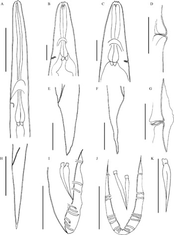
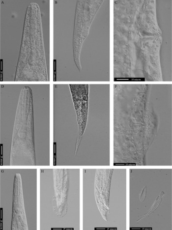
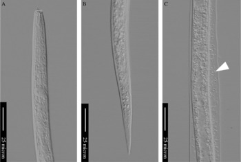
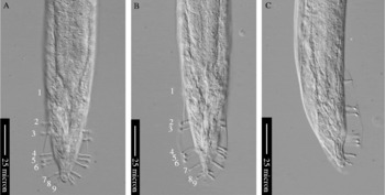
(figs 1–4)
Heterorhabditis atacamensis n. sp., line drawings. Infective juvenile (exsheathed): (A) pharyngeal region; (H) tail region. Hermaphrodite: (B) pharyngeal region; (D) vulva; (E) tail region. Adult female: (C) pharyngeal region; (F) tail region; (G) vulva showing exudates. Adult male: (I) tail in lateral view showing bursa and arrangement of genital papillae; (J) tail in ventral view showing bursa and arrangement of genital papillae; (K) spicule. Scale bars = 50 μm.

Heterorhabditis atacamensis n. sp., light micrographs of hermaphrodite and amphimictic adults. Hermaphrodite: (A) pharyngeal region; (B) tail region; (C) vulva showing slight protrusion. Adult female: (D) pharyngeal region; (E) tail region; (F) vulva showing exudates. Adult male: (G) pharyngeal region; (H) tail in ventral view showing arrangement of genital papillae; (I) tail region, lateral view; (J) spicule and gubernaculum.

Heterorhabditis atacamensis n. sp., light micrographs of infective juveniles. (A) Pharyngeal region (exsheathed); (B) tail region (exsheathed); (C) mid-body region (ensheathed) showing possible bacterial or fungal spores between cuticle and sheath (arrow).

Heterorhabditis atacamensis n. sp., light micrographs of adult male. (A–C) Tail region, showing bursa and position and orientation of nine pairs of genital papillae: (A) pair 7 grouped with pairs 8 and 9; (B) pair 7 somewhat separated from pairs 8 and 9; (C) lateral view.

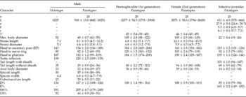
The morphometrics of adult stages and IJs are given in table 1.
Morphometrics of Heterorhabditis atacamensis n. sp. All measurements in μm and in the form: mean±SE (range).

n, Number of specimens observed; L, length; a, length/maximum body diameter; b, length/head to pharynx length; c, length/tail; V, head to vulva length (as percentage of body length); D%, (head to excretory pore length/head to pharynx length) × 100; E%, (head to excretory pore length/tail) × 100; SW%, (spicule length/anal body diameter) × 100; GS%, (gubernaculum length/spicule length) × 100.
Hermaphrodite
Body C-shaped when heat-relaxed, always with >15 eggs. Cuticle with slight annulation, more prominent at tail. Head low, relatively truncate, continuous with body contour. No lateral field observed. Cephalic region with six labial papillae, four cephalic papillae and two pore-like amphidial apertures. Posterior part of stoma funnel-shaped, well cuticularized. Cheilorhabdions prominent, sclerotized, other rhabdions not distinguishable. Procorpus cylindrical, slightly narrower isthmus and valvate, pyriform basal bulb. Nerve ring surrounding isthmus at anterior end of isthmus. Excretory pore usually located close to posterior level of basal bulb. Non-overlapping junction between pharynx and intestine, cardia present. Genital system didelphic, amphidelphic. Vulva a transverse slit, located approximately mid-body. Vulval lips somewhat protruding in all specimens observed, protruding 5 (3–8) μm above body contour, epiptygmata absent. Shallow folding of cuticle around vulva visible under light microscope. Vagina extending 16 (11–19) μm into body. Slight post-anal swelling present. Tail conoid, about 2–3 times longer than anal body diameter, with finely rounded terminus.
Male
Body J-shaped when heat-relaxed. Cuticle smooth under light microscope. Head low, truncate, sometimes slightly offset from body contour. No lateral fields observed. Six lips, each bearing one labial papilla. Cephalic papillae not observed. Amphidial apertures pore-like. Posterior part of stoma funnel-shaped, well cuticularized. Cheilorhabdions visible, sclerotized, other rhabdions not distinguishable. Pharynx with cylindrical procorpus, slightly narrower isthmus. Pyriform basal bulb with valve, occasional slight dorsal displacement by excretory cell. Nerve ring at about mid-isthmus level. Excretory pore located posterior to basal bulb, anterior portion of excretory duct cuticularized, posterior section short, debouching into excretory cell. Non-overlapping junction between pharynx and intestine, cardia present, extending into intestine. Testis monorchic, ventrally reflexed. Spicules paired, colourless. Manubrium shape somewhat square, offset from shaft; shaft short; blade almost straight; terminus of blade pointed; velum absent. Gubernaculum straight in lateral view. Bursa peloderan, papillae totalling nine pairs in all specimens observed, comprising (anterior to posterior): pair 1 well anterior to cloacal aperture; pairs 2 and 3 forming a group anterior to cloacal aperture; pairs 4, 5 and 6 forming a group posterior to cloacal aperture; and pairs 7, 8 and 9 forming another group around the tail tip, although pair 7 somewhat separated from pairs 8 and 9 on 60% of specimens observed. All pairs curved ventrally apart from pairs 4 and 7 which are curved laterally. Pairs 8 and 9 shorter than pair 7, particularly pair 8 which is substantially reduced on 90% of specimens observed. No obvious post-anal swelling. Tail conoid, ventrally curved, slightly longer than anal body diameter, with pointed tip.
Amphimictic female
Similar to hermaphrodite in general morphology, but slightly shorter. Body C-shaped when heat-relaxed. Vulva protruding on 60% of specimens observed, 2 (2–6) μm above body contour, epiptygmata absent. Exudates ( = copulatory plug?) around vulva on 70% of specimens observed. No cuticular folds around vulva when viewed under light microscope. Slight post-anal swelling present. Tail conoid, about three times longer than anal body diameter, tapering to a finely rounded terminus, occasional regular duplex termination observed.
Infective juvenile
Body slender, almost straight when heat-relaxed, tapering gently towards anterior and posterior ends. Cuticle finely annulated with delicate transverse striations. Cephalic region rounded not annulated, continuous with body contour. Prominent, dorsally placed tooth-like structure located near oral opening, observed in exsheathed specimens. Tooth dorsally directed at approximately 45° to body axis. Retained second-stage cuticle commonly observed after harvesting. Lateral amphidial apertures and four cephalic papillae visible in exsheathed specimens. Stoma closed. Pharynx with cylindrical procorpus, slightly swollen metacorpus, slender isthmus and distinct, valvate, basal bulb with somewhat truncated posterior end. Excretory pore prominent, situated between nerve ring and anterior end of basal bulb, excretory duct cuticularized. Deirid sometimes visible under light microscope, located between nerve ring and anterior end of basal bulb. Nerve ring surrounding isthmus about mid-way between posterior of metacorpus and anterior of basal bulb. Junction between pharynx and intestine non-overlapping, cardia prominent extending into intestine. Hemizonid present, lenticular in shape, located at anterior end of basal bulb, usually 3–4 annules anterior to excretory pore. Hemizonion not seen. Lateral field starting as two lines around nerve ring level, forming four lines (two ridges) at about basal bulb level, with the two ridges separated by 2–3 μm, continuing as such until approximately mid-tail level. Rod-shaped bacteria seen throughout anterior portion of intestine, observations further posterior obscured by body contents. Tail long, conoid, about five times as long as anal body diameter, with finely rounded tip. Length of exsheathed tail c. 70% of ensheathed tail. Spores (unknown origin) between the cuticle and sheath observed in 10% of specimens.
Type host and locality
The natural host of H. atacamensis n. sp. is unknown as the species was isolated by baiting a soil sample from the Atacama Desert, Chile (23°11′16.7″S, 67°59′31.6″W). This region, in the north of Chile, has very low rainfall throughout the year, and temperatures around 26°C during the day and 0°C at night. The sample was taken from a sandy bank, shaded by trees, at 2499 m above sea level. The temperature of the soil sample in situ was 21°C. The pH of the sample, as evaluated in the laboratory, was 6.9.
Type material
Holotype male isolated from haemocoel of G. mellonella, deposited in the CABI Nematode Collection, Europe-UK Centre, Egham, Surrey, UK. Five paratype males and hermaphroditic adults and ten paratype IJs, all as permanent slide mounts, together with 20 paratypes of each stage in TAF, deposited in the nematode collections of CABI, UK and INIA – Quilamapu, Chillán, Chile. Twenty paratypes of each stage in TAF, deposited in the United States Department of Agriculture Nematode Collection (USDANC), Beltsville, Maryland, USA. In addition, numerous paratype IJs and other stages preserved in 70% ethyl alcohol deposited in the CABI Nematode Collection, UK.
Etymology
Specific epithet derived from the Atacama Desert from where the species was collected.
Cross-breeding studies
No progeny were produced when H. atacamensis n. sp. and H. safricana were crossed. No progeny were produced from virgin females in the absence of males. When both species were self-crossed, progeny were produced.
Diagnosis and relationships
Heterorhabditis atacamensis n. sp. can be recognized by morphometrics of the IJ, with length (L) = 611 (578–666) μm, head to excretory pore length (EP) = 115 (101–126) μm, tail without sheath = 69 (62–79) μm long and L/maximum body diameter (ratio a) = 28 (25–31). The male is characterized by the length of spicules and gubernaculum, at 45 (40–49) μm and 20 (17–22) μm, respectively, and the number and position of genital papillae. The hermaphroditic female has a slight post-anal swelling and a conoid tail (88 (72–112) μm long) with a slightly rounded terminus.
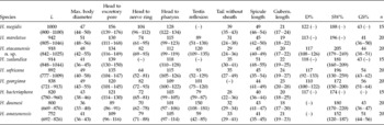
Based on the morphology of IJ, males and hermaphroditic females, H. atacamensis n. sp. is most similar to the following species: H. marelatus Liu & Berry, Reference Liu and Berry1996, H. downesi Stock, Griffin & Burnell, Reference Stock, Griffin and Burnell2002, H. amazonensis Andaló, Nguyen & Moino, Reference Andaló, Nguyen and Moino2006, H. bacteriophora, H. georgiana Nguyen, Shapiro-Ilan & Mbata, Reference Nguyen, Shapiro-Ilan and Mbata2008 and H. safricana. However, H. atacamensis n. sp. is separated from these species by a number of morphometric features (see tables 2 and 3) and by the following features: the genital papillae of males of H. atacamensis n. sp. consist of nine pairs with pairs 4 and 7 curved laterally, versus 7–8 pairs in H. amazonensis with only pair 4 curved laterally; the excretory pore of males of H. atacamensis n. sp. is located posterior to the basal bulb, versus anterior in H. downesi. There is equal protrusion of anterior and posterior vulval lips of hermaphroditic females of H. atacamensis n. sp., while the anterior lip protrudes more than the posterior lip in H. downesi; the tail terminus of H. atacamensis n. sp. is finely rounded and non-mucronate, versus blunt and mucronate in H. downesi. The hemizonid of IJ of H. atacamensis n. sp. is located anterior to the excretory pore, versus posterior in H. marelatus; the tail tip of H. atacamensis n. sp. is finely rounded, versus spike-like in the majority of H. downesi. The tail length of amphimictic females of H. atacamensis n. sp. is considerably longer than those of H. safricana, 94 (80–108) versus 69 (55–111) μm.
Comparative morphometrics of male of Heterorhabditis atacamensis n. sp. and other Heterorhabditis spp. (in descending order of body length). All measurements in μm and in the form: mean (range).

All measurements after original authors. (–) not available. L, length; n, number of specimens examined; D%, (head to excretory pore length/head to pharynx length) × 100; SW%, (spicule length/anal body diameter) × 100; GS%, (gubernaculum length/spicule length) × 100.
Comparative morphometrics of infective juveniles of Heterorhabditis atacamensis n. sp. and other Heterorhabditis spp. (in descending order of body length). All measurements in μm and in the form: mean (range).

All measurements after original authors. (–) not available. L, length; n, number of specimens examined; D%, (head to excretory pore length/head to pharynx length) × 100; E%, (head to excretory pore length/tail) × 100.
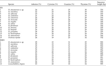
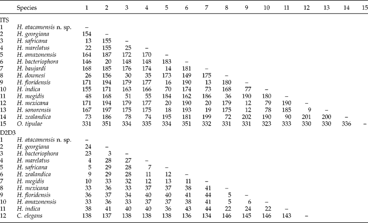
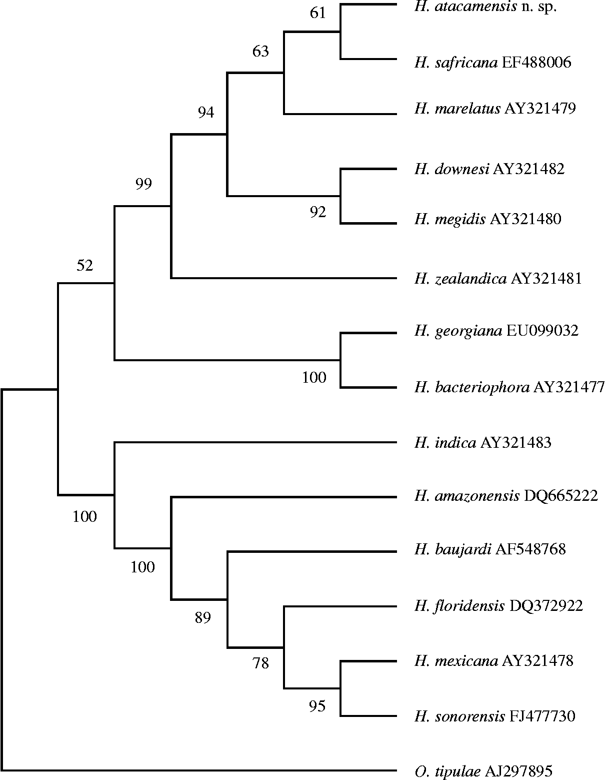
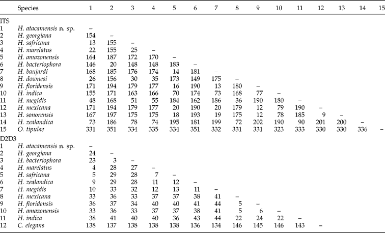
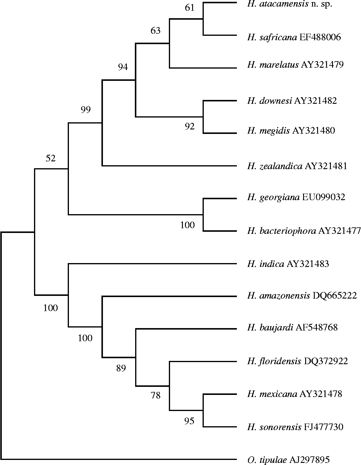
The longest contiguous ITS-containing rDNA sequence obtained for H. atacamensis n. sp. was 730 bp, with nucleotide composition A = 24%, C = 21%, G = 25% and T = 30% (table 4), although this did not contain the full ITS1 region, i.e. was not the complete ITS. Pairwise distances for the ITS region are shown in table 5. Heterorhabditis safricana is the least divergent species from H. atacamensis n. sp. (13 bp), H. floridensis Nguyen, Gozel, Koppenhöfer & Adams, Reference Nguyen, Gozel, Koppenhöfer and Adams2006b and H. mexicana Nguyen, Shapiro-Ilan, Stuart, McCoy, James & Adams, Reference Nguyen, Shapiro-Ilan, Stuart, McCoy, James and Adams2004 are the most divergent (171 bp). Among the Heterorhabditis species (excluding H. atacamensis n. sp.), the least divergent species are H. mexicana and H. sonorensis (9 bp), H. mexicana and H. floridensis (12 bp), and H. sonorensis and H. floridensis (12 bp), i.e. divergence between H. atacamensis n. sp. and its closest taxon (H. safricana) is greater. For ITS regions, MP analysis shows that the alignment resulted in 799 characters, 308 were constant, 268 variable characters were parsimony-uninformative and 223 were parsimony informative. The phylogenetic relationships among 14 species of Heterorhabditis are presented in fig. 5 (for MP, tree length = 799, Consistency Index (CI) = 0.834, Retention Index (RI) = 0.845, Rescaled Consistency Index (RCI) = 0.705 and Homoplasty Index (HI) = 0.166). Heterorhabditis atacamensis n. sp. stands separately and forms a sister group with H. safricana, supported by bootstrap proportion of 61%. In the phylogenetic tree there are two broad monophyletic groups, the megidis-group, with H. atacamensis n. sp., H. safricana, H. marelatus, H. downesi, H. megidis Poinar, Jackson & Klein, Reference Poinar, Jackson and Klein1987, H. zealandica Poinar, Reference Poinar, Gaugler and Kaya1990, H. georgiana and H. bacteriophora, and the indica-group, with H. indica Poinar, Karunakar & David, Reference Poinar, Karunakar and David1992, H. baujardi Phan, Subbotin, Nguyen & Moens, Reference Phan, Subbotin, Nguyen and Moens2003, H. amazonensis, H. floridensis, H. mexicana and H. sonorensis.
Sequence lengths and base pair composition of the ITS and D2D3 regions of Heterorhabditis species.

Pairwise distances between Heterorhabditis species. Oscheius tipulae and Caenorhabditis elegans were used as outgroups.
Phylogenetic relationships of Heterorhabditis atacamensis n. sp. based on analysis of ITS rDNA regions using maximum parsimony. Oscheius tipulae was used as an outgroup. Numbers at the nodes represent bootstrap proportion. Heterorhabditis atacamensis n. sp. stands separately and forms a sister group with H. safricana, supported by bootstrap proportion of 61%.
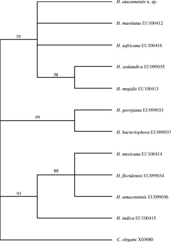
The D2D3 sequence obtained for H. atacamensis n. sp. (flanked by primers D2A and D3B) was 592 bp, with composition A = 25%, C = 19%, G = 31% and T = 25% (table 4). Pairwise distances for the D2D3 sequences (table 5) show H. marelatus is the least divergent species to H. atacamensis n. sp. (4 bp), followed by H. safricana (5 bp); the most divergent is H. indica (38 bp). Excluding H. atacamensis n. sp., the least divergent species are H. georgiana and H. bacteriophora (3 bp), i.e. divergence between H. atacamensis n. sp. and its closest taxa (H. marelatus and H. safricana) is greater. For D2D3 sequences, MP analysis shows that the alignment resulted in 619 characters, 450 were constant, 110 variable characters were parsimony-uninformative and 59 were parsimony informative. The phylogenetic relationships between 11 species of Heterorhabditis are presented in fig. 6 (for MP, tree length = 229, CI = 0.860, RI = 0.787, RC = 0.677, HI = 0.140). The phylogenetic tree has three monophyletic groups, the megidis-group with H. atacamensis n. sp., H. safricana, H. marelatus, H. megidis and H. zealandica, the indica-group with H. indica, H. amazonensis, H. floridensis and H. mexicana, and the bacteriophora-group, a group originally proposed by Nguyen et al. (Reference Nguyen, Shapiro-Ilan and Mbata2008), with H. bacteriophora and H. georgiana. The D2D3 sequences were not available for the species H. downesi and H. baujardi.
Phylogenetic relationships of Heterorhabditis atacamensis n. sp. based on analysis of D2D3 rDNA domains using maximum parsimony. Caenorhabditis elegans was used as an outgroup. Numbers at the nodes represent bootstrap proportion. The tree shows three monophyletic groups supported by robust bootstrap proportions (>70%).

Morphological and molecular information, supplemented by cross-breeding studies, are sufficient to confirm that H. atacamensis n. sp. is a new species.
Acknowledgements
This study was supported by the Darwin Initiative (project number 15/004), a programme coordinated by the UK Department for Environment, Food and Rural Affairs (Defra) and CABI Europe-UK. We are very grateful to Lukasz Tymo for his help with sequencing and to Khuong Nguyen for his helpful advice.