Coronary artery fistula draining into the left heart is rare, and reported in 8% of cases; 5% drain to the left atrium and 3% to the left ventricle. Reference Levin, Fellows and Abrams1 They cause left heart volume overload without pulmonary over-circulation. Reference Oztemir, Cemri and Dortlemez2 Fistulas to the left ventricle may be single and discrete or plexiform and diffuse; the latter may be seen with left ventricular non-compaction due to shared embryological morphogenesis. Reference Reddy, Gupta and Hamby3,Reference Nguyen, Rigsby, Gambetta and Kaushal4 The plexiform lesions are not amenable for any interventions. Reference Oztemir, Cemri and Dortlemez2,Reference Yokawa, Watanabe and Kurosaki5 Unlike most coronary fistulas, drainage to the left ventricle leads to may present with a diastolic murmur. Given the somewhat unique physiology, which is similar to a paravalvular aortic leak, fistulas draining to the left ventricle may present with only a diastolic murmur. Reference Yokawa, Watanabe and Kurosaki5
Large fistula causing heart failure in children warrants interventions. Reference Nguyen, Rigsby, Gambetta and Kaushal4 Device closures of the discrete large fistula to left heart are rarely reported in the literature. Techniques of closure differ from the closure of right-sided fistula by a need for arterio-arterial circuit formation or transseptal puncture for an arteriovenous circuit formation. Reference Li, Zhang, Wang, Xie, Zhang and Li6,Reference Perez-Brandao, Fiarresga, Sousa and Martins7 Anatomy and interventional techniques of three such fistulas are described to highlight the strategies followed in these rare fistulas.
Patient 1
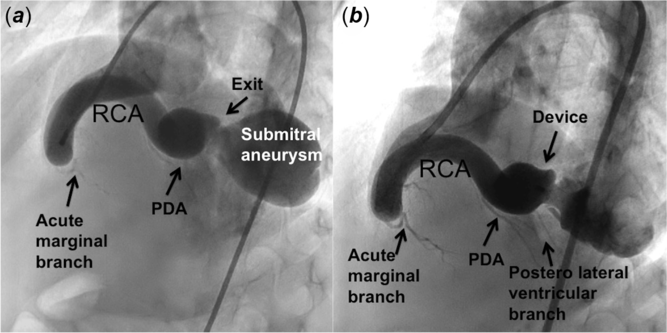
A 3-year-old boy weighing 10 kg presented with bounding pulses, cardiomegaly, hyperkinetic precordium, heart failure and a loud apical diastolic murmur. A large tortuous right coronary artery led to a fistula draining into the left ventricle. Its exit created a localised sub-mitral ventricular aneurysmal sac with normal mitral valve function. The left ventricle was dilated with normal contractility. An angiogram showed a large tortuous dominant right coronary artery that formed an end-artery fistula terminating near the left ventricular inflow after giving off the posterior descending and posterolateral ventricular branches. The large fistula measured 12–14 mm (Fig 1, Supplementary video S1).
A large end-artery fistula from the distal right coronary artery exits into the left ventricle where it forms a localised sub-mitral aneurysm. (a) Following the closure of the narrow distal exit with muscular ventricular septal occluder device and (b) the coronary branches fill well.

Surgical closure of the termination in the aneurysmal sac behind the posterior mitral leaflet was considered technically challenging. The narrowed 6.5-mm exit was closed with an 8-mm Amplatzer muscular VSD occluder (Abbott, Plymouth, MN, United States of America) through an 8F right coronary guide catheter advanced over the wire beyond the exit into the left ventricle. The distal coronary branches filled better after the fistula closure. At a 6-month follow-up, he is asymptomatic on aspirin and clopidogrel with no residual flows and normal ventricular function.
Patient 2
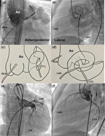
An 8-year-old girl weighing 20 kg had effort intolerance and growth failure. She had high-volume pulses, hyperkinetic cardiac impulse and continuous murmur in the left sternal border. A large fistula from the left main coronary artery had a tortuous course towards the left atrial appendage. After the transseptal puncture, the left atrium recorded v-waves of 40 mmHg and mean pressures of 24 mmHg. A large fistulous tract from the left main coronary artery to a dilated left atrial appendage had three parts with a spiral tortuous turn. The first proximal part from the left coronary artery measuring 5 mm turned to the second superior dilated part and turned again to the final third inferior more dilated distal part measuring 16 mm (Fig 2).
Angiogram in anteroposterior (a) and lateral (b) views show a large fistula from the left coronary artery to the left atrial appendage. Cartoon representation (c and d) shows a narrower first part that turns superiorly to a second broader part and finally again turns inferiorly to a third much larger part before exit into the left atrium. After closure (e and f) with two plugs, there is no residual flow.

A transseptal arteriovenous circuit was made over a floppy coronary guidewire and the transseptal sheath advanced into the exit. This exit that measured 16 mm was closed with a 22-mm Amplatzer vascular plug II (Abbott), which had a lower profile compared to the other devices. The entry to the fistula that measured 5 mm was closed with an 8-mm Amplatzer vascular plug IV (Abbott). She received aspirin for 3 years. A contrast computed tomography at a 3-year follow-up showed normal left coronary artery and branches, total occlusion of the fistulous tract and good remodelling.
Patient 3
A 9-month-old infant weighing 5 kg had persistent heart failure despite diuretics. There was tachypnoea, bounding pulses, prominent precordial pulsations and loud continuous precordial murmurs. The cardiothoracic ratio was 65% without a pulmonary plethora. A large 6-mm fistula connected the right coronary artery to the left atrium through a long intracardiac course alongside the atrial septum, which precluded a closed surgery (Supplementary figure 3).
The angiogram showed a large fistula from the proximal right coronary artery to the left atrium (Supplementary video S2). Transseptal puncture and arteriovenous circuit formation helped to advance a 6F braided venous sheath through the fistula to the right aortic sinus. The entry of the high-flow fistula was closed with a 6-4 Memopart duct occluder (Lepu Medical, Beijing, PRC). There was marked improvement of growth and relief of symptoms at a 3-month follow-up on aspirin therapy.
The fluoroscopic time in the three patients were 9.6, 32.8 and 10.8 minutes and radiation doses were 3116, 9130 and 2920 milligray.square centimetres.
Discussion
The coronary artery fistula to the left ventricle represents an embryological failure of the obliteration of inter-trabecular myocardial sinusoids causing persistent ventricular communications. Reference Nguyen, Rigsby, Gambetta and Kaushal4 While widespread developmental arrest leads to the plexiform fistula, localised arrest causes discrete fistula. Reference Li, Zhang, Wang, Xie, Zhang and Li8 Occurrence of the plexiform fistula with ventricular non-compaction is explained by similar morphogenesis of both the conditions. Reference Nguyen, Rigsby, Gambetta and Kaushal4 The sub-mitral aneurysmal sac beyond the exit of the fistula in the first patient is possibly due to a similar localised developmental arrest.
Catheter interventions for large fistula draining into the left heart causing heart failure are extremely rare; most reports demonstrate small fistulas in asymptomatic patients. Reference Perez-Brandao, Fiarresga, Sousa and Martins7–Reference Mahla, Mahimarangaiah, Sastry, Chikkaswamy, Bhat and Manjunath9 Transseptal arteriovenous circuit formation facilitated closure despite tortuous turns in the left atrial fistulas. The procedural tips to achieve safe closure in these left heart draining fistula are deep cannulation of fistula draining to left ventricle till their exit; transseptal circuit formation in left atrial fistula to facilitate closure near the entry; use devices with fabric to close the high-flow fistula and close both exit and entry whenever using vascular plugs as they have no fabric.
Closure at the distal termination beyond all the left ventricular branches was the only option in the high-flow end-artery fistula in the first patient. A muscular VSD occluder was chosen for the high-flow fistula in the first patient as the large 16-mm retention skirts with fabric facilitated occlusion and small delivery sheath profile permitted deep cannulation. Large delivery sheaths could not negotiate the tortuous course in the second patient leading to the choice of low-profile vascular plugs that could be delivered through small catheters and sheaths. As the plugs were devoid of fabrics that allow immediate closure, the fistula was closed at both the entrance and the exit. The size of the plugs should be at least 1.5 times the diameter of the target vessel. The small profile of duct occluder with fabric permitted its use in the infant with a large left atrial fistula.
Fistulas are classified as proximal fistula arising from a side branch or distal end-artery fistula to plan post-procedural anticoagulation strategies. Thrombus propagation in a dilated end-artery fistula endangers myocardial perfusion through all the proximal branches, justifying dual antiplatelet agents or Coumadin as in the first patient. Reference Zhang, Xu and Bai10 If the proximal side branch fistula is closed without any cul-de-sac to allow stagnation, aspirin would suffice.
Supplementary Material
To view supplementary material for this article, please visit https://doi.org/10.1017/S1047951120003364.
Acknowledgements
None.
Financial Support
This research received no specific grant from any funding agency, commercial or not-for-profit sectors.
Conflicts of Interest
None.
Ethical Standards
The authors assert that all procedures contributing to this work comply with the ethical standards of the Indian council of medical research and with the Helsinki Declaration of 1975, as revised in 2008, and has been approved by the Institutional Committee of Madras Medical Mission, Chennai, India.