I. INTRODUCTION
Due to their strictly function-oriented structures, and the unique combination of maximum performance with minimum strain on the structure, natural materials such as mammalian bones, mollusc shells, and crustacean exoskeletons can often be superior to their artificial counterpartsReference Mayer1, Reference Smith, Schäffer, Vlani, Thompson, Frederick, Klndt, Belcher, Stuckyll, Morse and Hansma2 and can be a very useful model for the design of advanced synthetic functional materials. The resulting bioinspired materials can be further optimized, and their properties and performance can be enhanced when fabricated under strictly controlled conditions to acquire unique target properties. A very recent example of the colossal potential borne by biogenic structural elements can be found in the innovation of artificial photonic crystals and light waveguides, which is a revolutionary bioinspired technology that is already being applied in the fields of optics and optoelectronics.Reference Lourtioz, Benisty, Chelnokov, David and Olivier3
Using fullerene-C60 as example, the potential of the interior of siliceous sponge spicules—natural animal skeletal elements with outstanding hierarchy in their structural organizationReference Holzhuter, Lakshminarayanan and Gerber4–Reference Weaver, Pietrasanta, Hedin, Chmelka, Hansma and Morse7 and notable optical propertiesReference Muller, Wendt, Geppert, Wiens, Reiber and Schroder8, Reference Sarikaya, Fong, Sunderland, Flinn, Mayer, Mescher and Gaino9—as templates for the deposition of materials, or for bringing about (photo)chemical reactions, is demonstrated. Using the nanostructured surface of the spicule silica as a natural substrate, an ensemble of C60 particles was produced through templated vapor deposition in the central spicule channel. The combination of high rigidity, limited space, and structured surface of the channel provides a unique environment, resulting in an unprecedented morphology of the deposited fullerene.
II. EXPERIMENTAL PROCEDURE
A. Spicule extraction and treatment
Long basalia (basal spicules) of Euplectella marshalli (“Venus’ basket”) were manually separated from the lower half of the animal skeleton of an air-dried specimen sourced from the Philippines. The organic parts and the central silicatein filament were removed through treatment of the washed spicules in a solution containing 30% H2O2 for 2 days, after which they were rinsed with water and dried in a vacuum (100 °C) for 5 h. The spicules were cut and treated with an H2SO4:HNO3 (4:1) mixture for 3 wk, after which they were rinsed thoroughly, dried in a vacuum (200 °C) for 16 days, and stored over desiccant.
B. Filling the spicules with C60
A mixture of crystals of pure C60 (37.8 mg) and spicules (7.4 mg) preactivated in a vacuum at 200 °C for 30 min were sealed in Pyrex ampoule under vacuum and heated at 600 °C for 4 days. After heat treatment, the spicules were quickly washed with CS2, sonicated to remove the excess C60 (prolonged washing or sonication results in the dissolution of the filled C60), filtered and dried in air. Attempts to fill the spicules with C60 using supercritical CO2 or organic solvents were unsuccessful.
C. Microstructural characterization
A microstructural observation was performed on uncoated samples attached to a conductive tape using a field-emission type scanning electron microscope (SEM) (S-4800; Hitachi High-Technologies, Tokyo, Japan). For transmission electron microscopy observation, the dried spicules were finely sliced using a focused ion beam (FIB) system equipped with a microprobe attachment (Hitachi FB-2100, Hitachi High-Technologies). The transmission electron microscope (TEM) micrographs were recorded using a field-emission type microscope (JEM-2100F, JEOL, Tokyo, Japan) providing lattice and particle resolutions of 0.1 and 0.19 nm, respectively.
D. Mechanical characterization
The effect of the applied treatments on the mechanical properties of the Euplectella spicules was assessed, applying flexural testing and micro- and nanoindentation measurements. The bulk mechanical properties of nontreated spicules (“original”), spicules with their central silicatein filament removed (“core removed”), and those treated to be filled with fullerene (“C60-treated”) were measured in a three-point bending test, using a tabletop screw-driven mechanical tester (EZ Test; Shimadzu Corporation, Kyoto, Japan) at a span length of 600 μm and a loading rate of 500 μm/min. The surface mechanical properties of the as-received and treated spicules were characterized by micro- and nanoindentation testing on polished cross sections of resin-mounted spicules. Microindentation tests were carried out using a conventional depth-sensing microhardness (MH) tester (DUH-W201S, Shimadzu Corporation) supplied with a Berkovich-type indenter. The MH values were measured with the application of maximum loads of 20, 30, and 40 mN. The indented cross-sectional spicule surfaces were observed using a confocal laser microscope, and the projected areas of the residual impressions were measured on the obtained images. MH values were obtained through the ratioReference Brotzen and Moore10:
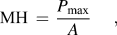 $$<![CDATA[$${\rm{MH}}\, = \,{{P_{\max } } \over A},$$
in terms of the maximum applied indentation load (P max) and the projected area of the measured residual impression (A). Reduced microindentation elastic moduli, ME, were obtained from the unloading stiffness, S, of the spicules, calculated from the initial region of the unloading curve of the indentation load–depth data fitted to the power lawReference Oliver and Pharr11:
 $$<![CDATA[$$P = B(h - h_{\rm{f}} )^m ,$$
where P is the indentation load, h is the indentation depth, B and m are empirical fitting parameters, and h f is the residual depth of the indentation, which was also determined by curve fitting. S was subsequently derived by differentiating Eq. (2) at the maximum indentation depth. Assuming that the used diamond indenter tip is perfectly rigid, the ME values of the spicules were determined as:
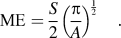 $$<![CDATA[$${\rm{ME}} = {S \over 2}\left( {{{\rm{\pi }} \over A}} \right)^{{1 \over 2}} .$$
Nanomechanical properties were measured using a nanohardness (NH) testing system (Ub1; Hysitron Inc., Minneapolis, MN) equipped with a Berkovich-type diamond tip, by applying 2000 and 3000 μN maximum loads. During the measurements, indentations were performed in various regular areal patterns on the polished cross sections to obtain average NH and reduced nanoindentation elastic modulus (EN) values.
The fracture toughness, K Ic, of the spicules was estimated according to the relation:
 $$<![CDATA[$$K_{{\rm{Ic}}} = Y{\rm{\sigma }}_{\rm{F}} ({\rm{\pi }}a)^{1/2} ,$$
derived through the Griffith’s approach,Reference Griffith12 where Y is a geometry-dependent constant, σF is the fracture strength as determined in the three-point bending test, and a is the critical crack length. Here, the value 200 nm was included for a, which is equal to the average thickness of the outermost layer of the Euplectella spicules.Reference Aizenberg, Weaver, Thanawala, Sundar, Morse and Fratzl13Y was set to ∼2,Reference Fett and Munz14 which represents a reasonable value considering the geometry of the spicule samples and the shallow surface cracks associated with the thickness of the outermost layers.
III. RESULTS AND DISCUSSION
A. Treatment of the spicules and deposition of fullerene-C60
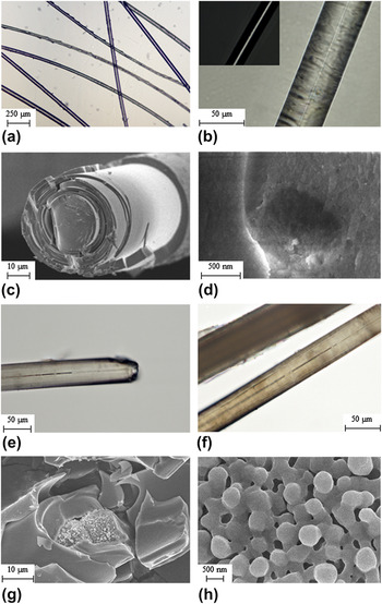
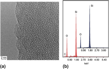
Nonpillared siliceous basalia, which feature only the lowest two of the seven hierarchical levels in the complex structural organizationReference Aizenberg, Weaver, Thanawala, Sundar, Morse and Fratzl13 of the skeleton of Euplectella, were selected [Fig. 1(a)]. The spicules are composed of concentric transparent siliceous layers, joined together by thin regions of organic tissue [Fig. 1(c)]. The resulting inorganic–organic composite structure accounts for the exceptional flexibility of the spicules, which, although being silica based, can still bend without breaking and serve as a tough yet flexible mechanical skeletal support for the animal. SEM micrographs [Fig. 1(d)] confirm earlier observationsReference Muller, Belikov, Tremel, Perry, Gieskes, Boreiko and Schroder5, Reference Aizenberg, Weaver, Thanawala, Sundar, Morse and Fratzl13 that the channel walls are composed of patterned silica, while TEM micrographs of finely FIB-sliced spicules show that the internal surface is finely structured with round protrusions, several nanometers in diameter, which provide a nanoscale roughness to the surface [Fig. 2(a)]. The interior part, on the other hand, is generally uniform, consisting of amorphous silica [Fig. 2(b)]. After the removal of the organic component and the central silicatein filament using a peroxide and acid treatment, hollow central channels with diameters larger than approximately 1 μm became clearly visible and permitted the passage of a laser beam by total internal reflection in a range of visible wave lengths [Fig. 1(b)], which is reminiscent of the properties of artificial optical fibers.
Optical (a, b, e, f) and scanning electron (c, d, g, h) micrographs of native and treated spicules from Euplectella: (a) appearance of basal spicules before treatment; (b) size of the central channel in peroxide-treated spicules (the inset shows light from a beam from a green continuous-wave laser passing by total reflection through the channel); (c) cross section of a spicule, showing the layered structure of silica interleaved with organic material; (d) view of the central channel at the cross section, removed of organic components; (e, f) spicules in which the central channel has been partially filled with C60; and (g, h) SEM micrographs showing the microporous structure and the appearance of the particles of the deposited particles of C60 at low (g) and high (h) magnifications.

(a) TEM micrograph of FIB-sliced peroxide-treated spicule, showing the shape and size of the surface structure elements in the central silicatein channel; (b) energy-dispersive x-ray spectra taken before and after the filling showing the presence of the carbon from the guest C60.

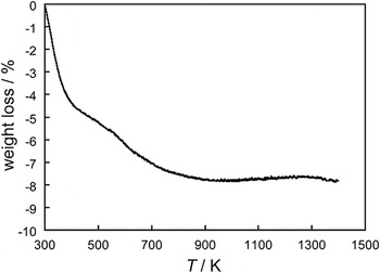
As a target material for deposition, fullerene-C60 was chosen on the basis of its notable thermal stability and based on previous attempts to include it in mesoporous silica through sublimation,Reference Rachdi, Hajji, Goze, Jones, Maireles-Torres and Roziere15 sol–gel processes,Reference Subbiah and Mokaya16 and reflux,Reference Govindaraj, Nath and Eswaramoorthy17 and on the well-established methods for the inclusion of C60 in nanotubes.Reference Khlobystov, Britz and Briggs18 The thermal analysis profile given in Fig. 3 indicates that heat-exposed powdered spicules are dehydrated in two steps, losing about 7.5% of their original weight. As shown in Figs. 1(e) and 1(f), the vacuum sublimation of C60 in sealed quartz ampules in the presence of pretreated spicules resulted in a partial filling of the central channels. Although the yield was relatively low, due to the transparency of the spicules and the contrasting dark color of the inserted material, the included fullerene is readily observable using optical microscopy, particularly in the narrow regions of the channels [Figs. 1(e) and 1(f)]. Cross-sectional SEM micrographs of broken spicules show that the fullerene molecules assemble in the form of round-shaped particles with a relatively uniform average diameter of 0.3–0.10 μm [Figs. 1(g) and 1(h)]. The energy-dispersive x-ray spectrum provided evidence of the significant presence of only carbon, silicon, and oxygen [Fig. 2(b)]. The most interesting feature of the filled channels is the porous network of cavities formed by the C60 particles. Such porosity is unexpected in the sublimation of C60 in the form of bulk crystals and may be attributed to the limited space and ascribed to the templated growth of the crystal on the nanostructured silica surface of the channels.
Thermogravimetry curve of Euplectella spicules, which were treated with peroxide and dried.

B. Mechanical properties of treated and C60 deposited spicules
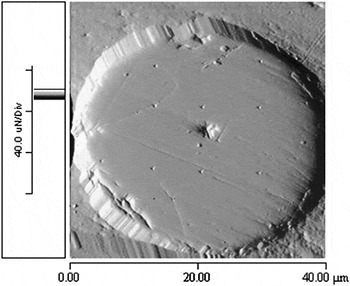
The effect of the chemical treatments applied to remove the central filament and the subsequent heating to deposit C60 in the channel of the spicule structure were comparatively assessed from the resulting mechanical properties. Table I summarizes the surface and bulk mechanical properties of the original and treated spicules characterized in the scope of this study. Figure 4 presents the polished cross section of the spicule patterned with the nanoindenter to determine the NH and nanoindentation moduli. The obtained average NH and nanoindentation modulus of the original spicules of ∼4.5 and 43.7 GPa, respectively, corroborate previously reported values.Reference Woesz, Weaver, Kazanci, Dauphin, Aizenberg, Morse and Fratzl19 While the silicatein core removal through peroxide oxidation did not have a considerable effect on these parameters, the heat treatment and infiltration of C60 changed the mechanical properties significantly. Namely, C60-treated spicules were around 50% harder and stiffer than the virgin (original) and core-removed spicules, and a similar trend was observed in the MH and microindentation modulus values. The agreement between the properties obtained through nano- and microindentation measurements demonstrates the fact that the average surface mechanical properties of the multilayer structured spicules are acceptable in relation to the applied measurement techniques and microstructural feature sizes involved.
Topographic image of the polished cross section of the spicule, patterned with the nanoindenter.

Measured mechanical properties of original, core-removed, and C60-treated spicules.

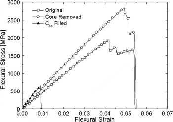
The effect of chemical treatments on the bulk mechanical properties of the spicules was determined using three-point bending tests, in which the flexural strengths and moduli of the original and core-removed spicules are around 2.2–2.5 GPa and 48–51 GPa, respectively. Considering the magnitude of the standard deviations of the measured values, it can be concluded that the core removal treatment does not have significant effect on the bulk mechanical properties of the spicules. Contrarily, the flexural strength and modulus of the C60-treated spicules amount approximately 700 MPa and 75 GPa, respectively, and thus are significantly different to those of the original and core-removed spicules. A similar effect of the chemical treatment can be seen in the strain-to-failure values of the spicules. While the original and core-removed spicules demonstrated a strain-to-failure value of around 5%, the C60-treated spicules failed at a strain of less than 1% (Fig. 5).
Flexural stress versus flexural strain data of the “original,” “core-removed,” and “C60-treated” spicules obtained through a three-point bending test.

The dramatic effect of the chemical treatment on the fracture behavior of the spicules can clearly be observed on the flexural stress versus flexural strain data presented in Fig. 5. After the linear increase in flexural stress with strain through the elastic deformation region, a sudden drop in stress from the maximum value is witnessed, indicating that C60-treated spicules fail catastrophically at a relatively low strain-to-failure. However, in the case of both the original and core-removed spicules, this flexural stress did not experience a sudden drop from the maximum values; instead, an incremental decrease in stress appears to occur up to a certain strain value. This behavior indicates the presence of stable crack propagation, requiring the action of energy absorbing mechanisms to retard failure.
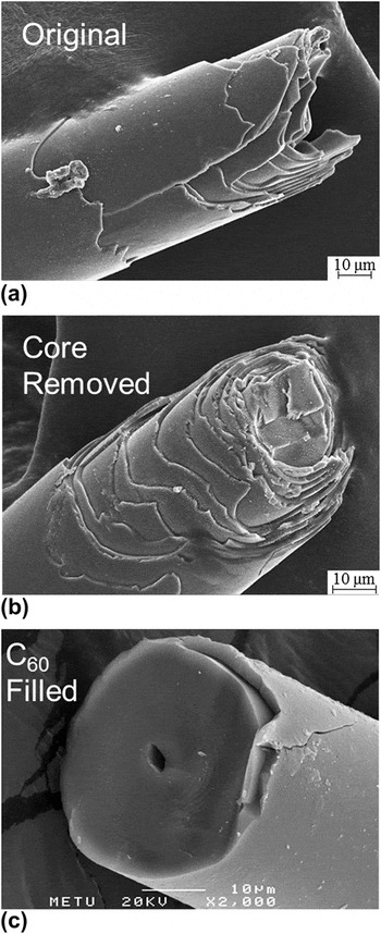
Figures 6(a) and 6(b) show evidence of the crack deflection in the original and core-removed spicules on the weak interlayer surfaces, which prevents a growing crack from propagating directly into the next layer, taking rather a “staggered” path, and thus extending its total length. Lacking the action of energy absorbing mechanisms found in the original and core-removed spicules, the clear flat fracture surface of the C60-treated spicule [Fig. 6(c)] resulted in a brittle fracture nature in the absence of mechanisms for energy dissipation. In the presence of operative mechanisms, such as crack deflection and branching, the amount of energy absorbed by the material before the failure occurs can be correlated with the area under the flexural stress–strain curve, which, even though not quantitatively exact, may also be used in a relative manner to compare fracture behaviors. In this respect, as can be ascertained from the small area underneath the stress–strain curve of C60-treated spicules, when compared to those of the other two samples, the applied treatment significantly changes the failure behavior of the spicules from “damage tolerant” to “brittle.”
Fracture surfaces of three-point bending tested “original,” “core-removed,” and “C60-treated” spicules.

At this point, for comparative purposes, the fracture toughness of the spicules is calculated, assuming that the maximum attained flexural stress is the fracture stress and assuming that the critical crack length is equal to the thickness of the outermost layer of the spicules, which is in the order of ∼200 nm. Here, it should be noted that although the calculated fracture toughness values are not absolute characteristic of the spicules, they still represent the worst-case scenario, where the critical crack length is taken to be the shortest possible dimension, leading to the estimation of the minimum fracture toughness for the subject materials.
According to this estimation, the original and core-removed spicules reveal fracture toughness values of between ∼3.5 and 4.0 MPa⋅m1/2 (Table I), which are comparable to those of commonly used bulk monolithic engineering ceramics such as Al2O3, Si3N4, and SiC.Reference Barsoum20 However, it must be mentioned that even though the estimated minimum fracture toughness of the spicules is similar to that of bulk monolithic ceramics, they are damage tolerant with stable crack propagation [evidenced by the extended areas underneath the stress–strain curves in Fig. 5 and the rough fracture surfaces in Figures 6(a) and 6(b)], unlike in the case of brittle ceramics, which are prone to catastrophic failure. The fracture toughness estimated for the C60-treated spicules is ∼1.1 MPa⋅m1/2(Table I), which is significantly lower than that of the original and core-removed spicules but still higher than that of a monolithic silica rod of similar diameter.Reference Sarikaya, Fong, Sunderland, Flinn, Mayer, Mescher and Gaino9 A similar finding has also been reported for the anchor spicule of Monorhaphis chuni, where laminated spicules show about 2.5 times higher fracture toughness compared to the monolithic core, which has been attributed to the process of crack deflection within the organic layers involved.Reference Miserez, Weaver, Thurner, Aizenberg, Dauphin, Fratzl, Morse and Zok21
As both the surface and bulk mechanical properties of the original and core-removed spicules are comparable, it can be concluded that the applied peroxide and acid treatments leading to removal of the central silicatein filament do not significantly affect the internal structure of the multilayered spicule. Contrarily, the C60 filling procedure conducted at 600 °C seems to alter the structure of the spicules considerably, which is evident from the increasing hardness and stiffness of the treated spicules at the expense of their damage tolerance and fracture toughness.
As previously mentioned, the damage tolerance in these spicules can be attributed to the presence of the organic constituent covering the amorphous silica particles with ∼3 nm diameterReference Woesz, Weaver, Kazanci, Dauphin, Aizenberg, Morse and Fratzl19 and bonding the individual layers of the multilayered structure formed by the same particles. This organic constituent not only provides an increase in the crack path within a single layer but also prevents the transmission of cracks from one layer to the next, providing a medium for efficient energy dissipation. The very low amount of highly elastic organic constituent (in the order of 2–3 vol%Reference Mayer1) in the spicule structure results in the unique combination of high hardness, stiffness, and strength, along with increased damage tolerance and fracture toughness.
In the case of the C60-treated spicules, the thermal treatment results in the removal of the labile organic component, and nanometer-sized silica particles may also have fused to each other, resulting in a thermally treated structure, composed of strongly bound coarser silica particles in a multilayered structure with enhanced interlayer adhesion. Consequently, such a structure is expected to behave similarly to monolithic bulk glass, being characterized by higher hardness and stiffness when compared to the original spicule, and without damage tolerance, ultimately resulting in brittle fracture behavior.
IV. CONCLUDING REMARKS
The confined space in the interior of the siliceous spicules from the sponge Euplectella was utilized for the first time as template and biogenic microvessel for the preparation of micrometer-sized clusters of fullerene-C60 through vacuum deposition. A unique new morphology of the C60 particles was produced through templated vapor deposition, using the nanostructured surface of the spicule channel as a natural substrate. The C60 particles were achieved in the form of a network of porous cavities. This morphology, which is not accessible through the sublimation of C60 as bulk crystals, provides testament to the uniqueness of the templated deposition process on the nanostructured silica surface of the spicule channel. The notably high temperature used during the filling procedure caused decomposition in the organic component, ultimately resulting in a drastic change in its microstructure and mechanical properties. Specifically, the filling treatment increased significantly the hardness and stiffness of the spicules, leading to a change in their failure behavior, from damage-tolerant to brittle.
ACKNOWLEDGMENTS
The authors would like to acknowledge the contribution of Mr. Tatsuya Hirabayashi to the mechanical characterization stages of the study. Part of this work was conducted in the AIST Nano-Processing Facility, supported by the “Nanotechnology Support Project” of the Ministry of Education, Culture, Sports, Science and Technology, Japan.