1. Introduction
ABC transporters hydrolyse ATP to power translocation across cell membranes of a vast array of allocrites. They are found in all living organisms, are involved in both import and export, and comprise one of the largest protein superfamilies (Ames et al. Reference Ames, Mimura, Holbrook and Shyamala1992; Higgins, Reference Higgins1992; Jones & George, Reference Jones and George2004; Jones et al. Reference Jones, O'mara and George2009; Kos & Ford, Reference Kos and Ford2009; Locher, Reference Locher2009; Oldham et al. Reference Oldham, Davidson and Chen2008; Rees et al. Reference Rees, Johnson and Lewinson2009; Saurin et al. Reference Saurin, Hofnung and Dassa1999). ABC transporters are engaged in multifarious physiological and pathological processes, including multidrug resistance (MDR) exhibited by cancers, parasites and pathogenic microbes, immune responses, stem cell differentiation (Huls et al. Reference Huls, Russel and Masereeuw2009), vision, lipid and cholesterol metabolism and degenerative proteopathies (Abuznait et al. Reference Abuznait, Qosa, Busnena, EL Sayed and Kaddoumi2013; Pahnke et al. Reference Pahnke, Frohlich, Krohn, Schumacher and Paarmann2013). Mutations in ABC genes are responsible for prevalent and serious inheritable diseases in humans. Understanding the molecular mechanism of these enzymes is requisite for the development of drugs to control their function, and thereby to new approaches in the treatment of a wide range of diseases, as well as to improvements in agriculture and food production.
1.1 ABC transporter structure
The core architecture of ABC transporters comprises two transmembrane domains (TMDs) that form the channel and bind and translocate the substrate, and two nucleotide-binding domains (NBDs) or ATP-binding cassettes, which bind and hydrolyse ATP to power the pump. Sequence conservation in the TMDs is low, their divergent nature likely reflecting the wide spectrum of their substrates. Crystallographic analyses have conformed to expectations that hydrophobic segments in the TMD sequences correspond to transmembrane (TM) α-helices, and have produced three radically different folds for ABC TMDs (Jones et al. Reference Jones, O'mara and George2009). Notably, a recently discovered bacterial ABC vitamin B12 importer was found to be related to mammalian ABC exporters (Gopinath et al. Reference Gopinath, Venclovas, Ioerger, Sacchettini, Mckinney, Mizrahi and Warner2013), suggesting the different folds observed in X-ray structures may not relate to either the nature of the substrate or the direction of transport. Intriguingly, recent bioinformatic analyses described a common evolutionary origin for ABC TMDs, with bacterial importers arising from a three TM progenitor, and eukaryote exporters originating from the central two TMs of a six TM repeat of this progenitor (Wang et al. Reference Wang, Dukarevich, Sun, Yen and Saier2009; Zheng et al. Reference Zheng, Vastermark, Shlykov, Reddy, Sun and Saier2013).
The NBDs in contrast are highly conserved and unambiguously monophyletic. They contain a number of characteristic sequence motifs involved in ATP binding and hydrolysis: the Walker A and B motifs common to the wider family of P-loop ATPases (Walker et al. Reference Walker, Saraste, Runswick and Gay1982), and the LSGGQ C-motif, diagnostic of the ABC ATPase (Ames et al. Reference Ames, Mimura, Holbrook and Shyamala1992; Bianchet et al. Reference Bianchet, Ko, Amzel and Pedersen1997). They also contain a number of flexible loops, designated according to conserved residues at their N- or C-termini, namely the D-, H-, Q-, and A-loops, and sequence motifs found in subgroups such as the ‘EAA’ motif in importers and the X-loop in MDR exporters (Dawson & Locher, Reference Dawson and Locher2006; Hunke et al. Reference Hunke, Mourez, Jehanno, Dassa and Schneider2000; Jones & George, Reference Jones and George2004).
The core structure of ABC transporters is thus a dimer of dimers. The TMD and NBD subunits are generally expressed separately for importers, and for exporters either as half transporters, which dimerize to form the complex, or as a single polypeptide comprising homologous halves. Crystal structures confirmed that the NBDs form a ‘sandwich’ dimer containing two ATP-binding sites, formed at the intermonomer interface by the Walker A and B sequences of one monomer and the C-motif of the opposite monomer (Jones & George, Reference Jones and George1999; Smith et al. Reference Smith, Karpowich, Millen, Moody, Rosen, Thomas and Hunt2002). Crystal structures also revealed that, despite their structural diversity, the TMDs form a putative TM channel that alternates between two conformations, which is accessible from only one or other side of the membrane. In structures of both importers and exporters, the TMDs and NBDs interact via intracytoplasmic loops (ICLs), which extend from, and are part of, the TMDs.
1.2 The switch model
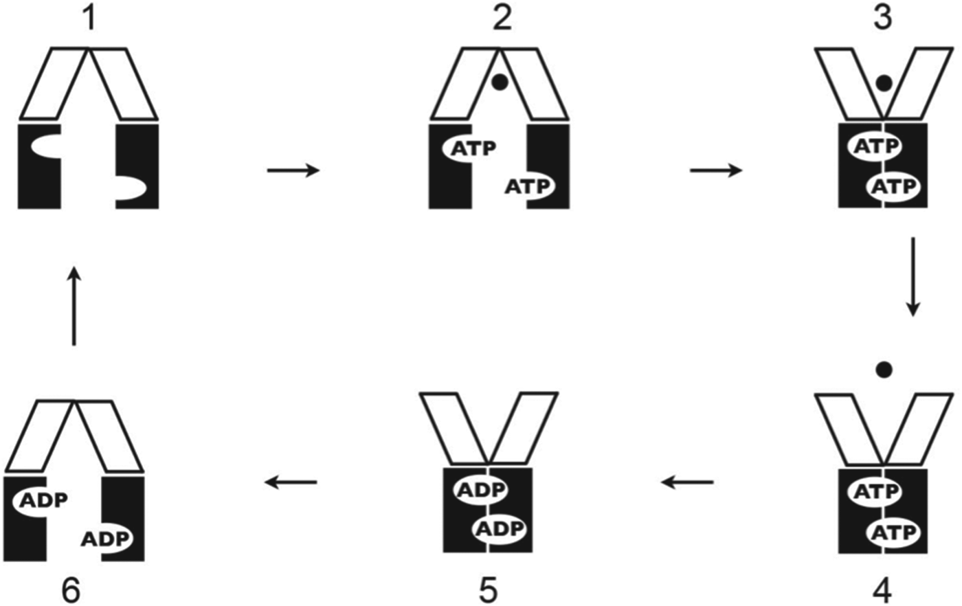
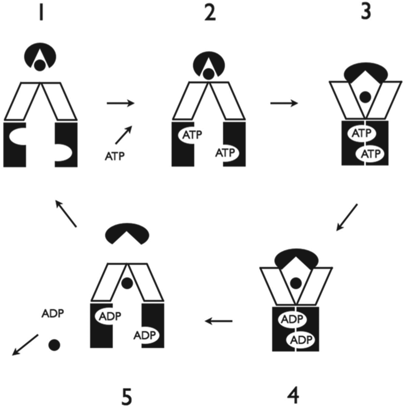
Currently, the generally accepted scheme for the function of ABC transporters is the Switch Model (Chen et al. Reference Chen, Lu, Lin, Davidson and Quiocho2003; Higgins & Linton, Reference Higgins and Linton2004; van der Does & Tampe, Reference van der Does and Tampe2004). According to this idea, the NBDs cycle between widely separated and dimerized states: Binding of two molecules of ATP to the dissociated NBD monomers induces formation of the sandwich dimer in a tweezers-like movement, whereupon sequential or processive hydrolysis of the two ATPs induces dimer separation. The two TMDs form a single channel which transitions from an inward-facing conformation accessible only from the cytoplasm in the dissociated state of the NBDs, to an outward-facing conformation accessible only from the extracellular space in the sandwich dimer state. Substrate binds to one or either state of the TMDs, depending on whether the transporter is an importer or exporter, and is presented to the opposite face of the membrane following the transition of the NBDs between states. The Switch Model scheme for exporters is shown in Fig. 1.
Switch Model for ABC Efflux Porters. TMDs are represented as unfilled parallelograms and the NBDs as black rectangles, nucleotides as text, and substrate as a black dot. (1) Nucleotide-free (apo) inward-facing conformation with wide apart NBDs. (2) Substrate binds to TMDs and two molecules of ATP bind to the NBDs. (3) NBDs dimerize to form a sandwich dimer with ATPs bound at their interface. TMDs are flipped to the outward-facing conformation and substrate is presented to the extracellular side. (4) Substrate disengages and ATP is hydrolysed. (5) ADP-bound state. May involve a semi-open conformation of the NBDs not depicted. (6) ADP induces separation of NBDs to a wide apart state, inducing reorientation of the TMDs to the inward-facing conformation, completing the cycle. Dissociation of ADP returns transporter to the apo state, to begin a new cycle.

Although crystallographic analysis has depicted both ABC importers and exporters in various conformations, each putatively representing different stages of the switch cycle, this has not resulted in significant articulation of the mechanism beyond that outlined in the original Switch Models. In particular, the mechanistic suite of events by which substrate binding stimulates ATP hydrolysis, and how ATP hydrolysis energizes and effects substrate translocation, remain as unresolved today as they were over a decade ago. Thus, while the Switch Model and the crystal structures seemingly correlate, they have not further illuminated each other, or the way further forward. This seems contrary to reasonable expectation. In view of this impasse, it is appropriate to question the foundations on which it is based and to investigate alternative possibilities, however speculative or divergent from current ideas.
1.3 A reciprocating twin channel model
In this review, an alternative model for the function of ABC transporters is proposed, in which two functionally separate substrate translocation pathways operate in an out of phase, or reciprocating manner in the TMDs, coupled to an alternating cycle of ATP hydrolysis in the NBDs.
The notion of a reciprocating function for the mechanism of ABC transporters was first suggested by Senior and co-workers (Senior et al. Reference Senior, Al-Shawi and Urbatsch1995), who proposed that ATP hydrolysis alternates between the two NBD active sites. Subsequently, Wang et al. (Reference Wang, Casciano, Clement and Johnson2000a) concluded that a half-of-the-sites reactivity model accounted well for the kinetics of inhibition of transport by various drug substrates for the ABC MDR P-glycoprotein (ABCB1). Half-of-the-sites reactivity is a type of negative cooperativity in which binding or activity at one site of a dimer suppresses or precludes activity at the other (Fersht, Reference Fersht1975; Levitzki & Koshland, Reference Levitzki and Koshland1976). The Wang et al. (Wang et al. Reference Wang, Casciano, Clement and Johnson2000a) study inferred that P-glycoprotein (P-gp) contained two different substrate-binding sites which function 180° out of phase in a reciprocating manner, such that ligand binding at one site renders the opposite unable to bind substrate. However, the sites, and the manner of the way in which the transporter might work, were not broached in this or any study to date.
The first proposal that the ABC transporter TMDs form two functionally separate substrate translocation pathways was the β-barrel model (Jones & George, Reference Jones and George1998), wherein each TMD was modelled as a TM β-barrel comprising a distinct substrate interaction site; this however is inconsistent with crystallographic analyses of detergent-solubilised ABC transporters. Based on studies of LmrA, the homodimeric ABC MDR from Lactococcus lactis, van Veen et al. (Reference van Veen, Margolles, Muller, Higgins and Konings2000) proposed a ‘twin-cylinder engine’ model for ABC MDRs, in which two functionally separate translocation channels operated in an alternating manner, broadly the same as the concept expounded here. A ‘double-barrelled’ model was suggested for the pore of the cystic fibrosis TM conductance regulator (Yue et al. Reference Yue, Devidas and Guggino2000), and distinct translocation pathways were inferred recently for human P-gp on the basis of biochemical investigation and homology models (Parveen et al. Reference Parveen, Stockner, Bentele, Pferschy, Kraupp, Freissmuth, Ecker and Chiba2011).
Previously, we examined the idea of 180° out-of-phase functioning in the context of the ABC NBDs. When applied to the required sequence of ATP binding and occlusion, hydrolysis, site opening, and product release, in each active site, this out-of-phase idea predicted a cycle of states for the NBDs that we referred to as the Constant Contact Model (Jones & George, Reference Jones and George2009). This scheme holds that nucleotide remains occluded in a closed site at all times, until release, which occurs mostly through rotation of one sub-domain of the NBD that opens the nucleotide site. This Constant Contact concept is now supported by a number of studies (George & Jones, Reference George and Jones2012; Jones & George, Reference Jones and George2013).
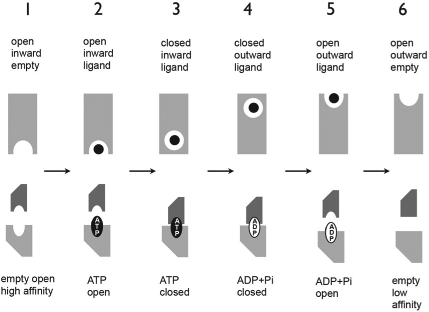
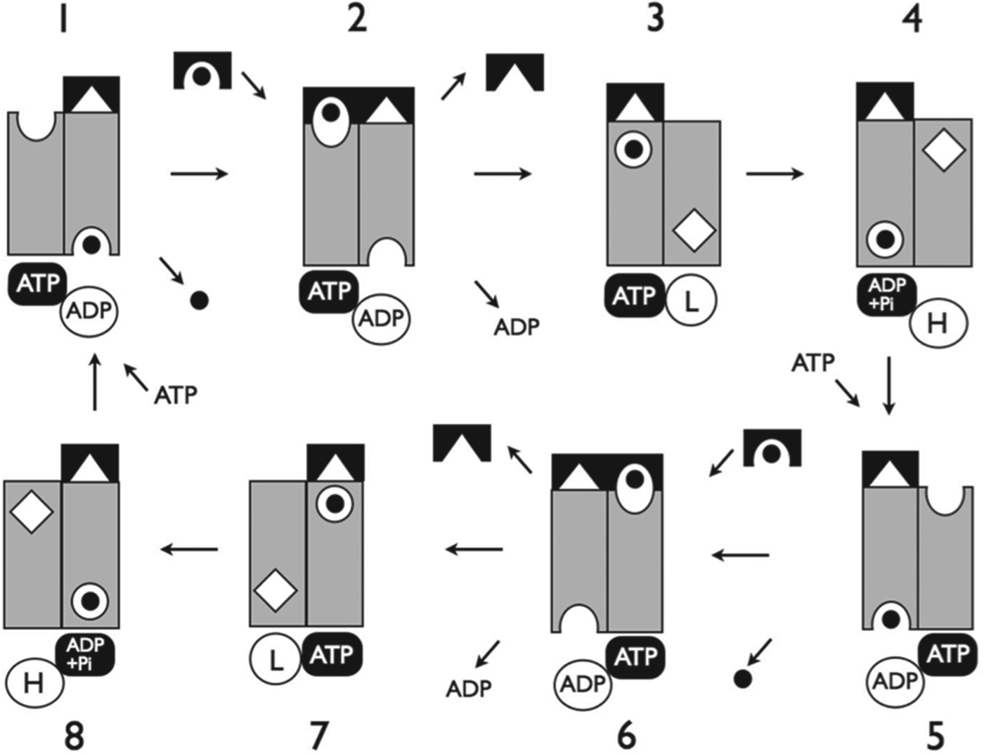
Here we extend the reciprocating idea to the whole transport cycle, proposing that the sequence of ATP binding, hydrolysis, and product release in each active site is directly coupled to the analogous sequence of substrate binding, translocation, and release in one of two functionally separate substrate translocation pathways (Fig. 2). A wide and diverse selection of data for both ABC importers and exporters is examined to show that, notwithstanding the simplistic and theoretical nature of its derivation, the Reciprocating Model explains the data with remarkable facility, clarity, and completeness. It is also able to embrace findings for a number of atypical ABC transporters, suggesting straightforward explanations for their seemingly idiosyncratic transport functions. This is a novel application of the reciprocating concept to the ABC transporter mechanism, and represents the most complete and coherent attempt at an all-encompassing scheme to explain how these important proteins work, that is consistent with sound biochemical and biophysical evidence.
Coupling between the substrate translocation and ATP hydrolysis cycles in the Reciprocating Model. Scheme for an ABC exporter is shown. The cycle of ATP binding, occlusion and hydrolysis, followed by site opening and product release is directly coupled to an analogous cycle of events in one of two functionally separate substrate translocation pathways. In exporters, the substrate binding site in each translocation pathway cycles in the order: inward-facing high-affinity empty; inward-facing substrate-bound; occluded substrate-bound cytosolic side; occluded substrate-bound extracellular side; outward-facing substrate-bound; outward-facing empty. Top half: A single translocation channel is depicted as a grey rectangle with the substrate-binding site as an inward- or outward-facing semicircle. The occluded substrate-binding site is depicted as a circle and the substrate as a black dot. Bottom half: One NBD active site is depicted by two shapes above and below representing the C-motif (dark grey) and Walker A and B motifs (light grey), respectively. (1) High-affinity inward-facing substrate-binding site is empty and active site is open and empty. (2) Substrate binds to the inward-facing site and ATP binds to active site. (3) Substrate is sequestered in an occluded site near the cytoplasmic side of the membrane; ATP is occluded in the active site. (4) ATP is hydrolysed, coupled to substrate translocation to an occluded site near the extracellular side of the membrane. (5) Opening of ADP-bound active site corresponds to substrate bound to extracellular-facing low-affinity site. (6) Substrate and nucleotide disengage; active site is in low-affinity state.
With respect to the interpretation of data in support of the Reciprocating Model, a number of assumptions are made: (a) the protein is freely in equilibrium between its possible conformational states, and ligand binding serves to stabilize particular states; (b) where two ligands can compete to stabilize mutually exclusive states, the complex with the lower free energy will be the more highly populated, and this generally will reflect the relative affinities of the transporter for the ligand(s); (c) the free-energy barriers to transitions between states vary according to the transition; some transitions have lower energy barriers than others and thus will occur spontaneously at higher rates; and (d) ligand binding can lower the free-energy barrier to a conformational transition.
The Reciprocating Model is a functional model; the word ‘channel’ does not refer to, nor is intended to imply, a physically distinct and exclusive conduit. Conversely, since in this model only one substrate is sequestered within the TMDs at any one time, a single conduit could embody physically the TM passage used by both translocation pathways; indeed this appears the most parsimonious scenario structurally.
2. ABC exporters
2.1 Allostery between substrate-binding sites in ABC MDRs
In the decade straddling the turn of the century, a large body of data was accrued showing that the mammalian ABC multidrug exporter ABCB1 contains at least three, and possibly as many as seven, distinct drug interaction sites, which display complex allosteric interactions (Martin et al. Reference Martin, Berridge, Higgins, Mistry, Charlton and Callaghan2000a; Safa, Reference Safa2004). Although a twin-channel concept was proposed to explain these findings (van Veen et al. Reference van Veen, Margolles, Muller, Higgins and Konings2000), the idea soon appeared obsolete, as the first whole ABC transporter structures led to the general adoption of the Switch Model. In this view, ABC MDRs contain multiple drug-binding loci within a large inward-facing cavity. However, a functional rationale that embraces the allosteric phenomena is yet to emerge. Despite the solved X-ray structures of ABCB1 (Aller et al. Reference Aller, Yu, Ward, Weng, Chittaboina, Zhuo, Harrell, Trinh, Zhang, Urbatsch and Chang2009; Ward et al. Reference Ward, Szewczyk, Grimard, Lee, Martinez, Doshi, Caya, Villaluz, Pardon, Cregger, Swartz, Falson, Urbatsch, Govaerts, Steyaert and Chang2013), a picture of the positions, numbers, and structural basis of allosteric interactions of substrate binding appears little clearer since the 1990s biochemical studies. Herein we show how a reciprocating mechanism provides a framework that readily renders coherence to these data that, from the perspective of the Switch Model, seem opaquely complex, anomalous, or confounding.
2.2 The reciprocating model for ABC exporters
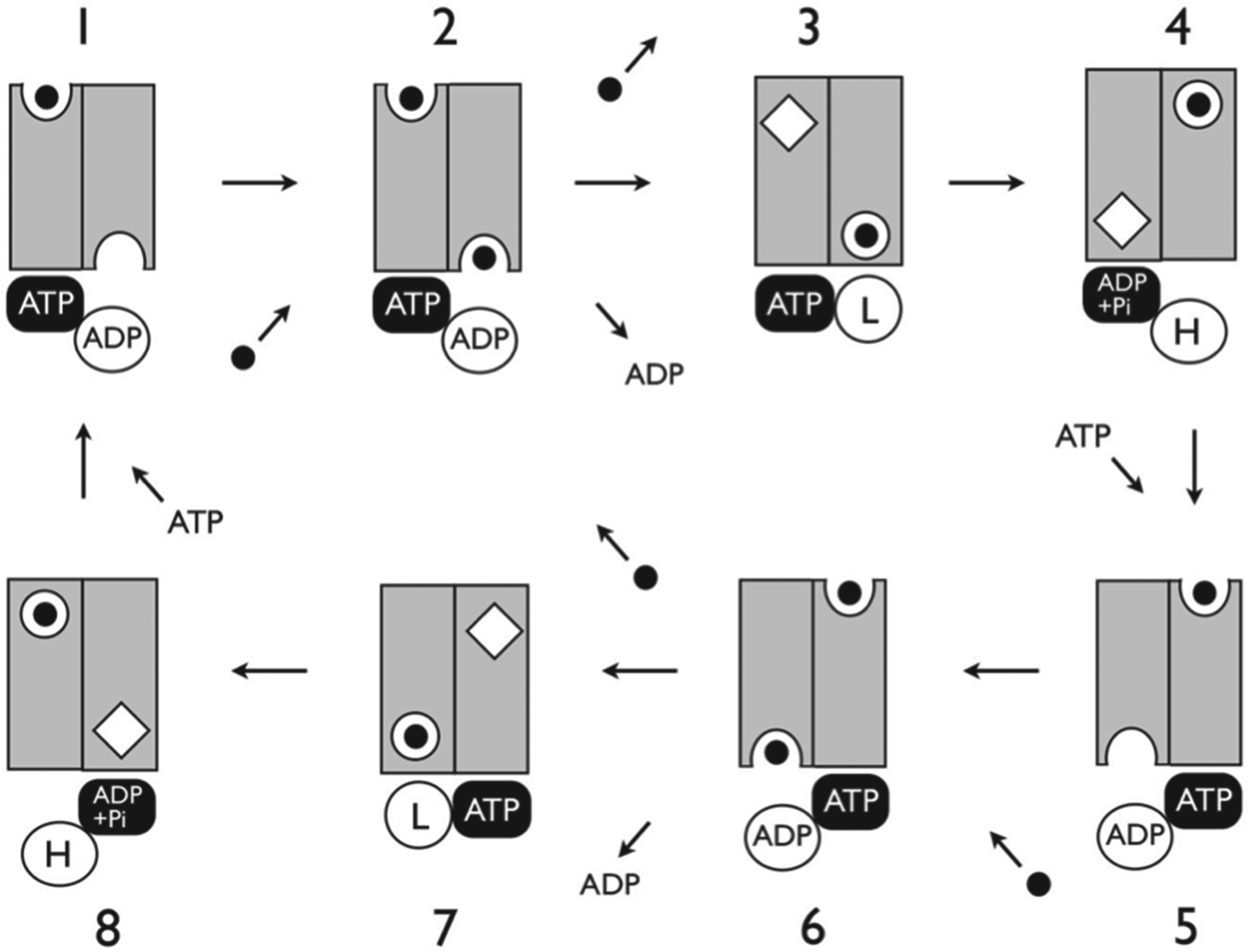
Allosteric interactions observed for ABC MDRs derive naturally from the Reciprocating Model illustrated for ABC exporters in Fig. 3. Thus, in equilibrium binding assays, in the absence of nucleotides, substrate binding at one inward-facing site will preclude binding at the other inward-facing site, and also at the outward-facing site associated with the same channel, manifesting as negative allostery between these sites and thus between substrates which preferentially bind to them. Conversely, substrate binding at one inward-facing site will promote binding of a second substrate molecule to the outward-facing site associated with the opposite channel, manifesting as positive cooperativity in binding. In kinetic assays, binding of drug to one inward-facing binding site will, by stimulating ATP hydrolysis, in turn stimulate the formation of the opposite/adjacent inward-facing binding site, thus accounting for the positive cooperativity frequently observed for ABC MDRs in transport of two different drug substrates or, indeed, for a single substrate.
Reciprocating model for an ABC Exporter. The ATP hydrolysis cycle occurs 180° out of phase between the two sites. The cycle in each active site is coupled to an equivalent sequence of events in one of two functionally separate substrate translocation pathways. The TMDs are depicted as grey rectangles, with the vertical midline demarking separate pathways, referred to as Channels 1 and 2 for the left and right pathways, respectively. The substrate is a black dot. Substrate-binding sites are depicted as semicircular for inward- or outward-facing, circular for substrate-bound sequestered states, and diamond shaped for unbound sequestered states. The active sites are depicted as white circles for open sites and black lozenges for occluded sites, referred to as Sites 1 and 2 for left and right, respectively. The physical nature of site opening, whereby the NBD core subdomain rotates away from the complex, is depicted by the position of the NBD circle downward. ‘L’ and ‘H’ refer to low and high affinity, respectively. The cycling scheme is as follows: (1) The exporter is poised to bind substrate to the high-affinity inward-facing site of Channel 2. Transported substrate awaits dissociation from Channel 1. (2) Substrate binds to the high-affinity inward-facing site in Channel 2, substrate is released from Channel 1. (3) ADP is released from Site 2, which closes to a low-affinity conformation. Substrate in Channel 2 is sequestered on the cytosolic side of the membrane. The transporter is competent for ATP hydrolysis. (4) ATP hydrolysis products (ADP + Pi) in Site 1 stabilize the configuration in which substrate in Channel 2 is in an occluded site on the extracellular side of the membrane. (5) ATP binding to site 2 causes an isomerization of the transporter that opens the ADP-bound Site 1, occludes the ATP in Site 2, regenerates the high-affinity inward-facing substrate-binding site in Channel 1, and presents the transported substrate in Channel 1 to the extracellular milieu. Stages (6)–(8) complete the cycle. Stages (1)–(4) and (5)–(8) are reciprocating phases of one another.
2.2.1 LmrA: the twin-cylinder engine
Alternating twin translocation pathways were proposed for ABC exporters based on studies of the ABCB1 homologue LmrA, a homodimeric MDR from L. lactis (van Veen et al. Reference van Veen, Margolles, Muller, Higgins and Konings2000). Equilibrium binding analysis using the transported drug vinblastine revealed two distinct, strongly cooperative substrate-binding sites. Transport assays showed vinblastine and the dye Hoechst 33 342 enhanced transport of each other at low concentrations and inhibited it at high concentrations, indicating two functionally coupled binding sites. Vinblastine bound with a stoichiometry of two molecules per transporter; one to an extracellular low-affinity binding site and the other to a high-affinity intracellular site.
Notably, in the Switch Model, substrate cannot bind to the inward- and outward-facing sites simultaneously; moreover, the model provides no apparent explanation for the allostery observed in this study of LmrA. In contrast, the Reciprocating Model suggests a mechanistic underpinning for these findings. Thus, cooperativity observed for equilibrium binding of vinblastine occurs since substrate binding to one site increases the stability of the complex, thereby promoting binding of a second substrate molecule, as depicted in Stage 2/6 in Fig. 3. In transport assays at low concentrations, vinblastine and Hoechst 33 342 stimulated transport of each other, by increasing the cycling rate relative to that induced by one drug alone at the concentration (van Veen et al. Reference van Veen, Margolles, Muller, Higgins and Konings2000). At higher concentrations they will compete directly for the high-affinity sites, thus inhibiting each other's transport.
2.2.2 ABCB1 has multiple drug-binding sites displaying complex interactions
Human ABCB1, also known as P-gp or MDR1, is the most well characterized of ABC MDRs. ABCB1 is a single polypeptide comprising pseudo-symmetrical, homologous halves, each consisting of a TMD followed by a canonical NBD. A large body of experimental data indicates the existence of multiple drug-binding sites in ABCB1. In a classic study of P-gp, Callaghan and co-workers (Martin et al. Reference Martin, Berridge, Higgins, Mistry, Charlton and Callaghan2000a) used equilibrium binding assays to show that the non-transported modulator XR9576 binds at a site distinct from the vinblastine binding site, and is able to reduce the number of these sites through a negative allosteric interaction.
The Reciprocating Model, as outlined above, can explain these data. For example, if XR9576 binds to the outward-facing site of Channel 1 as depicted in Stage 2 of Fig. 3, this would abrogate binding to the inward-facing site of Channel 1, which may be taken to be the vinblastine binding site. This also explains how XR9576 can modulate transport, but is itself not transported. In addition, since P-gp has a higher affinity for XR9576 than for vinblastine, this model also explains the finding that in equilibrium binding experiments, XR9576 could displace vinblastine, but not vice versa.
In this same study, the transported substrates rhodamine 123, paclitaxel, and Hoechst 33 342 decreased vinblastine binding via a negative allosteric effect, just as had been seen for the modulator XR9576. It was inferred from detailed analysis of the data that P-gp contains at least four distinct drug interaction sites, displaying complex allosteric interactions. In terms of the Reciprocating Model, due to its pseudo-symmetrical sequence structure, P-gp contains two different inward-facing high-affinity substrate-binding sites, with different substrate specificities, and similarly two different low-affinity outward-facing sites. This accounts generally for the number of sites and their observed allosteric connectedness.
A number of studies characterizing the effects of a wide range of drug substrates on the kinetics of transport or ATPase activity have demonstrated that P-gp contains distinct drug interaction sites (Bruggemann et al. Reference Bruggemann, Germann, Gottesman and Pastan1989; Garrigos et al. Reference Garrigos, Mir and Orlowski1997; Greenberger et al. Reference Greenberger, Lisanti, Silva and Horwitz1991; Orlowski et al. Reference Orlowski, Mir, Belehradek and Garrigos1996; Spoelstra et al. Reference Spoelstra, Dekker, Schuurhuis, Broxterman and Lankelma1991, Reference Spoelstra, Westerhoff, Pinedo, Dekker and Lankelma1994) and also demonstrated allosteric coupling between drug interaction sites (Ayesh et al. Reference Ayesh, Shao and Stein1996; Ferry et al. Reference Ferry, Malkhandi, Russell and Kerr1995, Reference Ferry, Russell and Cullen1992; Litman et al. Reference Litman, Zeuthen, Skovsgaard and Stein1997; Martin et al. Reference Martin, Berridge, Higgins and Callaghan1997; Pascaud et al. Reference Pascaud, Garrigos and Orlowski1998; Shapiro & Ling, Reference Shapiro and Ling1997; Sharom et al. Reference Sharom, Yu, Didiodato and Chu1996, Reference Sharom, Yu, Lu, Liu, Chu, Szabo, Muller, Hose, Monks, Varadi, Seprodi and Sarkadi1999; Tamai & Safa, Reference Tamai and Safa1991; Wang et al. Reference Wang, Casciano, Clement and Johnson2000a, Reference Wang, Casciano, Clement and Johnsonb). As an illustrative example of these transport assays, the drugs verapamil and daunorubicin were shown to interact non-competitively with P-gp (Litman et al. Reference Litman, Zeuthen, Skovsgaard and Stein1997; Pereira et al. Reference Pereira, Borrel, Fiallo and Garnier-Suillerot1994; Spoelstra et al. Reference Spoelstra, Westerhoff, Pinedo, Dekker and Lankelma1994), while inhibition of daunorubicin transport by verapamil, and by 12 other drugs, showed a sigmoidal response, indicating cooperativity in inhibition by these agents (Wang et al. Reference Wang, Casciano, Clement and Johnson2000b). This indicates that while verapamil and daunorubicin bind to distinct non-overlapping binding sites on P-gp, inhibition of daunorubicin transport by verapamil involves an allosteric affect.
In terms of the Reciprocating Model, the two high-affinity inward-facing drug-binding sites have opposite affinities for each drug: one has high affinity for daunorubicin and low affinity for verapamil, while the reverse is true for the other. This accounts for observations of non-competitive interactions between these drugs. Under conditions of high daunorubicin, verapamil at low concentrations will compete with daunorubicin for the high-affinity verapamil-binding site. As its concentration rises verapamil will increasingly compete directly with daunorubicin for the high-affinity daunorubicin site. This example illustrates a mechanism that accounts for the sigmoidal response commonly observed in transport assays for many, if not most P-gp substrates.
2.2.3 ABC exporters with two substrate interaction sites
The Type I secretion systems of Gram-negative bacteria are involved in export of a wide range of peptides (Fath & Kolter, Reference Fath and Kolter1993; Kanonenberg et al. Reference Kanonenberg, Schwarz and Schmitt2013). They comprise a homodimeric ABC transporter that in many cases includes an intracellular N-terminal extension, which interacts with the substrate, in some cases being involved in preprocessing of the peptide to be transported. The presence of two identical auxiliary substrate interaction domains per transporter is consistent with the Reciprocating Model, since each could couple to a separate pathway. However, it is less clear in terms of the Switch Model. In this context it suggests either the coordination of simultaneous transport of two peptides through the single channel, or that substrates are recruited singly from one or other substrate interaction domain. This latter scenario would clearly be asymmetric with respect to the symmetrical transporter, and it is difficult to see how this could be coordinated to operate efficiently. Thus, while the presence of two substrate interaction domains per homodimer transporter fits the Reciprocating Model straightforwardly, the explanation in terms of the Switch model is less clear.
2.2.4 ABC exporters with one non-consensus active site
A number of ABC exporters contain one NBD active site lacking one or more residues highly conserved amongst ABC transporters (Jones & George, Reference Jones and George1999). Where these transporters have been characterized, it is generally found that the non-consensus active site is able to bind but not hydrolyse ATP, or hydrolyse ATP at greatly reduced rates. Since the Switch Model requires hydrolysis of two molecules of ATP to energize the transition from the outward- to the inward-facing conformation, it seems at odds with asymmetric catalytic activity between the active sites.
In contrast, the Reciprocating Model is able to embrace these observations. Thus, cycling involving translocation through only one pathway could be achieved provided ATP can bind to, and induce the closure of, the non-catalytic site (Fig. 3, steps 1–5). This still requires an explanation as to why only one pathway is used, or else why translocation through only one pathway requires ATP hydrolysis. One possibility is that one channel mediates the passage of a substrate that moves down its concentration gradient, which therefore does not require energy; its passage serves a dual function both to facilitate the translocation of the allocrite and to enable the completion and continuation of the transport cycle.
Notably, all ABC transporters with one degenerate active site comprise heterodimeric TMDs, consistent with the idea that the TMD subunits could form two distinct pathways that each translocates a different substrate, or a different set of substrates. According to this idea, the passively translocated substrate could move in either direction. Theoretically, such a system could also be used for sets of substrates, which have a concentration gradient that favours their passive efflux, but that is too weak to sustain required levels of translocation; thus, the chemical free energy of one ATP molecule can be harnessed to translocate two substrates, in situations where it exceeds the free-energy cost of translocation.
2.2.5 The retina-specific ABC transporter ABCA4: an ABC antiporter?
Although to date no ABC exporters have been shown definitively to export substrates passively, there is nevertheless evidence consistent with such a scenario. The retina-specific ABC transporter, ABCA4 (Allikmets et al. Reference Allikmets, Shroyer, Singh, Seddon, Lewis, Bernstein, Peiffer, Zabriskie, Li, Hutchinson, Dean, Lupski and Leppert1997), is involved in retinal transport in rod outer segment discs. The accepted role of ABCA4 is in translocation of all-trans-retinal (ATR), the toxic product of light-induced isomerization of the visual chromophore 11-cis-retinal, from the disc lumen into the rod cell cytoplasm. ATR is then converted to 11-cis-retinal, thereby regenerating the chromophore, which is returned to the disc lumen.
The striking feature of this process is that, while ABCA4 is closely related to eukaryotic exporters, the direction of ATR transport is into the cell (Quazi et al. Reference Quazi, Lenevich and Molday2012). Notably thus, ABCA4 contains the ‘EAA’ motif characteristic of prokaryotic ABC importers (Mourez et al. Reference Mourez, Hofnung and Dassa1997; Tsybovsky et al. Reference Tsybovsky, Molday and Palczewski2010), as well as a large asymmetric extracellular (intraluminal) domain (Tsybovsky et al. Reference Tsybovsky, Orban, Molday, Taylor and Palczewski2013), conceivably functionally equivalent to the periplasmic-binding protein (BP) in ABC permeases. Interestingly, while a mechanism of 11-cis-retinal export across the disc membrane into the disc lumen has not been identified, ABCA4 has been implicated in this process (Biswas-Fiss et al. Reference Biswas-Fiss, Affet, Ha and Biswas2012; Boyer et al. Reference Boyer, Higbee, Currin, Blakeley, Chen, Ablonczy, Crouch and Koutalos2012). Indeed, 11-cis-retinal interacts with the non-catalytic NBD1 of ABCA4 and stimulates ATP hydrolysis in the catalytic NBD2 (Biswas-Fiss et al. Reference Biswas-Fiss, Affet, Ha and Biswas2012).
Clearly, these data uniquely can be understood from the perspective of the scenario discussed above. ABCA4 actively imports the toxic ATR adduct into the cell, while facilitating the export of 11-cis-retinal, which moves passively down the concentration gradient generated by the intracellular metabolism of ATR; 11-cis-retinal efflux by ABCA4 is regulated by its interaction with NBD1, possibly by affecting ATP binding.
2.2.6 The ABCC family: why do they only need one catalytic NBD?
The above explanation for ABC transporters with non-catalytic active sites is consistent with the function of other ABC transporters with one non-catalytic active site. One example is human multidrug resistance proteins (MRPs), part of the ABCC family, whose members contain non-canonical residues in one catalytic site. It was found for MRP1 that while the substrate leukotriene C 4 (LTC4) stimulated vanadate (VO4) trapping of ADP, another substrate, E217βG, did not, suggesting that E217βG does not stimulate ATP hydrolysis (Letourneau et al. Reference Letourneau, Nakajima, Deeley and Cole2008). In addition, some MRP1 substrates require the presence of glutathione to be transported, while some are co-transported with glutathione and/or their transport is stimulated by glutathione (Rothnie et al. Reference Rothnie, Callaghan, Deeley and Cole2006), consistent with the idea of two separate translocation pathways, and also with the possibility that translocation through one pathway is required to enable transport through another (Rothnie et al. Reference Rothnie, Callaghan, Deeley and Cole2006).
For MRP3 it was observed that many transported substrates stimulated ATPase activity, but one substrate, carboxyfluorescein, while being transported in a concentration-dependent manner, did not stimulate ATPase activity above the basal levels (Seelheim et al. Reference Seelheim, Wullner and Galla2013). These data are consistent with the idea that some MRP substrates may be passively transported. Notably, MRPs are involved in the efflux of metabolites of xenotoxins as well as physiological substrates produced within the cell, at least some of which will have higher concentrations inside the cell than outside. The mechanism of MRP1 is examined in greater detail below.
2.2.7 CFTR: does the exception prove the rule?
The Reciprocating Model is able to decipher the function of another member of the ABCC family, the cystic fibrosis transconductance regulator (CFTR; ABCC7), an atypical ABC transporter that acts as a nucleotide-gated chloride channel (Gadsby et al. Reference Gadsby, Vergani and Csanady2006; Riordan et al. Reference Riordan, Rommens, Kerem, Alon, Rozmahel, Grzelczak, Zielenski, Lok, Plavsic and Chou1989). CFTR is thought to have evolved from an ABCB1-like progenitor, representing a degenerate ‘leaky’ ABC exporter, in which the TMDs and NBDs are uncoupled to some extent (Bai et al. Reference Bai, Li and Hwang2011; Jin et al. Reference Jin, Lu, Li and Han2012). CFTR's NBD1 deviates significantly from the canonical sequence and fold, and the active site comprising the NBD1 Walker A and B motifs and the NBD2 C-motif (Site 1) is catalytically impaired due to non-canonical active site residues. Binding of two ATP molecules to CFTR is required for channel activity (Vergani et al. Reference Vergani, Lockless, Nairn and Gadsby2005). Channel opening and closing are correlated with ATP binding and hydrolysis, respectively, at the active site comprising the NBD2 Walker A and B motifs (Site 2). Multiple gating cycles occur while ATP remains bound, but not hydrolysed, at Site 1 (Szollosi et al. Reference Szollosi, Muallem, Csanady and Vergani2011; Tsai et al. Reference Tsai, Shimizu, Sohma, Li and Hwang2009). Channels are closed in the absence of ATP or when ATP is not bound at Site 1 (Basso et al. Reference Basso, Vergani, Nairn and Gadsby2003).
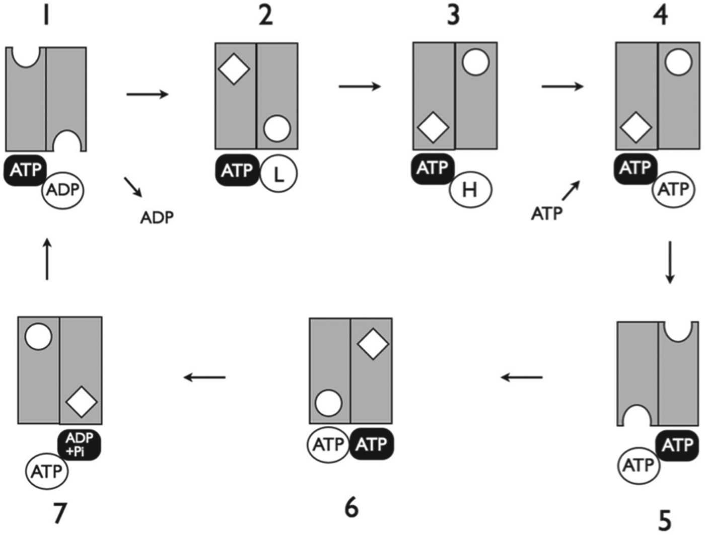
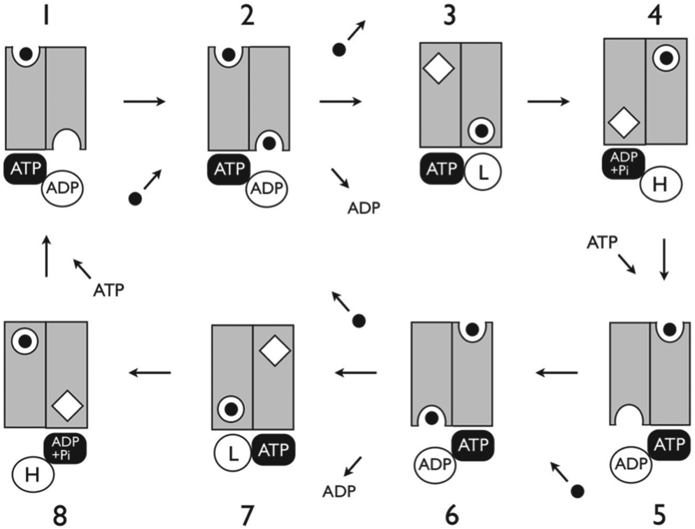
Figure 4 illustrates a proposed CFTR cycle derived from the scheme in Fig. 3. ADP release from the open Site 2 in Stage 1 results in closure of Site 2 and the formation of the prehydrolytic state in Stage 2. In CFTR, the isomerization in the TMDs that is normally coupled to ATP hydrolysis in Site 1 occurs without ATP hydrolysis (uncoupled), leading to Stage 3. ATP then binds to the open Site 2 (Stages 3 and 4); this induces closure of Site 2 and opening of Site 1 to form the configuration in Stage 5 (this is the same as Stage 5 in Fig. 3, but with ATP rather than ADP in Site 1).
Reciprocating model for CFTR. TMD channels, NBD active sites and nucleotides are depicted as in Fig. 3. (1) ATP is occluded in NBD Site 1. ADP bound in open Site 2 is released. (2) Release of ADP allows Site 2 to close and the TMDs move to an occluded state. This is equivalent to the pre-hydrolytic state in the normal cycle (Stages 3/7 in Fig. 3). (3) TMDs undergo the isomerization in which the occluded site in Channel 2 moves to the extracellular side of the membrane. ATP remains occluded in Site 1, and the empty Site 2 opens; this is equivalent to the post-hydrolytic state in the normal cycle (Stages 4/8 in Fig. 3). (4) ATP binds to open Site 2. (5) Site 2 closes, occluding ATP. Site 1 opens, retaining ATP. TMDs move to the conformation where the extracellular and intracellular-facing sites are accessible; this is the open channel. (6) Site 2 closes, switching the TMDs to an occluded state, in which the channel is closed. ATP in Site 2 is hydrolysed. (7) ADP + Pi in Site 2 induces the configuration with the occluded site in Channel 2 on the extracellular side of the membrane, and Site 1 opens.
Stage 5 in Fig. 4 corresponds to the open CFTR channel, which is formed by the inward-facing Channel 1. Thus, the CFTR open channel corresponds to an inward-facing channel that is leaky at the extracellular face. This is consistent with recent data indicating that in the open channel, residues lining the pore are accessible to bulky reagents from the intracellular side, but not from the extracellular side (Wang & Linsdell, Reference Wang and Linsdell2012). This is contrary to the prediction of the Switch Model, since open channels are expected to correspond to the outward-facing conformation (Gout, Reference Gout2012) (Fig. 1). A narrow configuration at the extracellular face of open channels is also consistent with data that showed that the size of the sidechain at a single position close to the extracellular surface had a profound effect on channel conductance (Linsdell et al. Reference Linsdell, Zheng and Hanrahan1998). In addition, since the channel is closed in Stage 6, and spontaneous transitions can occur between Stages 5 and 6, prior to hydrolytic closure (Stage 7), the model thus explains the flickering closures typically observed during open channel bursts.
In this CFTR scheme, the channel can only be open sustainably when ATP is bound and occluded at Site 2, as is observed experimentally (Vergani et al. Reference Vergani, Lockless, Nairn and Gadsby2005). It is also noteworthy that, if upon ADP release from Stage 1, ATP were to bind to open Site 2 before it closed to form Stage 2, the transporter could return to open channels by proceeding in a reverse direction directly to Stage 6 and thereby to Stage 5. This is consistent with data showing that the protein has two distinct routes to the open channel (Jih & Hwang, Reference Jih and Hwang2013). The proposed dependence of the cycle on spontaneous, or thermally driven isomerizations in the TMDs that are uncoupled from ATP hydrolysis, is also consistent with the observation that CFTR currents are highly temperature sensitive (Aleksandrov et al. Reference Aleksandrov, Kota, Aleksandrov, He, Jensen, Cui, Gentzsch, Dokholyan and Riordan2010).
We suggest that the above interpretation provides a clear and comprehensive template that can accommodate a considerable body of data for CFTR (Chong et al. Reference Chong, Kota, Dokholyan and Forman-Kay2013; Gadsby et al. Reference Gadsby, Vergani and Csanady2006), one that is also coherent within a more general scheme for ABC transporter function. It could also be of significance in the design of correctors of mutant CFTRs, an endeavour that has produced promising results in recent years (Becq & Chanson, Reference Becq and Chanson2013; Hanrahan et al. Reference Hanrahan, Sampson and Thomas2013; Hoffman & Ramsey, Reference Hoffman and Ramsey2013). Thus, if CFTR retains vestigial substrate-binding sites, as might be expected, these could be exploited to develop agents that stabilize specific conformations. For example, a drug that binds with high affinity to the outward-facing site in Stage 5 might assist in stabilizing open channels. Indeed, the findings of a recent study of the effects of the arylaminobenzoate NPPB on CFTR activity can be explained if it is assumed NPPB binds to the inward-facing site in Stage 5 and facilitates the transition to, and thence stabilizes, Stage 6 (Csanády & Töröcsik, Reference Csanády and Töröcsik2014).
2.2.8 Asymmetric functioning in the TMDs
A fundamental difference between the Reciprocating and Switch Models is with respect to asymmetry in the functional mechanism. The Switch Model only depicts symmetrical states (Fig. 1); no essential asymmetry is posited for the processes of NBD separation and dimerization, or the alternation of the TMDs between the inward- to outward-facing states. While asymmetry may exist, for example between the hydrolysis of the first and second ATPs, or where a singly transported substrate binds the TMDs, this would appear not germane to the overall mechanism. Nevertheless, there is long-standing evidence indicating asymmetric functioning of the NBDs (Senior & Bhagat, Reference Senior and Bhagat1998; Sharma & Davidson, Reference Sharma and Davidson2000; Shyamala et al. Reference Shyamala, Baichwal, Beall and Ames1991), and more recently of the TMDs (Koike et al. Reference Koike, Conseil, Leslie, Deeley and Cole2004; Letourneau et al. Reference Letourneau, Nakajima, Deeley and Cole2008; Mittal et al. Reference Mittal, Boehm, Grutter, Bordignon and Seeger2012; Ritchie et al. Reference Ritchie, Kwon and Atkins2011; Wang et al. Reference Wang, Fu, Pan, Wu, Gong, Wang and Shi2013).
2.2.9 Antibody binding to P-gp is consistent with a reciprocating mechanism
Ritchie et al. (Reference Ritchie, Kwon and Atkins2011) used surface plasmon resonance to examine the interaction of P-gp with two antibodies, known to bind to extracellular epitopes. The antibodies bound with high affinity in the presence or absence of nucleotides, and this was unaffected by the substrate vinblastine; thus it was inferred that the antibodies bind to multiple conformations. In contrast, neither antibody bound in the presence of ADP and VO4, which together stabilize a conformation near the transition state of the hydrolysis reaction, where the nucleotide is occluded in the ‘sandwich’ NBD configuration.
It appears incredible that the antibodies can bind equally well to the inward- and outward-facing conformations, since the extracellular loops comprising their respective epitopes, and required for binding (Georges et al. Reference Georges, Tsuruo and Ling1993; Schinkel et al. Reference Schinkel, Arceci, Smit, Wagenaar, Baas, Dolle, Tsuruo, Mechetner, Roninson and Borst1993; Zhou et al. Reference Zhou, Gottesman and Pastan1999), are predicted to be deployed radically differently in these two states, both according to the Switch Model and by inference from crystal structures of ABC MDRs. Seeming to confound this, the energy difference between the AMP–PNP-bound state (expected to mimic the ATP-bound state) and the trapped state indicated a very large conformational change between them. In terms of the Switch Model, no significant differences are predicted. Thus, the nature of, and explanation for, the observed antibody binding is not clear.
The Reciprocating Model can account for the above findings for P-gp. Thus, the antibodies bind to one conformation in which an extracellular-facing substrate-binding site is exposed. In the absence of substrates, but in the presence or absence of ATP or ADP, no particular state will be stabilized (Fig. 3), and so the conformation involving the extracellular-facing site will become available for the antibody to bind. Their very high affinities would also enable antibody binding to compete with the tendency of substrate and ATP to stabilize occluded states of the TMDs. However, neither antibody binds to the highly stable ADP + VO4 state, where according to our model, the transporter is in a conformation close to the post-hydrolytic Stage (4/8 in Fig. 3), where neither extracellular-facing site is available.
When ADP and VO4 were presented to preforme P-gp-antibody complexes, the dissociation kinetics were markedly biphasic, with a roughly 60/40 split between complexes dissociating more rapidly than the others. This otherwise puzzling finding can be explained by the Reciprocating Model. If antibody binds, for example, to Stage 1 in Fig. 3, while binding of ADP (+VO4) in Site 1 will require a large isomerization in the TMDs to reach Stage 4 (the trapped conformation) from Stage 1, in contrast, binding of ADP + VO4 in Site 2 will require a much smaller, and therefore more rapid change in the TMDs to reach Stage 8 from Stage 1. If nucleotide binds with approximately equal probability to either active site, this accounts roughly for the 60/40 split.
2.2.10 The CFTR pore is not symmetrical with respect to the TMDs
Data from studies mapping the pore in CFTR are at odds with symmetrical alignment and function of the TMDs. Whilst TM6 has been shown to play a significant role in determining the functional properties of the pore (Hwang & Kirk, Reference Hwang and Kirk2013), mutagenic analysis, guided by homology models based on whole ABC exporter structures, has failed to identify symmetrically located residues with similar functional effects (Wang et al. Reference Wang, Fu, Pan, Wu, Gong, Wang and Shi2013), and it remains unclear why one TM appears dominant in affecting pore function. Indeed, it is notable that for P-gp, TM6, which is equivalently located to TM6 in CFTR with respect to their sequence homology, was analogously found to have a central role in transport, particularly in the coupling of substrate transport to ATP hydrolysis (Devine et al. Reference Devine, Ling and Melera1992; Storm et al. Reference Storm, O'mara, Crowley, Peall, Tieleman, Kerr and Callaghan2007). However, in contrast to CFTR, for P-gp, TM12 in TMD2, which is equivalent to TM6 in TMD1 in terms of their homology, was also found to have an equivalent role to TM6. This further suggests that the CFTR TMDs do not contribute symmetrically to the pore, consistent with the Reciprocating Model (see above).
2.2.11 Two distinct translocation pathways in P-gp
A recent study of P-gp biochemically dissected the drug translocation pathway (Parveen et al. Reference Parveen, Stockner, Bentele, Pferschy, Kraupp, Freissmuth, Ecker and Chiba2011). A pair of residues, located symmetrically in the TMDs with respect to their sequence homology, and identified by photolabelling to be proximal to the substrate translocation pathway, were investigated using site-directed mutagenesis combined with transport assays. The results indicated that, although rhodamine123 has a preference for one translocation path, verapamil, propafenones, and vinblastine preferentially use another. The authors inferred the existence of two solute translocation pathways in P-gp, and selective but not exclusive use of one of these pathways by different P-gp solutes. Clearly, these data are in accord with the analyses of P-gp's interaction with substrates outlined above, and with the Reciprocating Model.
Although the translocation pathways were mapped to the inward-facing cavity using the mouse P-gp structure, it appears unclear to what extent the twin translocation pathway idea, as an articulation of the Switch Model, has gained currency. On the other hand, the Reciprocating Model, while being in accord with these biochemical data, is not so with their structural interpretation (Parveen et al. Reference Parveen, Stockner, Bentele, Pferschy, Kraupp, Freissmuth, Ecker and Chiba2011), because this requires wide separation of the NBDs.
2.3 NBD:TMD coupling and the transport cycle
The mechanism of coupling between the NBDs and TMDs in the transport cycle is one of the key unanswered questions in ABC transporter research. As depicted in Fig. 3, the coupling process is integral to the Reciprocating Model and is clearly articulated at the functional level. In contrast, in the Switch Model, the mechanistic steps by which substrate binding triggers the transition between the two states that delivers substrate to the opposite face of the membrane are not depicted to an equivalent resolution.
2.3.1 Coupled changes between the substrate binding and active sites in P-gp
Liu & Sharom (Reference Liu and Sharom1996) studied the effects of substrates on the accessibility of the active sites using fluorescent probes attached to native cysteines in the P-loops of P-gp. Substrates caused a reduction in the accessibility of the probes, in the presence or absence of ATP. In terms of the Reciprocating Model, substrate binding will tend to stabilize a conformation around Stages 2–3/6–7 in Fig. 3 in which, due to coupling between the NBDs and TMDs, one active site is more occluded, thus explaining an overall drop in accessibility of the probes.
Martin et al. (Reference Martin, Berridge, Mistry, Higgins, Charlton and Callaghan2000b) used the substrate vinblastine and the slowly hydrolysable ATP analogue ATPγS, in equilibrium radioligand-binding assays, to investigate the coupling process in P-gp. ATP binding induced a conformational change in the substrate-binding site that reduced the binding capacity for the substrate, suggesting the site has reduced accessibility. A later study by Qu et al. (Reference Qu, Russell and Sharom2003) found that substrate binding increased the affinity of P-gp for ATP to a degree that was dependent on the substrate. These data are consistent with the Reciprocating Model. Thus, ATP alone will tend to stabilize Stage 3/7 (Fig. 3) in which the substrate-binding sites are occluded, thereby reducing their overall accessibility relative to the ligand-free state. As discussed in the previous paragraph, substrate binding will tend to close one ATP site, thereby increasing the overall measured affinity for nucleotide; this will occur to an extent dependent on the strength with which the substrate binds.
From the perspective of the Switch mechanism, the interpretation of these data characterizing the interaction between the substrate-binding and catalytic sites in P-gp appears less certain. Reduced binding capacity for the substrate might correspond, for example, to partial closure of the inward-facing conformation. However, it is difficult to see what function it would serve for ATP-binding to induce this, relative to the nucleotide-free state, and how this fits into the model (Fig. 1). Conversely, substrate binding might reduce the accessibility of the ATP-binding sites by inducing partial or full dimerization of the NBDs, for example. This seems possible, but the NBDs would probably have to be relatively near to closed for this to occur, and this is not part of the proposed scheme. Of course, there are other possible interpretations, but the internal logic of the model does not appear to provide any clear direction or insight into what they might be.
2.3.2 How do mutations in the NBDs cause altered substrate specificity in ABC MDRs?
A number of studies have found that mutations in the NBDs cause an alteration in the substrate specificity of ABC MDRs. Beaudet & Gros (Reference Beaudet and Gros1995) and Beaudet et al. (Reference Beaudet, Urbatsch and Gros1998) reported that alteration of residues in the X-loop of mouse P-gp (mdr3) caused a dramatic alteration of substrate specificity, due to an alteration in the ability of various drugs to stimulate ATPase activity. These effects were also found for the T578C mutation (Beaudet et al. Reference Beaudet, Urbatsch and Gros1998), which occurs at the C-terminus of the α-helix, immediately downstream of the D-loop (Jones & George, Reference Jones and George2012). Hoof et al. (Reference Hoof, Demmer, Hadam, Riordan and Tummler1994) found that a conservative lysine-to-arginine mutation at position 6 of the C-motif in P-gp NBD1 (LSGGQK/LSGGQN) resulted in an altered profile of cross-resistance and chemosensitization in cells expressing the mutant P-gp. Finally, a recent study of the fungal ABC MDR Pdr5 observed that mutation of the D-loop aspartate within the non-canonical N-terminal active site, altered Pdr5-mediated drug resistance in a substrate-dependent manner (Furman et al. Reference Furman, Mehla, Ananthaswamy, Arya, Kulesh, Kovach, Ambudkar and Golin2013).
It was concluded in the studies of P-gp and Pdr5, that the NBD mutations most likely affect communication between the drug-binding and active sites. In other words, the respective wild-type residue performs a function distinct from that of direct interaction with, and thereby detection of, the substrate; rather it is involved in propagating an ‘on’ or ‘off’ signal, and it is the threshold and/or the bias for the propagation of this signal that is altered by mutation.
The Reciprocating Model can explain the data showing altered substrate specificity for P-gp and Pdr5 from NBD mutations. Thus, as discussed above, due to their heterodimeric nature, these transporters will have two different inward-facing high-affinity drug-binding sites, with complementary but overlapping substrate specificities. Since each of the two substrate-binding sites interact asymmetrically with the NBD dimer, an alteration of the coupling in one NBD will alter the overall balance in the substrate transport profile by affecting transport differentially between the two halves. In contrast, there is no published explanation for this phenomenon from the perspective of the Switch Model.
2.3.3 Asymmetric responses in the NBDs to MRP1 substrates
Koike et al. (Reference Koike, Conseil, Leslie, Deeley and Cole2004) investigated the effects of a mutation in the human ABC exporter ABCC1 (MRP1). Position 1150 is located within a TMD cytoplasmic loop, expected – by analogy with bacterial ABC MDRs – to engage the NBDs. The P1150A mutant showed increased transport of the glucuronidated Oestrogen E217βG due to a decreased K m, but conversely, showed diminished efficiency of LTC4 transport. Intriguingly, the P1150A mutation showed greater affinity for ATP and an increase in V max when supporting E217βG transport, whereas for LTC4 transport, the K m(ATP) of P1150A was unchanged from the wild-type, and a decrease in V max was observed. The authors concluded that the P1150A mutation altered signalling between the NBDs and some substrate-binding sites, but not others.
The Reciprocating Model can explain the asymmetric response in the NBDs to different substrates by P1150A MRP1. Thus, with reference to Fig. 3, the primary translocation pathways for LTC4 and E217βG are assigned to Channels 1 and 2, respectively, and the left and right NBDs, corresponding to the MRP1 NBD Sites 1 and 2, respectively. In line with the authors’ interpretation (Koike et al. Reference Koike, Conseil, Leslie, Deeley and Cole2004), we postulate that the P1150A mutation partially uncouples substrate binding from the inward- to outward-facing transition in Channel 1, thereby increasing spontaneous cycling in Channel 1. Thus, relative to wild-type, in the presence of E217βG alone, the V max for E217βG transport through Channel 2 will be increased due to the increased cycling of Channel 1, which will stimulate ATP hydrolysis in NBD Site 2 and increase the rate of cycling to Stage 2. This will result in the presence of more inward-facing substrate-binding sites in Channel 2 and more open NBD2 sites (Stage 2); this in turn will lower the K m for substrate and ATP for E217βG transport. Conversely, in the presence of LTC4 alone, uncoupled cycling in Channel 1 will reduce the efficiency of LTC4 transport.
2.3.4 P-gp modulators: the intriguing case of cis-(Z)-flupentixol
Dey and co-workers have extensively characterized the mode of action of cis-(Z)-flupentixol, a thioxanthene-based non-transported modulator of P-gp (Dey et al. Reference Dey, Ramachandra, Pastan, Gottesman and Ambudkar1997; Maki et al. Reference Maki, Hafkemeyer and Dey2003, Reference Maki, Moitra, Ghosh and Dey2006a, Reference Maki, Moitra, Silver, Ghosh, Chattopadhyay and Deyb). While inhibiting Pgp-mediated drug transport, flupentixol nevertheless facilitates recognition and binding of a transportable photoactive derivative of the substrate prozosin. As a modulator, flupentixol does not affect ATP binding, or ADP release, but interferes with substrate translocation and release by interrupting the hydrolytic step. Most intriguing was the observation that while flupentixol and prozosin stimulate ATP hydrolysis independently, together they reduce the stimulatory effect of the other. It was concluded that modulation by flupentixol involves allosteric disruption of cross-talk between the substrate and active sites, without any direct interference with their individual functions.
From the perspective of the Reciprocating Model, flupentixol binds with high affinity to one of the two extracellular-facing substrate-binding sites. Either in the presence or absence of ATP, high-affinity binding to an outward-facing site would enhance binding of a substrate to the complementary inward-facing high-affinity site, explaining its ability to enhance labelling of a transported substrate.
Although substrate stimulates hydrolysis by stabilizing Stage 3/7, the transporter can still enter the pre-hydrolytic Stage 3/7 and hydrolyse ATP in the absence of bound substrate; this is consistent with basal ATP hydrolysis observed for many ABC transporters. In the presence of ATP, flupentixol stabilizes Stage 1 in Fig. 3. When the modulator dissociates (due to its equilibrium with the unbound state), the transporter is preconfigured to enter Stage 3, and hydrolyse ATP. This accounts for flupentixol's ability to stimulate ATP hydrolysis; as a function of flupentixol concentration and the rate at which the transition to Stage 3 occurs. Consistent with this, flupentixol stimulated ATPase activity decreases at high modulator concentrations, displaying a ‘bell-shaped’ characteristic (Maki et al. Reference Maki, Moitra, Silver, Ghosh, Chattopadhyay and Dey2006b). When prozosin is bound to the intracellular site, the transition from Stages 2 to 3 is slowed, such that it cannot occur in the flupentixol unbinding/binding interval; thereby flupentixol interferes in the process of substrate stimulated ATP hydrolysis and substrate translocation.
Although the interpretation of these data in terms of the Reciprocating Model is somewhat speculative, the model nevertheless provides a plausible, coherent and detailed account of the underlying mechanistic and physiological basis. In contrast, to date, we know of no interpretation from the viewpoint of the Switch Model that provides a similarly comprehensive account. These studies represent primary examples of biochemical evidence supporting the Reciprocating Model's proposed mechanism that – together with other examples discussed for both ABC exporters and importers throughout this paper – reinforces this ‘whole of transporter’ reciprocating scheme.
2.4 Models of the complete coupling mechanism
Finally, we compare and contrast the proposed coupling mechanisms in the Switch and Reciprocating Models for ABC exporters. In consideration of the Switch Model in Fig. 1, it can be seen that for the complex to progress from Stages 1 to 4, each stage must be energetically lower than those previous. It also seems clear that ATP hydrolysis in Stage 4 must follow substrate release; otherwise there appears nothing to ensure that the substrate does not become trapped when the TMDs revert to the inward-facing state. How does the transporter detect that the substrate has disengaged from the outward-facing site and cleared the channel? If Stage 4 is energetically lower than Stage 1, what is to stop the transporter going directly from Stages 1 to 4 without binding substrate, and then cycling from Stage 4 back to Stage 1 via Stages 5 and 6, thereby futilely hydrolysing ATP?
From the perspective of the Reciprocating Model (Fig. 3), it can be seen that the transporter is only able to remain stable in the hydrolysis-competent state in Stages 3/7, after the substrate translocated in the previous cycle has disengaged, and substrate bound at the intracellular face is sequestered. This ensures that translocated substrate is released, and that ATP hydrolysis will lead to substrate translocation. In this scheme, the cycle could proceed in both forward and reverse directions. Thus, the vectorial nature of transport is determined by the energy gradient for ATP hydrolysis, and the close coupling of the hydrolysis reaction to the process of substrate efflux by the transporter.
3. ABC importers
ABC influx porters or permeases are found predominantly in bacteria, where they mediate the uptake of a wide range of substrates, generally nutrients (Davidson et al. Reference Davidson, Dassa, Orelle and Chen2008; Lewis et al. Reference Lewis, Ween and Mcdevitt2012). The subunits of the ABC importer complex are expressed separately, and include a soluble periplasmic substrate-BP. The BPs gather and deliver substrate or solute ligands to the TMDs; substrate is bound in a cleft between the two lobes of the BP, which undergo a transition between closed and open conformations to bind and release substrate, the so-called Venus flytrap mechanism (Felder et al. Reference Felder, Graul, Lee, Merkle and Sadee1999). BPs associated with many ABC importers in Gram-negative bacteria are free to diffuse throughout the periplasmic space, whereas in other cases they are covalently attached to the TMDs, or are anchored to the membrane (van der Heide & Poolman, Reference van der Heide and Poolman2002).
3.1 The switch model for ABC importers
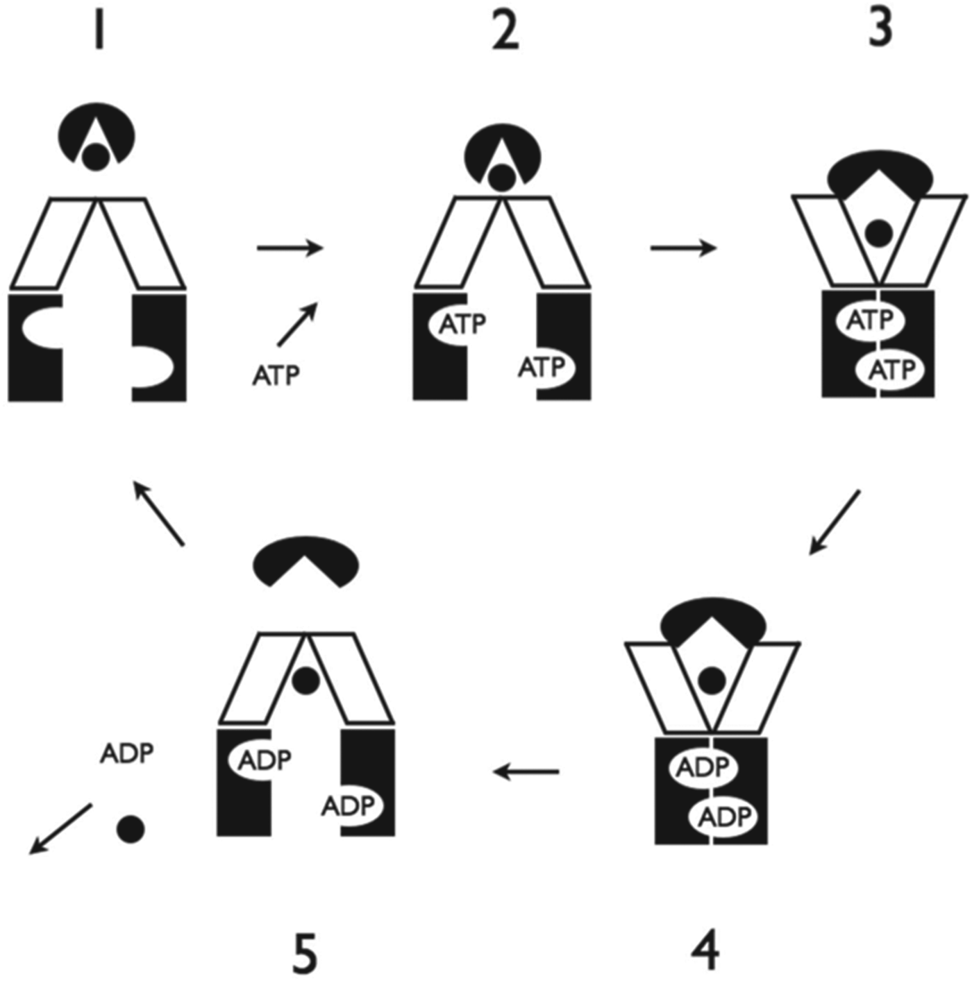
Currently, the dominant hypothesis for the mechanism by which ABC permeases (importers) function is the Switch Model, shown in Fig. 5. In this view, the closed substrate-loaded BP attaches to the inward-facing conformation of the TMDs; this together with the binding of two ATP molecules in the NBDs triggers the conformational switch to the outward facing conformation, driven by movement of the NBDs together to the sandwich dimer state. In this process, the BP lobes are opened and substrate released into the TMD channel. This is then followed by hydrolysis of both ATPs; the instability of the resultant ADP-bound sandwich dimer drives the NBDs apart, resetting the TMDs to the inward facing conformation and releasing the open BP. Release of substrate and ADP into the cytosol resets the transporter for the next cycle.
Switch Model for ABC Importers. TMDs, NBDs, nucleotides and substrate are depicted as in Fig. 1. BP is shown as a black circle with a narrow ‘V’-shaped notch (closed) or as a black ellipse with a wide ‘V’-shaped notch (open). (1) Nucleotide free (apo) or resting state. TMDs are in the inward-facing conformation and NBDs are wide apart. (2) Closed substrate-loaded BP attaches to the closed extracellular face of TMDs. Two ATPs bind to the NBDs. (3) Binding of ligands triggers the conformational switch to the outward facing conformation, driven by movement of the NBDs together to the sandwich dimer state. In this process, the BP lobes are opened and substrate is released into the TMD channel. (4) ATPs are hydrolysed sequentially. (5) ADP-bound in sandwich dimer induces NBD separation, resetting the TMDs to the inward-facing conformation and releasing the open BP. Release of substrate and ADP into the cytosol resets the transporter for the next cycle.
3.2 The reciprocating model explains data at odds with the switch model
Despite general acceptance of the Switch mechanism, a significant body of data from a number of well-characterized ABC importers is clearly at odds with key aspects of the general scheme shown in Fig. 5.
3.2.1 Ligand-bound BP has low affinity for the transport complex regardless of ATP binding
In a recent study of the maltose permease, Bao & Duong (Reference Bao and Duong2012) showed that in both the nucleotide-free and the ATP-bound states, the closed ligand-bound BP has low affinity for the transport complex. Similarly for the molybdate/tungstate importer ModBC-A reconstituted in liposomes, no binding of the BP ModA to the transport complex was detected in the presence of substrate, in either the nucleotide free or the ATP-bound states (Hollenstein et al. Reference Hollenstein, Frei and Locher2007; Vigonsky et al. Reference Vigonsky, Ovcharenko and Lewinson2013).
These observations, strikingly anomalous in terms of the Switch Model, may nevertheless be explained by the Reciprocating Model for ABC importers as depicted in Fig. 6. In this scheme, in the absence of nucleotides to stabilize a particular conformation, the substrate-bound BP would have low affinity for the transport complex. In the presence of ATP under non-hydrolysis conditions, the transporter would be stabilized at Stage 3, to which the closed ligand-bound BP cannot bind. The model thus explains the low affinity of the liganded BP for the transport complex in the ATP-bound state.
Reciprocating model for an ABC Importer. TMD channels, substrate-binding sites, substrate, NBD active sites, and nucleotides are depicted as in Fig. 3. The BPs are above the TMDs and are depicted as a black rectangle with either a triangular (ligand-free) or semicircular (ligand-bound) binding site. (1) The exporter is poised to release substrate from the low-affinity inward-facing site of Channel 2 and ADP from open Site 2. (2) Substrate-loaded BP binds to the high-affinity outward-facing site in Channel 1; substrate is released from Channel 2. (3) ADP is released from Site 2, which closes to a low-affinity conformation. BP disengages from Channel 2. Substrate in Channel 1 is sequestered on the extracellular side of the membrane. The transporter is competent for ATP hydrolysis. (4) ATP hydrolysis in Site 1 results in translocation of the substrate in Channel 1 to an occluded site on the intracellular side of the membrane. NBD Site 2 opens to a high-affinity state, ready to bind ATP. (5) ATP binding to site 2 causes an isomerization of the transporter that opens the ADP-bound Site 1, occludes the ATP in Site 2, regenerates the high-affinity outward-facing substrate binding site in Channel 2, and presents the transported substrate in Channel 1 to the intracellular milieu. Stages (6)–(8) complete the cycle. Stages (1)–(4) and (5)–(8) are reciprocating phases of one another.
3.2.2 Unliganded BP has high affinity for the ATP-bound transport complex
The Reciprocating Model is also consistent with the otherwise inexplicable finding that, in the absence of substrate, the BP binds to the ATP-bound transport complex, which has been observed for the maltose permease (Bao & Duong, Reference Bao and Duong2012), ModBC-A (Vigonsky et al. Reference Vigonsky, Ovcharenko and Lewinson2013) and the histidine permease (Ames et al. Reference Ames, Liu, Joshi and Nikaido1996). As can be seen in Fig. 6, in Stage 3 the open BP binds to the ATP-bound state; this can still occur in the absence of substrate since under non-hydrolysis conditions ATP would stabilize Stage 3. This aspect of the model also explains the observation that the BP can stimulate ATP hydrolysis, even in the absence of substrate. Experimentally, this effect was observed to occur at around 10% of BP-stimulated ATPase in the presence of substrate; this is also consistent with the model since the presence of bound substrate in Stage 3 would provide significantly greater stability to the pre-hydrolytic conformation and thereby further facilitate hydrolysis.
3.2.3 Unliganded BP in excess inhibits transport: how and why?
Other puzzling findings can be explained by the Reciprocating Model. A high molar excess of the MalE BP in the presence of sub-stoichiometric levels of substrate inhibits transport in the maltose permease (Merino et al. Reference Merino, Boos, Shuman and Bohl1995) and a similar effect was observed for the histidine permease (Prossnitz et al. Reference Prossnitz, Gee and Ames1989). This phenomenon occurs, according to the reciprocating scheme, because a high concentration of unliganded BP would inhibit release of the BP from the complex between Stages 2 and 3, and thereby slow progress to the hydrolysis-competent Stage 3 (Fig. 6). Findings in another study that the BP remains bound throughout the catalytic cycle (Daus et al. Reference Daus, Berendt, Wuttge and Schneider2007) are also explained by the model, since although individual BPs engage and disengage during the cycle, at least one BP remains bound at all times.
3.2.4 ADP and ATP are both required for high-affinity binding of the substrate-loaded BP
The Reciprocating Model predicts that the ligand-bound BP will bind optimally to the transporter in the presence of both ATP and ADP, which together will stabilize Stage 2. Notably, for ABC transporters, the ATP-bound state is rarely characterized in the presence of ADP, even though this is closer to physiological conditions, since the ADP/ATP-bound state is considered transitory and not mechanistically significant in the Switch Model.
For the histidine permease, it was found that, in vivo, the concentration of substrate required for half maximal binding of the BP HisJ to the TMD was 56 nM, close to the K d for histidine binding to HisJ (30 nM) (Ames et al. Reference Ames, Liu, Joshi and Nikaido1996). However, in proteoliposomes, in the absence of nucleotides, the equivalent K d for HisJ binding was found to be nearly three orders of magnitude higher (30 μM histidine). Thus, under physiological conditions, and at low substrate concentrations, HisJ binds to the transport complex with an affinity only limited by its affinity for the substrate ligand, yet binds with very much lower affinity to the nucleotide-free state. Since the Switch Model predicts high-affinity binding of the liganded BP in the absence of nucleotides, these observations appear unexplained. In contrast, these data are consistent with the Reciprocating Model, since it predicts that the highest affinity binding of the ligand-bound BP occurs at Stage 2, with ATP and ADP bound, as available in vivo.
3.3 ABC solute BP dimers
As depicted in Fig. 6, the Reciprocating Model proposes that at two points in each cycle (Stages 2 and 6), two BPs are bound to the transporter complex simultaneously. Each bound BP will function in coordination with the channel with which it is directly associated, and thus simultaneously bound BPs will be out of phase with respect to their conformational cycle. Since direct contact between bound BPs could facilitate the overall coordination between the two channels, the Reciprocating Model is consistent with the possibility that the BPs manifest allosteric coupling between their ligand-binding sites mediated through their direct physical contact.
3.3.1 Biochemical and biophysical evidence for allosterically coupled BP dimers
For some ABC importers, the BP is covalently attached to the TMD, and thereby two BPs are associated with each transport complex. One of the best characterized of such importers is the osmoregulated betaine importer OpuA (van der Heide & Poolman, Reference van der Heide and Poolman2002). For OpuA it was found that the rate of transport increased in a sigmoidal manner with increasing substrate concentration, indicating positive cooperativity; this was lost in OpuA transporters with only one BP, which showed a hyperbolic response. Moreover, when a substrate molecule was covalently attached to one of the two OpuA BPs, rendering it unable to bind or release substrate, the transporter was nonetheless twice as active as those with only one BP, and nearly as active as the wild-type. These data are consistent with the idea that the two BPs interact in a cooperative manner to stimulate transport.
Other data further support the notion of functional BP dimers. A study of the maltose permease MalFGK-E found transport exhibited a sigmoidal dependence on the BP concentration, indicating that MalE is involved in a cooperative interaction in the transport process (Manson et al. Reference Manson, Boos, Bassford and Rasmussen1985). For the molybdate/tungstate importer MolBC-A, the stoichiometry of substrate binding, as determined by isothermal calorimetry, was closer to 0·5 than to 1 (Tirado-Lee et al. Reference Tirado-Lee, Lee, Rees and Pinkett2011), clearly in accord with a reciprocating BP dimer. Also of relevance, the arginine-binding BP, TM0593 from Thermotoga maritima, was found to exist as a homodimer even under strongly denaturing conditions (Ausili et al. Reference Ausili, Pennacchio, Staiano, Dattelbaum, Fessas, Schiraldi and D'auria2013; Luchansky et al. Reference Luchansky, Der, D'auria, Pocsfalvi, Iozzino, Marasco and Dattelbaum2010). For the arginine, lysine, and histidine importer ArtMP-J from Geobacillus stearothermophilus (Vahedi-Faridi et al. Reference Vahedi-Faridi, Eckey, Scheffel, Alings, Landmesser, Schneider and Saenger2008), Scatchard plot analysis of substrate-binding data for the BP ArtJ yielded curves with two slopes, indicating low- and high-affinity sites, consistent with the existence of a reciprocating dimer.
3.3.2 Crystal structures of BP dimers
Although there is biochemical evidence indicating cooperativity between BPs, the crystallized structures of most ABC transporter BPs nevertheless reveal a monomeric state. From the perspective of the Reciprocating Model, this is not surprising, since the BP's principal interaction is with the complex, from which it must disengage, while another BP remains bound. BPs whose crystallized forms are suggested below to reveal a physiological interface between the BPs fall into two groups, and their ability to be crystallized in their dimeric form, while most do not, is explained as follows. The first group includes BPs from hyperthermophilic Achaea, where the dimer may have heightened stability to withstand the extreme environments in which these organisms typically exist. The second group includes BPs associated with Type II ABC permeases (Jones et al. Reference Jones, O'mara and George2009). For two Type II ABCs, namely the vitamin B12 importer BtuCD-F and the molybdate/tungstate importer MolAB-C, enzymological data suggest that the BP is bound to the transporter complex in the absence of substrate, and binds substrate from that state (Lewinson et al. Reference Lewinson, Lee, Locher and Rees2010; Vigonsky et al. Reference Vigonsky, Ovcharenko and Lewinson2013). Thus, these BPs may retain their dimeric state more strongly because they predominantly remain bound to the transport complex (George & Jones, Reference George and Jones2011).
For the arginine, lysine, and histidine importer ArtMP-J from G. stearothermophilus (Vahedi-Faridi et al. Reference Vahedi-Faridi, Eckey, Scheffel, Alings, Landmesser, Schneider and Saenger2008), the BP ArtJ was found to form dimers with identical intermonomer interfaces in crystal structures of the BP bound to each of its substrates (Fig. 7A). Of numerous different intermonomer contacts observed amongst three distinct crystal lattices, only one was repeated, and this occurred in all three cases. As discussed above, substrate-binding data for ArtJ suggested low- and high-affinity sites, further supporting the notion that the structural dimer reflects a physiological interaction.
Ribbon diagrams of crystallographic BP dimers discussed in the text. Monomers are coloured green and blue. Substrates are in space filling representation. (A) ArtJ (PDB 2Q2A). ArtJ dimer with the substrate arginine bound in the central cleft of each monomer. (B) MaModA (PDB 3K6Z). MaModA dimer showing an interfacial β-barrel in the centre. Molybdate (red) is bound in each monomer. (C) Archaeal ModA shares a half β-barrel feature. Overlay of archaeal ModA (Hollenstein et al. 2009) with the region equivalent to β-barrel-forming interface in MaModA coloured. C-terminal region truncated in four of the five constructs can be seen at bottom left. Structures were aligned in SwissPDBViewer using Cα coordinates and the ‘magic best fit’ facility. PDB 3CIJ (AfModA), 3CFX (MaModA), 3CG1 (PfModA), 3CFZ (MjModA) and 3CG3 (PhModA). (D) TroA (PDB 1TOA) dimer with zinc (red) bound in the monomers. (E) BtuF (PDB 1N4A) dimer with vitamin B12 bound in the monomers.
Crystallographic analysis revealed that the BP of the molybdate importer from Methanosarcina acetivorans, ModA (MaModA) formed dimers via an interface comprising an eight-stranded β-barrel (Fig. 7B) (Chan et al. Reference Chan, Giuroiu, Chernishof, Sawaya, Chiang, Gunsalus, Arbing and Perry2010); this occurred in two distinct crystal forms, arguing against the dimers being crystal packing artefacts. In a separate study (Hollenstein et al. Reference Hollenstein, Comellas-Bigler, Bevers, Feiters, Meyer-Klaucke, Hagedoorn and Locher2009) (Fig. 7C), MaModA and four archaeal ModA orthologues were all found to contain a four-stranded β-sheet, structurally closely similar and equivalently located to that which formed the β-barrel interface in the study by Chan et al. (Reference Chan, Giuroiu, Chernishof, Sawaya, Chiang, Gunsalus, Arbing and Perry2010). Significantly, this β-barrel feature is not located in a position to affect substrate binding or the interaction of ModA with its cognate ABC transporter; it is an addition not found in prokaryotic ModA homologues, which are otherwise identical (Chan et al. Reference Chan, Giuroiu, Chernishof, Sawaya, Chiang, Gunsalus, Arbing and Perry2010).
While the β-barrel dimer interface was not observed in the ModA structures reported by Hollenstein et al. (Reference Hollenstein, Comellas-Bigler, Bevers, Feiters, Meyer-Klaucke, Hagedoorn and Locher2009), four of the five expression constructs in that study were truncated in order to ‘facilitate better lattice contacts’. Notably, these facilitated contacts involved hydrophobic residues unnaturally exposed by the truncation (Chan et al. Reference Chan, Giuroiu, Chernishof, Sawaya, Chiang, Gunsalus, Arbing and Perry2010; Hollenstein et al. Reference Hollenstein, Comellas-Bigler, Bevers, Feiters, Meyer-Klaucke, Hagedoorn and Locher2009), which could account for why the β-barrel interface was not reported. It is thus possible that the β-barrel dimer interface observed for MaModA could be formed similarly by its archaeal orthologues: indeed, no alternative function for the conserved half β-barrel structure is either known, suggested or apparent. In addition, there appears nothing to indicate that the observed MaModA BP dimer could not form in vivo; therefore it may well represent a functional state.
Crystallographic analyses have found that a number of BPs from Type II ABC permeases formed dimers, namely TroA (Lee et al. Reference Lee, Deka, Norgard, Radolf and Hasemann1999), AdcAII (Loisel et al. Reference Loisel, Jacquamet, Serre, Bauvois, Ferrer, Vernet, Di Guilmi and Durmort2008), and BtuF (Borths et al. Reference Borths, Locher, Lee and Rees2002; Karpowich et al. Reference Karpowich, Martsinkevich, Millen, Yuan, Dai, Macvey, Thomas and Hunt2001) (see Fig. 7D and E). In each instance, the dimer interface involved equivalent regions of the respective sequences (George & Jones, Reference George and Jones2011), as expected of a functional dimer-forming interface. Moreover, in separate studies, substrate-bound BtuF (Borths et al. Reference Borths, Locher, Lee and Rees2002; Karpowich et al. Reference Karpowich, Martsinkevich, Millen, Yuan, Dai, Macvey, Thomas and Hunt2001) and apo BtuF (Karpowich et al. Reference Karpowich, Martsinkevich, Millen, Yuan, Dai, Macvey, Thomas and Hunt2001) were observed to form closely similar dimers in three separate crystals, each with distinct crystal lattice arrangements, with the root-mean-square deviation between Cα atoms of residues involved in the interface (residues 90–141 from both monomers) being <1 Å between all three dimers. As in the case of the identical ArtJ BP dimers discussed above, this correctly should be viewed as solid if not compelling evidence that a specific intermonomer interface is predisposed to form.
Most notably, for all the BP dimers observed in crystal structures, and suggested above to reflect a physiological interaction, the dimer interface involves only one of the two lobes, which would allow the opposite lobe to open and close to deliver or bind the substrate ligand. Moreover, in all cases, in the putative dimer configurations, either BP can disgorge its substrate towards the TMDs (Fig. 7).
In conclusion, we suggest that the enzymological and structural data discussed above provide strong support for the idea that the BPs have the capacity to function as coupled alternating dimers, consistent with the existence of two equivalent but distinct BP interaction sites on the transporter complex. This is clearly consistent with the Reciprocating Model. Conversely, it appears clear that the notion of reciprocally functioning BP dimers is fundamentally irreconcilable with the symmetrically operating single channel described by the Switch Model. In addition, crystal structures of ABC importers clearly show that bound BPs straddle the extracellular entrance to the putative translocation pathway, exactly as depicted in the Switch Model, with no logical place or function evident for a second BP, far less for one in a reciprocal conformation to the first. Thus, while the Reciprocating Model is in accord with the above data, it appears, in this respect at least, at odds with the crystal structures.
3.4 The TMD:NBD allosteric mechanism
A central and long-standing question is how binding of substrate-loaded BP at the periplasmic face of the TMDs is communicated to the NBDs to coordinate the transport cycle. Although crystallographic analyses have produced conformations of the maltose permease suggested to embody key stages of the cycle (Chen, Reference Chen2013), and biochemical studies have identified residues involved in the communication pathway (Daus et al. Reference Daus, Landmesser, Schlosser, Muller, Herrmann and Schneider2006; Schmees et al. Reference Schmees, Stein, Hunke, Landmesser and Schneider1999; Speiser & Ames, Reference Speiser and Ames1991; Walter et al. Reference Walter, Wilken and Schneider1992), the exact signalling mechanism remains unclear. Indeed, since the structures suggest that the role of the NBD:TMD interface is essentially to ensure that the two domains move in concert, they provide no insight into key findings that single point mutations at the NBD:TMD interface, or even in regions of the NBDs most distal to the BP-binding site, enable cycles of ATP hydrolysis that are uncoupled from BP binding.
Whilst neither the Switch Model nor the crystal structures convincingly or even adequately illuminate the structural or dynamic basis of signal propagation between the BP-binding site and the NBDs, the Reciprocating Model provides significant and clear insights into this mechanism. As depicted in Fig. 6, binding of the substrate-loaded BP in Stage 2 induces an isomerization of the transporter to the configuration depicted in Stage 3. Thus, due to allosteric coupling between the two channels, which will tend them towards reciprocal conformations with respect to the cycle at all times, binding of BP to one channel results in a change at the intracytoplasmic face of the opposite channel, thereby providing a mechanistic route by which substrate binding is communicated to the NBDs. Thus, mutations in the NBDs, which interrupt communication and coupling between the TMDs and the NBDs, and/or the reciprocal functioning between the active sites, could allow ATP hydrolysis in the absence of BP binding, thus explaining such observations (Daus et al. Reference Daus, Landmesser, Schlosser, Muller, Herrmann and Schneider2006; Schmees et al. Reference Schmees, Stein, Hunke, Landmesser and Schneider1999; Speiser & Ames, Reference Speiser and Ames1991; Walter et al. Reference Walter, Wilken and Schneider1992).
Finally, another notable feature of the Reciprocating Model is that ATP hydrolysis occurs when incoming substrate has disengaged from the BP and is sequestered within a defined TMD-binding site, after translocated substrate in the opposite channel is released into the cytosol, thus ensuring tight coupling and coordination (Fig. 6; Stages 3 and 7). In contrast, for the Switch Model (Fig. 5), there is a lack of clarity regarding how, in the outward-facing conformation, the NBDs detect whether substrate has disengaged from the BP and the TMD location of substrate before hydrolysis occurs and resets the transporter; and how, in the inward-facing conformation, the transporter detects whether substrate has disengaged and cleared the channel prior to BP and ATP binding initiating a new cycle.
4. Conclusion
Here we have expounded a model for the functional cycle of ABC transporters based on three simple premises. The first is the necessary sequence of events in the active site: ATP binding to an open site, site closure, ATP hydrolysis, site opening and product release. The second is that the molecule functions as a reciprocating dimer, in which two equivalent halves perform identical cycles, 180° out of phase. The third is that the events of ligand binding (substrate, ATP), translocation/hydrolysis, and release in the TMDs and NBDs are directly coupled, such that analogous events occur in step. The first premise appears obligate, the second was inferred previously from the data, and the third is a simple and direct interpretation of the notion of coupling.
The whole transporter mechanism deduced from these premises involves four distinct processes, which are integrated and contingent, in such a way that provides new insights into a large and diverse range of data. The Reciprocal Model's fit with the data is remarkable considering its paradigmatic relativity to the Switch Model. For a model so conceived, the possibility appears remote that its unique power and ability to illuminate the data is merely coincidental. Thus, the premises above may be inferred to reflect the underlying principles that govern the ABC transporter function, and the Reciprocating Model to present a plausible scheme of its mechanistic cycle.
One aspect of the Reciprocating Model that warrants comment is the apparent difficulty in reconciling it with the crystal structures of whole ABC transporters. In respect of this, it is important to note that the crystallographic data ought not to be regarded as having a preeminent authority. Clearly, the arbiter of mechanistic models, as of the structures, should be the wider body of biochemical and biophysical data.
The notion put in this review of a half-of-sites reactivity and reciprocating function is a crucial insight and novel key concept in the understanding of the ABC transporter mechanism. Importantly, the reciprocating scheme readily suggests novel experimental approaches to investigating the transport mechanism, whilst we await the arrival of the complete structural record.