Introduction
Tourette syndrome (TS) is characterized by involuntary movements and vocalizations, which are commonly preceded by sensory-psychological urges termed premonitory sensations. Tics are thought to reflect dysfunction within motor regions including the basal ganglia (Albin & Mink, Reference Albin and Mink2006; Wang et al. Reference Wang, Maia, Marsh, Colibazzi, Gerber and Peterson2011). However, the behavioural spectrum of TS frequently involves complex tics or compulsions including swearing tics (coprolalia), mirroring behaviours (echophenomena) and context-dependent socially inappropriate urges (Kurlan et al. Reference Kurlan, Daragjati, Como, McDermott, Trinidad, Roddy, Brower and Robertson1996; Eddy & Cavanna, Reference Eddy and Cavanna2013a ). The neural mechanisms underlying these latter features are yet to be determined.
Urges to carry out socially inappropriate behaviours could be linked to reasoning about people's mental states such as beliefs and emotions. For example, a socially inappropriate remark (e.g. ‘big nose’) is likely to prompt a negative emotional reaction in the subject, and inferences about the beliefs or intentions of the speaker. The speaker may also experience a negative emotional reaction on realizing the consequences of their remark. Adults with TS can respond differently to healthy controls on tasks involving social cognition, such that their interpretations of socially inappropriate faux pas, humour, sarcasm and emotional facial expressions can be unconventional (Eddy et al. Reference Eddy, Mitchell, Beck, Cavanna and Rickards2010a , Reference Eddy, Mitchell, Beck, Cavanna and Rickards b , Reference Eddy, Mitchell, Beck, Cavanna and Rickards2011). People with TS do not demonstrate a ‘lack’ of Theory of Mind (ToM, i.e. the ability to reason about mental states), but rather subtle differences to controls when drawing inferences relating to social interaction (Eddy & Cavanna, Reference Eddy and Cavanna2013b ). One study (Eddy & Cavanna, Reference Eddy and Cavanna2015) examined patients’ behavioural responses to a task that required them to describe the ambiguous movements of animated shapes. When shown videos featuring random movement, patients with TS were more likely than controls to attribute emotions and intentions to the shapes. This raises the possibility of hyper-mentalizing in TS, i.e. a tendency towards excessive attribution of mental states, which may be associated with inaccurate conclusions about social behaviour (e.g. Sharp et al. Reference Sharp, Pane, Ha, Venta, Patel, Sturek and Fonagy2011). Hyper-mentalizing may also help to explain why patients can report the occurrence of socially inappropriate actions in benign control stories containing no faux pas (Eddy et al. Reference Eddy, Mitchell, Beck, Cavanna and Rickards2010a , Reference Eddy, Mitchell, Beck, Cavanna and Rickards2011), and attribute more negative intentions to imaginary conspecifics during cooperative games (Eddy et al. Reference Eddy, Mitchell, Beck, Cavanna and Rickards2011). Furthermore, people with TS report increased personal distress during every-day social interactions, indicating increased susceptibility to negative emotions in the self when witnessing other people in distress, i.e. enhanced empathy or emotional contagion (Eddy et al. Reference Eddy, Macerollo, Martino and Cavanna2015). Hyper-mentalizing or increased personal distress could help explain social anxiety in TS (e.g. Thibert et al. Reference Thibert, Day and Sandor1995) or insecure attachment styles involving relationship anxiety and avoidance (e.g. Dehning et al. Reference Dehning, Burger, Krause, Jobst, Yundina, Müller, Meyer, Zill and Buchheim2015).
Previous research has raised the possibility of dissociation between different aspects of social cognition in TS. For example, despite evidence of hyper-mentalizing, people with TS can self-report doing less everyday cognitive perspective taking than healthy controls (Eddy et al. Reference Eddy, Macerollo, Martino and Cavanna2015). This finding could help explain why one study found that activity in the posterior cingulate, right amygdala and right temporo-parietal junction (TPJ) increased to a lesser degree in patients with TS than in healthy controls when these patients were asked to reason about story characters’ beliefs (Eddy et al. Reference Eddy, Cavanna, Rickards and Hansen2016). The aim of the current study was to further investigate social cognition in TS using functional magnetic resonance imaging (fMRI) and a different measure: the Reading the Mind in the Eyes Task (RMET; Baron-Cohen et al. Reference Baron-Cohen, Wheelwright, Hill, Raste and Plumb2001). This task was selected as it is quite different to the story-based false-belief task used by Eddy et al. (Reference Eddy, Cavanna, Rickards and Hansen2016), and relies upon the use of visual cues to infer mental state. Furthermore, the RMET may more accurately be described as a test of emotion recognition rather than ToM (Oakley et al. Reference Oakley, Brewer, Bird and Catmur2016), suggesting it may offer greater insight into affective aspects of social cognition, such as empathy, or the ability to relate to others’ emotions. The standard version of the RMET can elicit mild behavioural differences in TS (Eddy et al. Reference Eddy, Mitchell, Beck, Cavanna and Rickards2011), which could reflect dysfunction in regions typically activated by this task such as the orbitofrontal cortex (OFC), amygdala or temporal lobe (see Schurz et al. Reference Schurz, Radua, Aichhorn, Richlan and Perner2014). Indeed, some of these neural regions may be activated by facial expressions even when mental state recognition is not relevant to the task goal, as a result of implicit ToM (Tseng et al. Reference Tseng, Tseng, Liang, Hung, Tzeng, Muggleton and Juan2014). In the current study, a newly developed version of the eyes task which required judgements about age rather than mental states was included for comparison. We hypothesized that patients with TS would show neural differences to controls during both versions of the task (given that salient emotional expressions were present in both), but that this would vary according to whether they were prompted to consider mental or physical states.
Materials and method
Participants
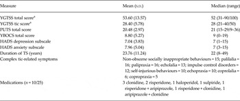
Twenty-five outpatients with uncomplicated TS and 25 healthy controls matched one-to-one for gender (6 females, 19 males) age (TS: mean = 31.48 years, s.d. = 11.50, median = 29, range = 17–59; controls: mean = 31.76, s.d. = 12.65, median = 28, range = 18–59) and education (TS: mean = 14.68 years, s.d. = 2.06, median = 15, range = 11–19; controls: mean = 14.64, s.d. = 1.95, median = 14, range = 11–19) took part. All were English first-language speakers with no history of head injury, seizure or substance abuse. Patients were recruited through a specialist outpatient clinic in Birmingham, UK, and Tourettes Action UK, and healthy controls were recruited through the Queen Elizabeth Hospital, Birmingham and University of Birmingham, UK. Controls had no psychiatric or neurological diagnoses and were not taking psychoactive medication. Patients (Table 1) had TS diagnosed by an experienced neurologist (A.E.C.) using DSM-IV-TR (APA, 2000) criteria, but no co-morbid psychiatric or neurological disorders as screened for using the National Hospital Interview Schedule for Tourette syndrome (Robertson & Eapen, Reference Robertson and Eapen1996), therefore the sample may be considered to comprise participants with ‘uncomplicated’ TS. There was no significant difference in age between the two groups (Wilcoxon–Mann–Whitney value = 274, p = 0.386). Group comparisons for performance on neuropsychological tests, which indicated limited differences on some tests assessing set-shifting, are reported in an earlier study (Eddy et al. Reference Eddy, Cavanna, Rickards and Hansen2016).
Patient group clinical characteristics

HADS, Hospital Anxiety and Depression Scale; PUTS, Premonitory Urge for Tics Scale; YBOCS, Yale–Brown Obsessive Compulsive Scale; YGTSS, Yale Global Tic Severity Scale.
a Measure was clinician rated. All other scales were self-report.
Protocol
The protocol received NHS approvals and all participants gave written informed consent. After neuropsychological assessment (see Eddy et al. Reference Eddy, Cavanna, Rickards and Hansen2016), participants were shown task instructions and example stimuli, before being made comfortable in the scanner.
Eyes tasks
The in-scanner task was based on the RMET (Baron-Cohen et al. Reference Baron-Cohen, Wheelwright, Hill, Raste and Plumb2001). This task contains 36 photographs of eyes surrounded by four mental state terms (e.g. interested, flirtatious, doubtful, preoccupied). One option is the conventional ‘correct’ answer as based on healthy control responses. We included a matched version that required judgements about physical rather than mental states (i.e. the age of the eyes), similar to previous studies (e.g. Moor et al. Reference Moor, Macks, Güroglu, Rombouts, Molen and Crone2012). However, our comparison stimuli differed from those used in the Moor et al. study as that study used a combination of age (older/younger) and gender (male/female) to create four options. The age version used in the current study was developed through testing 135 healthy participants (77 females, 58 males; mean age = 22.5 years, s.d. = 10.72; mean education = 13.5 years, s.d. = 1.47). First, 60 healthy participants estimated the approximate age of each pair of eyes. Sample means and modes were used to generate a ‘conventional’ answer for each photo. The three other forced-choice options were similar to the original task: one age option was close to the conventional answer (e.g. within 5 years) and two others were more different (e.g. within 8–20 years). The pilot age task was tested on a further 75 healthy participants along with the mental state version. Responses were reviewed periodically and the multiple choice options for the age version were adjusted, until the matched trials for age and mental state yielded a very similar number of errors. The 21 trial photographs with the most closely matched mean errors for the two versions comprised the final task (one practice item; original stimuli numbered 1, 2, 3, 5, 6, 7, 11, 13, 14, 15, 17, 18, 23, 25, 26, 27, 30, 31, 33, 34, 36; The stimuli set with age options is available on request). Participants completed four blocks containing ten trials (age; mental state; age; mental state). Instructions at the start of each block told participants to consider each photo and respond to the question mark cue by pressing one of four buttons, selecting the age/mental state that best matched the image. Each photo with four answer options was on screen for 10 s, and then replaced by a question mark cue for 2 s. Each trial was followed by a blank period with a fixation point in the middle of the screen (15.5 s) before the next photo appeared.
Data acquisition protocol
Sponge pads were used to reduce head movement, and patients were told the best time to tic was in between trials and scanning phases. Data were acquired during a single scanning session in a Philips Achieva 3.0 T MRI scanner (Philips Medical Systems, Eindhoven, The Netherlands) using an eight-channel head coil. Stimuli were presented using Presentation software version 14.9 (Neurobehavioral Systems, USA) which also recorded behavioural responses. One hundred and ten T2*-weighted gradient echo-planar imaging volumes were obtained for each of the four acquisition runs of the task. Scan protocol parameters were selected to achieve whole brain coverage (42 axial slices, obtained consecutively in a bottom up sequence) with TR = 2.5 s, TE = 35 ms, flip angle = 79°, SENSE factor = t, FOV 240 × 240 mm, acquisition matrix = 80 × 80, reconstructed to give isotropic voxels of size = 3 × 3 × 3 mm3. On completion of the task, high resolution T1-weighted gradient echo anatomical images were collected with 175 × 1 mm sagittal slices (TE = 3.8 ms, FOV = 288 × 232 × 175 mm, reconstructed to 1 × 1 × 1 mm3 isotropic voxels).
Neuroimaging analysis
Movement artefact (mean absolute movement across each run) was examined and participants were excluded if they had more than one run where they moved more than 1.5 mm (i.e. half a voxel or more). Exclusions left data from 23 patients and 24 controls. A comparison of maximum movement per block showed no significant group difference (p = 0.210).
Raw structural and functional data were converted from Philips PAR/REC format into NIfTI format. All data processing was carried out using FEAT v. 6.00, part of FSL v. 5.0.9 (Smith et al. Reference Smith, Jenkinson, Woolrich, Beckmann, Behrens, Johansen-Berg, Bannister, De Luca, Drobnjak, Flitney, Niazy, Saunders, Vickers, Zhang, De Stefano, Brady and Matthews2004). Processing steps included slice timing correction and MCFLIRT inter-volume motion correction using rigid-body transformations (Jenkinson et al. Reference Jenkinson, Bannister, Brady and Smith2002). Data were high-pass filtered using a Gaussian-weighted least-squares filter (sigma = 24 s), spatially smoothed using a 3D Gaussian kernel (FWHM = 5 mm) and grand-mean intensity normalized across the 4D dataset. Using FLIRT, the functional data were registered to their respective participant's T1 structural images using a 12-DOF linear transformation and to the standard template Montreal Neurological Institute (MNI) reference brain using a boundary-based registration transformation. A nonlinear FNIRT transformation with a warp resolution of 10 mm was used to register between participants’ T1 image and MNI space.
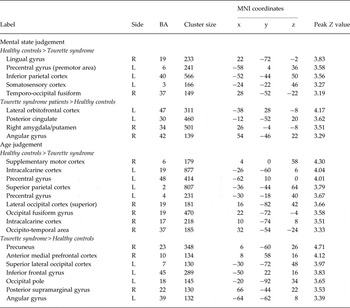
Button responses were modeled into the neuroimaging data as a covariate of no interest. The time-series for when each principal condition was active (10 s epochs) were convolved with a standard gamma-derived hemodynamic response function and high pass temporal filtering (sigma = 24 s) was applied to the model. The temporal derivatives of each of the two principal conditions were additionally added to the GLM in order to create a better fit for the overall model and reduce unexplained noise. Finally, the motion parameters generated by MCFLIRT were added to the overall GLM as separate regressors of no interest, in order to help reduce any residual uncorrected motion-related artifacts (Johnstone et al. Reference Johnstone, Ores Walsh, Greischar, Alexander, Fox, Davidson and Oakes2006). This model was used to generate the data for results (Tables 2 and 3) below, which show group activation differences for each version of the task.
Eyes task mental state judgements group activation differences

BA, Brodmann area; MNI, Montreal Neurological Institute.
Threshold z > 2.1; cluster size ≥130; p < 0.05 corrected.
Brodmann areas are approximate.
Group Z statistic images from these models were subsequently corrected for multiple comparisons by means of a two-step family-wise error (FWE) correction process in order to control for false positives. The AlphaSim program, part of the AFNI toolkit (Cox, Reference Cox1996), was used to control the FWE rate. A particular voxel-wise threshold was chosen and, together with the voxel dimensions and spatial smoothing kernel size used in the fMRI analysis, the probability of a cluster of specific size arising by chance was estimated by means of a Monte Carlo simulation. All data are reported here with FWE corrected p < 0.05, equivalent to a voxel-wise threshold of Z > 2.1 and cluster size ≥130.
Finally, we previously scanned this sample of patients during a ToM task involving false belief, and found that right TPJ activity covaried with echophenomena, impulsivity ratings and urges to tic; left TPJ activity covaried with socially inappropriate urges; and amygdala activity covaried with premonitory urges (Eddy et al. Reference Eddy, Cavanna, Rickards and Hansen2016). We therefore examined whether similar relationships were apparent for the RMET, using the same TPJ masks based on healthy control data from this previous study, and masks for the amygdalae based on the Harvard-Oxford Atlas. Symptom ratings were for lifetime tic severity on the Yale Global Tic Severity Scale (YGTSS; Leckman et al. Reference Leckman, Riddle, Hardin, Ort, Swartz, Stevenson and Cohen1989); tic urge severity on the Premonitory Urge for Tics Scale (PUTS; Woods et al. Reference Woods, Piacentini, Himle and Chang2005); obsessive-compulsive behaviours on the Yale–Brown Obsessive Compulsive Scale (YBOCS; Storch et al. Reference Storch, Larson, Goodman, Rasmussen, Price and Murphy2010); non-obscene socially inappropriate symptoms ratings (scored 0–3 based on 0 = absent or 1/2/3 of insults; other remarks; actions); impulse control disorders according to Minnesota Impulsive Disorders Interview (MIDI total count; Christenson et al. Reference Christenson, Faber and deZwaan1994) and echophenomena ratings (scored 0–2 based on 0 = absent or 1/2 of echolalia; echopraxia). Using the nlme package in R (https://CRAN.R-project.org/package=nlme), one mixed-effects model was fitted for each of the four masks, with percentage blood oxygen-level-dependent signal change as the dependent variable, participant identity as a random factor, and the six symptom measures as initial covariates in a stepwise backwards elimination to create the minimal adequate model where all surviving symptom measures were significant.
Ethical standards
The authors assert that all procedures contributing to this work comply with the ethical standards of the relevant national and institutional committees on human experimentation and with the Helsinki Declaration of 1975, as revised in 2008.
Results
Behavioural performance
Behavioural data are shown in Supplementary Table S1. Each of the 47 participants included in neuroimaging analysis completed 20 trials for each version of the eyes task. There were four missing responses. All responses within 5 s following the question mark cue were included, yielding 1790 (95.2%) valid responses out of 1880. Patients and controls showed no significant differences on either the mental state version (χ2 = 2.784, df = 1, p = 0.0952) or the age judgement version (χ2 = 0.0125, df = 1, p = 0.911) of the task in terms of accuracy. For reaction times, a mixed-effects model was run with reaction time regressed against dependent variables of population group, task condition and error status (whether the trial was correct or not). This indicated no significant effect of group (F 45,1 = 1.4657, p = 0.232) or task (F 1737,1 = 0.3586, p = 0.549) but correct responses were faster (F 1737,1 = 37.964, p < 0.0001) by approximately 131 ms on average.
Neuroimaging data
In healthy controls, the standard (mental state judgement) eyes task activated similar brain regions (Supplementary Table S2) to those reported in previous studies, including posterior superior temporal gyrus (STG) and inferior frontal gyrus (IFG) (e.g. Moor et al. Reference Moor, Macks, Güroglu, Rombouts, Molen and Crone2012; Schurz et al. Reference Schurz, Radua, Aichhorn, Richlan and Perner2014). The age judgement task led to activity in many regions overlapping with the mental state version (e.g. occipital, inferior frontal and anterior cingulate cortices), plus additional activity in areas such as middle frontal and dorsolateral prefrontal cortex (Supplementary Table S3). The general effects of task appear in line with those of Harris & Fiske (Reference Harris and Fiske2007), who used a different picture-based task in healthy participants and observed more superior frontal, middle temporal and parahippocampal activation for judging mental state (preference) and greater activity in bilateral precentral gyrus, middle frontal gyrus and insula for age judgements.
For the standard version of the eyes task (Table 2, Supplementary Fig. S1), patients with TS showed greater activity than controls in the left lateral OFC, posterior cingulate, right angular gyrus, and a cluster spanning the right amygdala and putamen. The opposite contrast revealed reduced activation (to baseline) in areas including the lingual gyrus, precentral gyrus and left inferior parietal cortex in TS.
During the age judgement version of the task (Table 2, Supplementary Fig. S1), patients with TS exhibited more activity than controls in precuneus, medial prefrontal cortex, left IFG, angular and supramaginal gyri, and regions within left occipital cortex. Healthy controls showed greater activation than patients in areas including left precentral gyrus, right supplementary motor cortex, left superior parietal cortex, left and right intracalcarine cortex and right occipital areas.
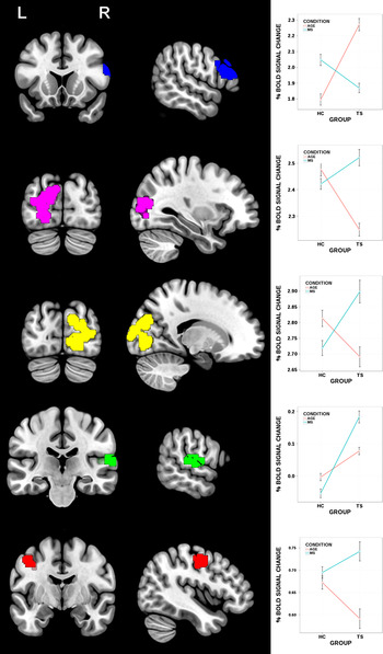
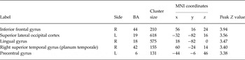
Statistically significant differences relating to the interaction between group and version of the eyes task are shown in Table 3. Differential activity was apparent in TS in right IFG, right STG (planum temporale), left precentral gyrus and bilateral occipital areas. Interaction plots are shown in Fig. 1. For the right IFG, brain activity increased more in TS for age judgement than mental state judgement, which is the opposite pattern to controls. For the other four regions, activity increases in TS are greater for the mental state version and smaller in the age version, with less difference between the two versions of the task in controls.
Interaction plots for the five areas showing an interaction between type of eyes task (mental state judgement or age judgement) and group. Brain regions correspond to Table 3. From the top downwards: right inferior frontal gyrus in blue; left superior lateral occipital cortex in pink; right lingual gyrus in yellow; right superior temporal gyrus (plaum temporale) in green; left precentral gyrus in red. AGE, Age judgement task (red line); HC, healthy controls; MS, mental state judgement task (blue line); TS, Tourette syndrome.

Interaction between task (mental state/age judgement) and group (Tourette syndrome; healthy controls)

BA, Brodmann area; MNI, Montreal Neurological Institute.
Threshold z > 2.1, cluster size ≥130; p < 0.05 corrected.
Brodmann areas are approximate.
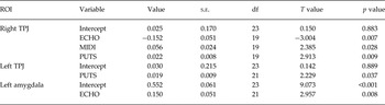
Finally, covariate analysis was performed on the left and right TPJ, and left and right amygdalae, based on previous findings (Eddy et al. Reference Eddy, Cavanna, Rickards and Hansen2016). One mixed-effects model was generated for each of the four masks for activity across both versions of the eyes task, with six symptom ratings as listed previously. Three regions (right TPJ, left TPJ and left amygdala) showed significant covariation between at least one symptom measure and activity across the eyes task (Table 4). Right TPJ activity covaried negatively with echophenomena, but positively covaried with premonitory urges and impulse control disorders. Activity in left TPJ covaried positively only with urges to tic, and left amygdala activity covaried positively with echophenomena ratings.
Significant models from covariate analysis for the Tourette syndrome group

ECHO, Echo-phenomena rating, number of types; MIDI, Minnesota Impulsive Disorders Interview, number of disorders; PUTS, Premonitory Urge for Tics Scale total score; TPJ, Temporo-parietal junction.
Discussion
During mental state recognition on the standard version of the eyes task, adults with TS exhibited greater activity than healthy controls in left OFC, posterior cingulate, right amygdala and putamen, and right TPJ. Reduced activity was apparent in TS in areas including left inferior parietal cortex. Age judgement elicited other group differences, including greater activity in TS within precuneus, medial prefrontal and temporal regions frequently implicated in ToM. The exact abilities and processes assessed by the RMET are still debated. For example, one recent study (Oakley et al. Reference Oakley, Brewer, Bird and Catmur2016) found that alexithymia (i.e. difficulties in interpreting and explaining one's own emotions) can be more closely related to impairments on the RMET than a diagnosis of autistic spectrum disorder, emphasizing the importance of emotional processes. However, as a previous study found no evidence of elevated rates of alexithymia in TS (Eddy et al. Reference Eddy, Macerollo, Martino and Cavanna2015), differential neural activity in TS in response to the eyes stimuli may be related to group differences in empathy or emotional contagion, complementing existing reports of neural differences on traditional tests of ToM (e.g. Eddy et al. Reference Eddy, Cavanna, Rickards and Hansen2016).
Both the OFC and amygdala are associated with operant conditioning and learning about aversive outcomes (e.g. Schoenbaum et al. Reference Schoenbaum, Chiba and Gallagher1998). More specifically, the left OFC is linked to recognition of negative emotions such as fear, anger and disgust (e.g. Sprengelmeyer et al. Reference Sprengelmeyer, Rausch, Eysel and Przuntek1998; Wicker et al. Reference Wicker, Keysers, Plailly, Royet, Gallese and Rizzolatti2003) and awareness of threatening social interactions (e.g. Sugiura et al. Reference Sugiura, Wakusawa, Sekiguchi, Sassa, Jeong, Horie, Sato and Kawashima2009). Greater OFC activity is seen in healthy adults when they control behavioural responses which are naturally incompatible with emotional facial expressions, e.g. approach responses towards angry faces (Roelofs et al. Reference Roelofs, Minelli, Mars, van Peer and Toni2009). More precisely, the left OFC peak in the current study matches that differentially activated for deceptive v. cooperative interactions in healthy participants (Lissek et al. Reference Lissek, Peters, Fuchs, Witthaus, Nicolas, Tegenthoff, Juckel and Brüne2008). This is notable given that patients with TS are more likely than controls to report accidental anti-social remarks as being motivated by negative intent (Eddy et al. Reference Eddy, Mitchell, Beck, Cavanna and Rickards2010a , Reference Eddy, Mitchell, Beck, Cavanna and Rickards2011). Increased activity in a brain area linked to the processing of negative emotional interactions could imply that being prompted to consider mental states has aversive associations in TS, perhaps because this could make patients more aware of unpleasant social attention due to tics. However, patients with TS also experience more personal distress than controls when witnessing other people's negative emotional experiences (Eddy et al. Reference Eddy, Macerollo, Martino and Cavanna2015), so increased reactivity to other people's emotions in general may be a more parsimonious interpretation of our findings.
Patients with TS also exhibited differential activity in a cluster encompassing right amygdala and putamen during prompted mental state recognition. Previous studies have reported amygdala dysfunction during emotional face processing in TS (Neuner et al. Reference Neuner, Kellermann, Stöcker, Kircher, Habel, Shah and Schneider2010). The right amygdala has been linked to subconscious processing of emotional expressions (Pegna et al. Reference Pegna, Khateb, Lazeyras and Seghier2005) and shows enhanced activation when social phobics view emotional expressions (Bertolino et al. Reference Bertolino, Arciero, Rubino, Latorre, De Candia, Mazzola, Blasi, Caforio, Hariri, Kolachana, Nardini, Weinberger and Scarabino2005). As well as supporting the suggestion that tics could involve orbitofrontal or amygdala reactivity to environmental cues (Eddy & Cavanna, Reference Eddy and Cavanna2013b ), these findings further raise the possibility that associated putamen activation could link emotional cues to motor responses, or perhaps prompt tics. In other words, the cardinal motor signs of TS (i.e. tics), may at least partly reflect underlying changes in limbic activity. However, as tic occurrence was not measured during scanning further research is needed to confirm this possibility.
Activity in the left amygdala was found to covary strongly with echophenomena ratings, while activity in left and right TPJ covaried with ratings of premonitory urges, and right TPJ activity was also linked to echophenomena and impulsive behaviours (similar findings for right TPJ were recently reported using a story-based ToM task; Eddy et al. Reference Eddy, Cavanna, Rickards and Hansen2016). More specifically, activity in right TPJ covaried negatively with echophenomena ratings, but positively with tic urges and impulse control problems. Right TPJ activity is reduced in association with greater self-other blending (Cheng et al. Reference Cheng, Chen, Lin, Chou and Decety2010). Therefore our findings are in line with an association between increased self-other blending and echophenomena. The positive relationship between right TPJ activity, tic urges and impulsive behaviours is harder to interpret, but may suggest these symptoms are linked to achieving a state of self-other distinction (Eddy, Reference Eddy2016). In summary, during tasks involving social cognition, activity within medial and lateral temporal regions in TS covaries with core symptoms (i.e. premonitory urges) and characteristics that may be related to social cognition (i.e. echophenomena).
Individuals with TS exhibited less activity than controls in left inferior parietal cortex during prompted mental state recognition. This region may contribute to motor simulation when understanding action goals and related mental states (e.g. Cerri et al. Reference Cerri, Cabinio, Blasi, Borroni, Iadanza, Fava, Fornia, Ferpozzi, Riva, Casarotti, Martinelli Boneschi, Falini and Bello2015). Moreover, there is stronger left inferior parietal activation when healthy participants imagine themself carrying out an action, and reduced activity in this area when imagining another person as the acting agent (Ruby & Decety, Reference Ruby and Decety2001). Differential inferior parietal activity in patients during mental state recognition could therefore further reflect differences in attributing agency for the observed actions or emotions. Problems disentangling the actions of one-self and another person could help to explain echophenomena. Furthermore, difficulties in disciminating between actor and observer when perceiving other people's negative emotions could lead to unpleasant emotions being over-attributed to the self, helping to explain increased personal distress in TS (Eddy et al. Reference Eddy, Macerollo, Martino and Cavanna2015).
The precuneus is frequently associated with social cognition (Cavanna & Trimble, Reference Cavanna and Trimble2006) and exhibits increased activation for third-person v. first-person simulation in healthy participants (Ruby & Decety, Reference Ruby and Decety2001). The posterior cingulate and precuneus showed greater activity in TS during both versions of the eyes task, perhaps indicating a predisposition towards ToM. Patients with TS also exhibited more activity than controls in medial prefrontal and posterior temporal areas frequently implicated in ToM specifically during age judgements. These findings support the suggestion that people with TS spontaneously hyper-mentalize (Eddy & Cavanna, Reference Eddy and Cavanna2015).
Group and task interactions indicated that while controls showed little difference in premotor activity across each eyes task, activity in left precentral gyrus increased more in TS during mental state judgement, but increased less than in controls for age judgement. This further links social cognition to motor activity in TS. In addition, controls showed similar increases in occipital activity for each task version, whereas in TS a greater increase was seen during mental state recognition, with less of an increase for age judgement. This could reflect comparatively greater attention and visual processing in TS specifically during mental state recognition.
The right IFG exhibited more of an increase in activity in TS during age judgement than mental state judgement, whereas the opposite pattern was apparent in controls. Right IFG has been implicated in the control of impulsive motor responses (Aron et al. Reference Aron, Robbins and Poldrack2004, Reference Aron, Robbins and Poldrack2014) and inhibition of distracting emotional stimuli (Mitchell et al. Reference Mitchell, Luo, Mondillo, Vythilingam, Finger and Blair2008). Indeed, response inhibition and aversive emotional stimulus processing may combine additively in IFG (Brown et al. Reference Brown, Lebel, Dolcos, Wilman, Silverstone, Pazderka, Fujiwara, Wild, Carroll, Hodlevskyy, Zedkova, Zwaigenbaum, Thompson, Greenshaw and Dursun2012). Therefore differential activity in this area in TS could reflect efforts to control emotional or motor responses elicited by the eyes stimuli. For example, perhaps greater right IFG engagement during age judgement may underpin patients’ efforts to reduce interference linked to emotion processing. As there were no significant behavioural differences between the groups for either version of the eyes task, at least some of our findings could reflect activation patterns which enabled adults with TS to maintain good task performance. If the pattern of right IFG activity across the two versions of the eyes test in TS did relate to attempts to control hyper-mentalizing or emotion processing, one interpretation is that this reflects a compensation mechanism.
The right STG (planum temporale) also showed in the condition and group interaction, with greater activity in TS, especially during mental state recognition. The right STG is activated by the standard version of the eyes task (e.g. Gallagher & Frith, Reference Gallagher and Frith2003) and may play a role in inferring meaning from gaze direction and facial expression (Allison et al. Reference Allison, Puce and McCarthy2000). However, the left planum temporale is implicated in speech and language comprehension (e.g. Sommer et al. Reference Sommer, Aleman, Somers, Boks and Kahn2008). In the current study, mental state options were presented as words whereas age options were numbers. Therefore the possibility that this interaction effect could be related to a group difference in hemisphere specialization for language cannot be ruled out. Indeed, the contribution of the right hemisphere to language processing in TS, and a possible relationship with symptoms such as coprolalia, is worthy of investigation.
Limitations of the current study include that the age judgement task was newly developed, and although some of our findings are in accordance with previous studies using a similarly designed task (e.g. Moor et al. Reference Moor, Macks, Güroglu, Rombouts, Molen and Crone2012), caution is needed in interpretation. For example, it would be naive to assume that emotional expressions would definitely not elicit some degree of automatic emotional processing even when judging age (e.g. Wagenbreth et al. Reference Wagenbreth, Rieger, Heinze and Zaehle2014). In addition, our ability to assess the link between tics, brain activity and social cognition is limited because we did not assess tics or urges during scanning. Furthermore, our patient sample was restricted to adults with moderate tic severity and no diagnosed co-morbid disorders. The prevalence of co-morbidities in TS can be as high as 90% (Cavanna & Rickards, Reference Cavanna and Rickards2013), although the rate may be lower in adults, or in community samples containing less severe cases (Scharf et al. Reference Scharf, Miller, Mathews and Ben-Shlomo2012). Despite limiting generalizability, studying patients without co-morbid disorders may help determine whether TS per se is likely to explain differences to healthy controls. One important point is that studies of social cognition in children with TS are lacking, and would offer further insight into the role of social cognition in this disorder.
In conclusion, TS is associated with greater activity in neural regions important for ToM when visual cues to mental state reasoning are available but are not required by the task. Furthermore, during prompted mental state recognition these patients exhibit reduced activity in brain regions involved in attributing agency alongside greater activity than healthy controls in areas that process negative emotion. Increased susceptibility to emotive social cues could help explain why patients experience elevated personal distress in interpersonal situations, and why tics worsen with negative emotions and social stress. Right IFG activations may reflect patients’ attempts to control their emotional reactions and related motor responses. Our findings once again highlight the right TPJ as an area of interest in relation to core symptoms of TS. Future studies seeking to clarify the precise relationship between tics, social cognition and emotional reactivity will make a unique and important contribution to our understanding of this neurodevelopmental condition.
Supplementary material
The supplementary material for this article can be found at https://doi.org/10.1017/S0033291716002725.
Acknowledgements
This study was unfunded. Scanning costs were met in house by Birmingham University Imaging Centre, UK: This was a BUIC development project. We are grateful to Nina Salman for assistance with MRI data collection, to Tourettes Action UK for publicizing the study, and to all of our participants. We also thank Birmingham University Imaging Centre for facilitating this study.
Declaration of Interest
None.