Introduction
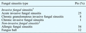
Orbital involvement is a feature of both invasive and non-invasive fungal sinusitis. Invasive fungal sinusitis, which occurs in both acute (i.e. fulminant) and chronic forms, is commonly caused by Aspergillus or Mucor species. Chronic invasive fungal sinusitis includes chronic invasive (non-granulomatous) and chronic granulomatous fungal sinusitis. The non-invasive form includes allergic fungal sinusitis and fungus ball. Histopathology and fungal culture results usually enable a conclusive diagnosis of the type of fungal sinusitis (Table I).
Characteristic histological features of fungal sinusitis types

Orbital involvement in both invasive and non-invasive fungal sinusitis can range from mere asymptomatic erosion of the ipsilateral lamina papyraceae to complete and permanent visual loss.Reference Dunlop and Billson1–Reference Aribandi, McCoy and Bazan20 Among patients with non-invasive fungal sinusitis, orbital involvement is far more common in those with allergic fungal sinusitis compared with fungus ball.
In patients with allergic fungal sinusitis, the prevalence of orbital involvement ranges from 14.7 per centReference Nussenbaum, Marple and Schwade8 to 60 per cent.Reference Manning, Merkel, Kriesel, Vuitch and Marple5 Proptosis, periorbital swelling, hypertelorism, epiphora and, rarely, visual loss may be encountered in patients with allergic fungal sinusitis. In patients with a fungus ball, orbital involvement is rare, and not often discussed in the literature.Reference Ferguson9, Reference Klossek, Serrano, Peloquin, Percodani, Fontanel and Pessey10
Studies of invasive fungal sinusitis cases have shown a high prevalence of clinical evidence of orbital involvement, up to 100 per cent in some series.Reference Bhansali, Bhadada, Sharma, Suresh, Gupta and Singh11, Reference Ferry and Abedi15, Reference Yohai, Bullock, Aziz and Markert16, Reference Dhiwakar, Thakar and Bahadur18 These are invariably cases of acute invasive fungal sinusitis with zygomycosis. The clinical features of orbital involvement in these patients include periorbital swelling, proptosis and limitation of extra-ocular muscle movement. Similar findings may occur in acute invasive fungal sinusitis due to Aspergillosis and in chronic invasive fungal sinusitis.Reference Veress, Malik, el-Tayeb, el-Daoud, Mahgoub and el-Hassan19, Reference deShazo, O Brien, Chapin, Soto-Aquilar, Gardner and Swain21
Orbital involvement is frequently encountered in different types of fungal sinusitis, but has not been extensively studied. Most previously published case reports or series have described orbital involvement related to only one particular type of fungal sinusitis.Reference Dunlop and Billson1–Reference Meyer and Nagi3, Reference Ghegan, Lee and Schlosser7, Reference Nussenbaum, Marple and Schwade8, Reference Klossek, Serrano, Peloquin, Percodani, Fontanel and Pessey10, Reference Bhansali, Bhadada, Sharma, Suresh, Gupta and Singh11, Reference Nithyanandam, Jacob, Battu, Thomas, Correa and D'Souza13–Reference Yohai, Bullock, Aziz and Markert16, Reference Dhiwakar, Thakar and Bahadur18, Reference Veress, Malik, el-Tayeb, el-Daoud, Mahgoub and el-Hassan19
To the best of our knowledge, this is the first report offering a comparative analysis of orbital involvement in a series of patients with invasive and non-invasive fungal sinusitis, and documenting the relative prevalence and extent of orbital involvement in these two groups.
Patients and methods
Patients
A total of 100 consecutive patients with biopsy-proven and culture-positive fungal sinusitis seen between 2005 and 2009, for whom complete data were available, were included in the study. Standard histological criteria were applied to establish a conclusive diagnosis (Table I). All patients were evaluated by, and treated either primarily or secondarily in, our ENT department.
Institutional review board approval was obtained prior to commencing the study.
Clinical and pathological evaluation
All patients had a detailed ENT examination, including rigid nasal endoscopy. Clinical evidence of orbital involvement was assessed by testing for visual acuity and fields, fundus examination, proptosis measurement, and testing of extra-ocular muscle movements. In acutely sick and non-ambulant patients, testing for ocular involvement was performed at the bedside as follows. Visual acuity was tested by finger counting at 1 m, perception of light was checked using a torchlight, the range and extent of extra-ocular muscle movements were checked with adequate lighting and by an experienced examiner, and the presence of proptosis and periorbital swelling was noted.
The presence of comorbidities, such as diabetes mellitus, immunosuppression and bronchial asthma, was noted. Intra-operatively or pre-operatively obtained specimens of diseased nasal mucosa, polyps and debris were sent for histopathological examination and fungal smear and culture. For patients with suspected allergic fungal sinusitis, polyps and allergic mucin were sent as two separate specimens, as detailed previously.Reference Rupa, Jacob and Mathews22 Specimens were stained with haematoxylin and eosin as well as Grocott's methenamine silver fungal stain to delineate fungal hyphae.
Radiological evaluation
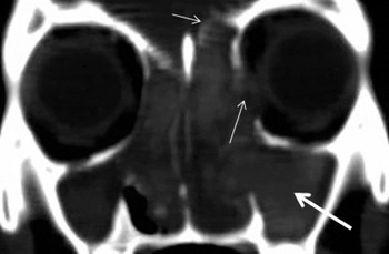
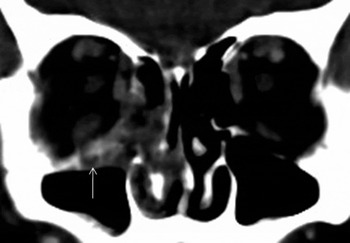
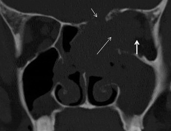
In patients with suspected allergic fungal sinusitis or fungus ball, a computed tomography (CT) scan of the paranasal sinuses (osteomeatal complex view) with coronal cuts was performed (Figures 1–3). In patients with suspected invasive fungal sinusitis, a contrast-enhanced CT scan of the paranasal sinuses with axial and coronal cuts was performed. All CT images of the paranasal sinuses of each patient were reviewed and the presence or absence of specific features noted.
Coronal computed tomography scan of patient with acute fungal sinusitis (AFS), showing erosion of the roof of the orbit (short, thin arrow) and lamina papyracea (long, thin arrow); note hyperdense areas (thick arrow) amid soft tissue thickening of nasal cavity and sinuses bilaterally, typical of AFS.

Coronal computed tomography scan of patient with acute invasive fungal sinusitis, showing soft tissue thickening of the right ethmoid sinus with erosion of the floor of the orbit (arrow); note infiltration of adjacent right inferior rectus and septal perforation.

Coronal computed tomography scan of patient with chronic granulomatous fungal sinusitis, showing large, homogeneous, infiltrative mass involving the left nasal cavity, middle turbinate and ethmoids and eroding the lamina papyracea (long, thin arrow), roof of the nose (short, thin arrow), septum and floor of the orbit, with invasion of extra-ocular muscles and compression of the optic nerve (short, thick arrow).

In some cases of invasive fungal sinusitis, magnetic resonance imaging (MRI) was performed to detect any involvement of the dura, cavernous sinus and other vascular structures. Patients with invasive and non-invasive fungal sinusitis were compared and orbital involvement noted both clinically and radiologically. Similar analysis was also carried out for patients with acute invasive fungal sinusitis and chronic invasive fungal sinusitis.
Statistical analysis
Frequencies and percentages of categorical variables were noted. Association between categorical variables was assessed using the chi-square and Fisher's exact tests. A p value of less than 0.05 was considered statistically significant. All statistical analyses were performed using the Statistical Package for the Social Sciences version 16.0 software program.
Results
Patient demographics
Of a total of 100 patients, 34 had invasive fungal sinusitis and 66 had non-invasive fungal sinusitis (Table II). Allergic fungal sinusitis was the most common diagnosis (54 per cent). The majority of patients were aged over 25 years in both the invasive (88.2 per cent) and non-invasive (87.8 per cent) fungal sinusitis groups. There were more female (69.7 per cent) than male (30.3 per cent) patients in the non-invasive compared with the invasive fungal sinusitis group, which had 73.5 per cent males and 26.5 per cent females. This difference was statistically significant (p < 0.001).
Distribution of invasive and non-invasive fungal sinusitis*

* n = 100; †n = 34; ‡n = 66. Pts = patients
Comorbidities
Patients with invasive fungal sinusitis were found to have comorbidities such as diabetes mellitus (70.5 per cent) and immunosuppression (14.7 per cent). In contrast, of those with non-invasive fungal sinusitis, only 11 (16.7 per cent) had diabetes mellitus and two (3 per cent) were immunosuppressed.
Clinical orbital involvement
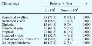
Orbital involvement was more common among patients with invasive fungal sinusitis (73.5 per cent) compared with those with non-invasive fungal sinusitis (12.1 per cent), and this difference was statistically significant (p = 0.000) (Table III). More patients with chronic than with acute invasive fungal sinusitis had orbital involvement clinically, although visual loss was seen in only 25 per cent of those with chronic invasive fungal sinusitis compared with 48 per cent of those with acute invasive fungal sinusitis.
Orbital involvement in invasive vs non-invasive fungal sinusitis: clinical evidence*

* n = 100; †n = 34; ‡n = 66. Inv = invasive; FS = fungal sinusitis; Non-inv = non-invasive; EOM = extra-ocular muscle; OA = optic atrophy
In patients with allergic fungal sinusitis, in whom the prevalence of orbital involvement was 14.8 per cent, periorbital swelling and proptosis were the commonest manifestations. Visual loss was seen in only three patients. The clinical presentation and ocular function in each of these three patients following treatment was unique. Two patients presented with visual loss of 7 and 8 days' duration recovered their vision over a period of 2 weeks and 3 months respectively following surgery and steroid therapy. The third patient, who presented one month after rapidly progressive visual loss, did not recover following similar therapy.
Patients with a fungus ball did not have any orbital involvement, except for one patient who presented with orbital apex syndrome. This was a 72-year-old man with diabetes who experienced progressive deterioration of vision in the left eye over one month. He was found to have a soft tissue mass in the left posterior ethmoid, adjacent sphenoid and orbital apex on CT scanning and MRI. Endoscopic excision of the mass showed polypoid mucosa covering a fungus ball. The histopathological appearance was of a mass of fungal filaments (characterised on fungal culture as Aspergillus flavus) with polypoid, inflamed sinus mucosa but without invasion. He developed rapidly progressive, diffuse pneumonia post-operatively and died soon after.
Radiological orbital involvement
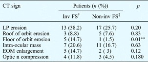
Comparing radiological findings in patients with invasive and non-invasive fungal sinusitis, it was found that erosion of the lamina papyraceae and the roof of the orbit was more prevalent in invasive (38.2 per cent) than non- invasive (25.7 per cent) fungal sinusitis, but this was not statistically significant (p = 0.20). (Table IV and Figures 1 and 3). Erosion of the floor of the orbit, however, was more common in patients with invasive fungal sinusitis (p = 0.01) (Figure 2). Overall, radiological differences between invasive and non-invasive fungal sinusitis cases were less remarkable compared with their clinical and histopathological differences.
Orbital involvement in invasive vs non-invasive fungal sinusitis: radiological evidence*

* n = 100; †n = 34; ‡n = 66. **Significant. CT = computed tomography; Inv = invasive; FS = fungal sinusitis; Non-inv = non-invasive; LP = lamina papyracea; EOM = extra-ocular muscle; n = nerve
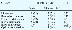
When cases of acute invasive fungal sinusitis and chronic invasive fungal sinusitis were compared radiologically, intra-ocular muscle enlargement (p = 0.01) and optic nerve compression (p = 0.003) were more commonly seen in those with chronic disease (Table V, Figure 3). Radiological evidence of orbital involvement was seen in all but one patient with chronic invasive fungal sinusitis and ocular symptoms. In contrast, no radiological evidence was found in four (16 per cent) patients with acute invasive fungal sinusitis who had clinical evidence of optic atrophy and/or papilloedema.

* Chronic invasive fungal sinusitis includes chronic granulomatous fungal sinusitis.
† n = 34; ‡n = 25; **n = 9. §Significant. CT = computed tomography; IFS = invasive fungal sinusitis; LP = lamina papyracea; EOM = extra-ocular muscle; n = nerve
Discussion
When proposing a classification for invasive fungal sinusitis, deShazo et al. Reference deShazo, O Brien, Chapin, Soto-Aquilar, Gardner and Swain21 noted that invasive and non-invasive fungal sinusitis had a number of features in common. Included among these were the facts that both diseases could occur in immunocompetent and immunodeficient individuals, both could have an acute or chronic course, and both could extend to the orbit and brain. However, the authors did not specifically comment on orbital involvement in both types of disease.
The results of our study show that patients with non-invasive fungal sinusitis had a lesser degree of clinically evident orbital involvement than those with invasive fungal sinusitis, and that this difference was statistically significant (p = 0.00).
Clinical orbital involvement in non-invasive vs invasive fungal sinusitis
Proptosis, periorbital swelling, reduced vision, diplopia, restriction of extra-ocular muscle movement and optic atrophy were seen with much greater frequency in those with invasive compared with non-invasive fungal sinusitis in the present study.
The low prevalence of orbital involvement in our non-invasive fungal sinusitis patients (12.1 per cent) is comparable with the figures from other case series of allergic fungal sinusitis.Reference Mukherji, Figueroa, Ginsberg, Zeifer, Marple and Alley6, Reference Nussenbaum, Marple and Schwade8 The high prevalence of orbital involvement (73.5 per cent) in our invasive fungal sinusitis patients is also similar to that of other series.Reference Bhansali, Bhadada, Sharma, Suresh, Gupta and Singh11, Reference Nithyanandam, Jacob, Battu, Thomas, Correa and D'Souza13, Reference Dhiwakar, Thakar and Bahadur18, Reference Veress, Malik, el-Tayeb, el-Daoud, Mahgoub and el-Hassan19
In our study, when specific subcategories of fungal sinusitis were evaluated, the prevalence of orbital involvement appeared to be greatest for those with chronic granulomatous fungal sinusitis (88.9 per cent). In contrast, Panda et al. Reference Panda, Balaji, Chakrabarti, Sharma and Reddy12 found that proptosis was found with a comparable frequency among patients with non-invasive aspergillus sinusitis (41.6 per cent) and those with chronic granulomatous fungal sinusitis (50 per cent). This is probably because the authors subcategorised the non-invasive variety into a non-invasive destructive group, in which bone erosion (without histological invasion) was present, and a group without bone erosion.
Radiological involvement in non-invasive vs invasive fungal sinusitis
Some authors have reported that up to 56 per cent of patients with allergic fungal sinusitis show orbital or skull base erosion.Reference Ghegan, Lee and Schlosser7 Others, studying individuals with invasive fungal sinusitis, have described erosion of the lamina papyraceae in 100 per cent of patients.Reference Dhiwakar, Thakar and Bahadur18
In the present study, comparing invasive with non-invasive fungal sinusitis, we found that radiological evidence of orbital involvement did not differ significantly between these two categories, with the exception of erosion of the floor of the orbit, which was more common in invasive fungal sinusitis (p = 0.01) (Table IV, Figure 2).
Bony erosion in invasive fungal sinusitis occurs through actual invasion or vascular necrosis of underlying bone. In contrast, in non-invasive fungal sinusitis, bony erosion occurs only in long-standing cases through a process of expansion and progressive thinning of the underlying bone.
Clinical orbital involvement in different categories of non-invasive fungal sinusitis
Among patients with non-invasive fungal sinusitis, those with allergic fungal sinusitis tend to have greater orbital involvement than those with a fungus ball. In a large series of 82 patients with allergic fungal sinusitis, Marple et al. Reference Marple, Gibbs, Newcomer and Mabry2 found an overall incidence of ocular involvement of 18.3 per cent. Telecanthus was the most common symptom (found in 7.3 per cent), followed by diplopia. Three patients had visual loss (3.7 per cent).
In the present study we found a comparable overall prevalence of orbital involvement in allergic fungal sinusitis of 14.8 per cent. The most common features of orbital involvement in these patients were periorbital swelling (14.8 per cent), impaired vision (5.5 per cent) and proptosis (9.1 per cent).
Proptosis and periorbital swelling are common features of allergic fungal sinusitis and have been reported in as many as 50 per cent of patients in some series.Reference Manning, Merkel, Kriesel, Vuitch and Marple5 In our study, the second most common orbital sign, on examination of patients with allergic fungal sinusitis, was proptosis (11.1 per cent). In an earlier study we found the prevalence of proptosis to be higher at 37.5 per cent.Reference Rupa, Jacob, Mathews, Kurian, Job and Chandi4 Proptosis, which is due to a mass effect produced by the expanding polyps and allergic mucin, resolves with surgical debridement of the sinuses and post-operative oral steroid therapy.
Visual loss is an unusual feature of non-invasive fungal sinusitis. The prevalence of visual loss in some series varies from 3.7 to 10 per cent.Reference Marple, Gibbs, Newcomer and Mabry2, Reference Manning, Merkel, Kriesel, Vuitch and Marple5 Visual loss in allergic fungal sinusitis is most often due to pressure on the optic nerve by expanding polyposis and allergic mucin. It is therefore reversible and often resolves with appropriate decompression along with oral steroid therapy. The addition of systemic steroid therapy has an additive effect to that of decompressive surgery in some cases.Reference Dunlop and Billson1 In others, the timing of surgery may influence the final outcome.
Marple et al. Reference Marple, Gibbs, Newcomer and Mabry2 described three patients with visual loss in their series of 82 patients with allergic fungal sinusitis. These authors rightly suggested that delayed presentation often led to incomplete recovery because of delayed intervention. This was also amply illustrated by one of the patients in our series.
Rarer presentations of allergic fungal sinusitis include restriction of ocular movementsReference Dunlop and Billson1 and subperiosteal abscess formation.Reference Meyer and Nagi3 Only two patients in our series had restriction of ocular movements. The probable cause for this phenomenon is mechanical or inflammatory oedema of the extra-ocular muscles, as invasion of extra-ocular muscles does not occur in allergic fungal sinusitis.
Visual impairment in patients with a fungus ball is unusual.Reference Ferguson9, Reference Klossek, Serrano, Peloquin, Percodani, Fontanel and Pessey10 In our study of patients with a fungus ball, the sole patient with ocular symptoms had a very unusual presentation, with orbital apex syndrome. The only other case report of fungus ball causing visual loss was that of Thiagalingam et al. Reference Thiagalingam, Fernando, Tan, O'Donnell, Weeks and Branley14 who reported a case of a fungus ball with associated acute invasive fungal sinusitis in the sphenoid sinus causing orbital apex syndrome.
Radiological orbital involvement in different categories of non-invasive fungal sinusitis
Radiological evidence of orbital involvement in non-invasive fungal sinusitis may manifest early as erosion of the lamina papyracea or as an intra-ocular mass. The roof and floor of the orbit are less commonly eroded (Table IV). As the ethmoid sinus is the most commonly involved sinus in allergic fungal sinusitis, erosion of the lamina papyracea may occur with expansion of the contents of the sinuses into the orbit.
In our 54 cases of allergic fungal sinusitis, we noted lamina papyracea erosion in up to 27.8 per cent of patients and an intra-ocular mass in 18.5 per cent. Other authors have noted CT evidence of orbital erosion in 15 per cent,Reference Nussenbaum, Marple and Schwade8 60 per centReference Manning, Merkel, Kriesel, Vuitch and Marple5 and 56 per cent,Reference Ghegan, Lee and Schlosser7 variously. None of these studies specified which part of the orbit was involved.
We found very few reports of radiological evidence of orbital erosion in patients with a fungus ball. In a large series of 109 cases of patients with fungus ball, Klossek et al. Reference Klossek, Serrano, Peloquin, Percodani, Fontanel and Pessey10 reported bone erosion in only four patients (3.6 per cent). The exact bones that were seen to be eroded on the CT scan were not mentioned. Aribandi et al. Reference Aribandi, McCoy and Bazan20 suggest that on the CT scan the bony sinus walls may be thickened, expanded and thinned, with focal areas of erosion, in these patients. In our study, of the 12 patients with fungus ball, two (16.7 per cent) had erosion of the lamina papyracea and one had an intra-ocular mass.
Clinical orbital involvement in invasive fungal sinusitis
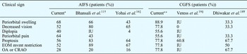
Compared with non-invasive fungal sinusitis, orbital involvement is more frequently seen in invasive fungal sinusitis (Table VI), and is well documented.Reference Bhansali, Bhadada, Sharma, Suresh, Gupta and Singh11, Reference Nithyanandam, Jacob, Battu, Thomas, Correa and D'Souza13, Reference Ferry and Abedi15–Reference Veress, Malik, el-Tayeb, el-Daoud, Mahgoub and el-Hassan19 In some of the larger, more recent series, a number of patients have presented with advanced disease similar to that of our patients.Reference Bhansali, Bhadada, Sharma, Suresh, Gupta and Singh11–Reference Nithyanandam, Jacob, Battu, Thomas, Correa and D'Souza13 Proptosis and ophthalmoplegia have been the commonest ocular findings.Reference Bhansali, Bhadada, Sharma, Suresh, Gupta and Singh11, Reference Ferry and Abedi15, Reference Yohai, Bullock, Aziz and Markert16, Reference Dhiwakar, Thakar and Bahadur18 In some reports, visual loss was more common than proptosis.Reference Nithyanandam, Jacob, Battu, Thomas, Correa and D'Souza13, Reference Yohai, Bullock, Aziz and Markert16 Yohai et al. Reference Yohai, Bullock, Aziz and Markert16 found that the severity of proptosis was greater in those with advanced disease. The orbit is particularly vulnerable to involvement in invasive fungal sinusitis, because of its close proximity to all four groups of paranasal sinuses.
Orbital involvement in acute invasive vs chronic granulomatous fungal sinusitis: reported clinical evidence

*n = 25; †n = 35 (cases of rhino-orbito-cerebral mucormycosis; ‡n = 80 (cases of rhino-orbito-cerebral mucormycosis taken from literature review of cases seen 1970–1993); **n = 8; §n = 46; ¥n = 6. AIFS = acute invasive fungal sinusitis; CGFS = chronic granulomatous fungal sinusitis; IU = information unavailable; EOM mvmt = extra-ocular muscle movement; OA = optic atrophy; CRAO = central retinal artery occlusion
Radiological orbital involvement in invasive fungal sinusitis
Radiological evidence of orbital involvement in our invasive fungal sinusitis cases was similar to that seen in other series (Table VI).Reference Dunlop and Billson1, Reference Panda, Balaji, Chakrabarti, Sharma and Reddy12, Reference Nithyanandam, Jacob, Battu, Thomas, Correa and D'Souza13 In chronic invasive fungal sinusitis, non-enhanced CT scans may show a soft tissue mass invading the affected orbit, with considerable bone destruction and opacification of the involved sinuses (Figure 3).
In acute invasive fungal sinusitis, findings are variable. DelGaudio et al. Reference DelGaudio, Swain, Kingdom, Muller and Hudgins23 studied patients with acute invasive fungal sinusitis who presented early in the disease course, and found CT scanning to be highly sensitive when 3 mm axial and coronal cuts with soft tissue and bone windows were used. These authors also found CT to lack specificity in early acute invasive fungal sinusitis, especially when the scans were compared with those of immunocompromised patients with probable bacterial sinusitis.
In the present study, in which patients usually presented late in the course of the disease, erosion of the lamina papyraceae and the presence of an intra-ocular mass were the most common radiological features in acute invasive fungal sinusitis cases, whereas lamina papyracea erosion, extra-ocular muscle involvement and optic nerve compression were more common in chronic invasive fungal sinusitis cases (Figure 3).
• Orbital involvement in invasive and non-invasive fungal sinusitis has been poorly studied
• This study found greater orbital involvement in invasive than non-invasive fungal sinusitis, more evident clinically than radiologically
• Radiological evidence of orbital involvement, although more common in invasive than non-invasive disease, was significantly greater only for orbital floor erosion
• Radiologically, there was less extra-ocular muscle enlargement and optic atrophy in invasive disease
• Up to 16 per cent of patients with acute invasive disease may show little or no radiological evidence of orbital invasion
Although similar clinical findings are seen for acute and chronic invasive fungal sinusitis, there are some radiological differences between the two conditions, as described above. Previous authors have noted a paucity of radiological findings in some individuals with acute invasive fungal sinusitis with advanced ocular involvement.Reference Nithyanandam, Jacob, Battu, Thomas, Correa and D'Souza13 In our series, four patients (16 per cent) with acute invasive fungal sinusitis and optic atrophy had negative scans. Occasionally, distinguishing fungal from bacterial sinusitis on the basis of clinical and radiological findings is difficult, particularly in immunosuppressed patients. Park et al. Reference Park, Muntz, Smith, Afify, Pysher and Pavia17 studied 17 immunosuppressed children with neutropenic fever to ascertain risk factors for acute invasive fungal sinusitis, and found no difference in radiological features between those with fungal or another type of sinusitis, as CT scans showed either mild sinus opacification or pansinusitis in both groups of patients.
Conclusion
Orbital involvement occurs to a much greater degree in invasive fungal sinusitis compared with non-invasive fungal sinusitis. This is more evident clinically than radiologically. Cases of chronic invasive fungal sinusitis show a greater degree of extra-ocular muscle enlargement and optic nerve compression than cases of acute invasive fungal sinusitis. There is a paucity of radiological signs in some cases of acute invasive fungal sinusitis with advanced disease. Early assessment of intra-orbital spread by both clinical and radiological evaluation enables timely medical and surgical intervention to limit the spread of disease.
Acknowledgement
The authors would like to thank Ms T Sebastian for providing assistance for the statistical calculations.