Implications
We studied skatole metabolism in hepatic S9 fractions from male and female pigs. Generally, skatole levels are higher in entire male than in female pigs. Until recently, we believed that this is because of the differences in skatole metabolism between male and female pigs. The present study suggests that production of skatole metabolites is similar in both male and female pigs. We found only minor differences in skatole metabolism, particularly the involvement of CYP2C and CYP3A in indole-3-carbinol formation. These results contribute to the knowledge on skatole metabolism by CYP450 and are of interest for animal and meat scientists.
Introduction
The sensory quality of meat from some sexually mature male pigs may be negatively affected by high concentrations of androstenone and/or skatole, often referred to as boar taint. Androstenone (5α-androst-16-ene-3one) is a pheromone produced in the testis and exhibits a urine-like and perspiration odour (Patterson, Reference Patterson1968). Skatole (3-methylindole) is synthesised in the large intestine by bacterial degradation of tryptophan, exhibiting a faecal-like and naphthalene odour (Vold, Reference Vold1970). A part of the skatole is excreted with faeces, whereas the remaining part is absorbed through the gut-wall. Hepatic skatole metabolism is mainly mediated by the cytochrome P450 (CYP450) superfamily of enzymes (Babol et al., Reference Babol, Squires and Lundström1998; Diaz and Squires, Reference Diaz and Squires2000). The activities of CYP1A, 2A and 2E1 are known to be of importance in the metabolism of skatole, although other CYP450 isoforms might also be involved (Terner et al., Reference Terner, Gilmore, Lou and Squires2006; Matal et al., Reference Matal, Matuskova, Tunkova, Anzenbacherova and Anzenbacher2009; Zamaratskaia and Squires, Reference Zamaratskaia and Squires2009). Pigs with high rate of skatole synthesis and low activity of CYP450 enzymes may accumulate high skatole in the fat due to decreased hepatic clearance. In the pigs with low synthesis of skatole, hepatic metabolism is of lower importance.
Usually, the activities of skatole-metabolising enzymes are higher in female and castrated pigs compared with entire male pigs (Skaanild and Friis, Reference Skaanild and Friis1999; Zamaratskaia et al., Reference Zamaratskaia, Zlabek, Chen and Madej2009). These differences are suggested to be because of the higher concentrations of sex steroids in male pigs (Skaanild and Friis, Reference Skaanild and Friis1999; Zamaratskaia et al., Reference Zamaratskaia, Zlabek, Chen and Madej2009; Rasmussen et al., Reference Rasmussen, Zamaratskaia and Ekstrand2011a). Until recently, we believed that this is because of the inhibition of skatole metabolism by high oestrogen levels found in uncastrated male pigs (Zamaratskaia et al., Reference Zamaratskaia, Gilmore, Lundström and Squires2007; Rasmussen et al., Reference Rasmussen, Zamaratskaia and Ekstrand2011b). However, a recent in vivo study showed no differences in CYP1A, CYP2A and CYP2E1 activities in male pigs with physiological and artificially reduced oestrogen levels (Zamaratskaia and Berger, Reference Zamaratskaia and Berger2014). It is also believed that androstenone may inhibit skatole metabolism, thus increasing its accumulation in the fat (Doran et al., Reference Doran, Whittington, Wood and McGivan2002, Tambyrajah et al., Reference Tambyrajah, Doran, Wood and McGivan2004). However, the results on androstenone involvement in skatole metabolism are conflicting, which may be due to the different age and breeds of animals used in the different studies, as well as different concentrations of androstenone used in vivo and in vitro studies (Doran et al., Reference Doran, Whittington, Wood and McGivan2002; Tambyrajah et al., Reference Tambyrajah, Doran, Wood and McGivan2004; Zamaratskaia and Squires, Reference Zamaratskaia and Squires2009). Another possible explanation for gender-related difference in skatole accumulation in the fat might be because of the involvement of different or a larger number of CYP450 isoforms in skatole metabolism in female pigs. Thus, in this study, we determined skatole metabolism in hepatic S9 fractions obtained from male and female pigs in the presence of specific inhibitors of several CYP450 isoforms. We chose S9 fractions, because they contain some cytosolic components that render the metabolism closer to physiological conditions (Brandon et al., Reference Brandon, Raap, Meijerman, Beijnen and Schellens2003). We used known specific inhibitors with documented effect on individual CYP450 isoforms.
Material and methods
Chemicals and reagents
Skatole, 3-methyloxindole (3MOI), indole-3-carbinol (I3C), 2-aminoacetophenone, reduced β-nicotinamide adenine dinucleotide phosphate (NADPH), oestradiol, quercetin, ellipticine, furafylline, 8-methoxypsoralen (8-MOP), ketoconazole and diallyl sulphide (DAS) were obtained from Sigma-Aldrich (Steinheim, Germany). 3-hydroxy-3-methyloxindole (HMOI) was synthesised as described by England et al. (Reference England, Merey and Padwa2007). Methanol and acetonitrile of HPLC grade were obtained from Merck (Darmstadt, Germany).
Animals
A total of six male and four female pigs (Landrace×Yorkshire dam and Duroc boar) were used in this study. All the animals were treated in accordance with the guidelines from the Danish inspectorate of animal experimentation. Animals were raised under the same conditions and slaughtered at the same age. For details about the animals and specific CYP450 expression and activity, see Rasmussen et al. (Reference Rasmussen, Zamaratskaia and Ekstrand2011b) and Rasmussen et al. (Reference Rasmussen, Zamaratskaia, Andersen and Ekstrand2012).
Preparation of S9 fractions and protein measurement
The S9 fractions (supernatants fraction of the 9000×g spin) were prepared. Frozen liver samples, ∼1 g, were cut into small pieces and homogenised in 1.2 ml of buffer (50 mM Tris, 150 mM KCl, 2 mM EDTA, pH 7.4). The homogenates were centrifuged at 9000×g for 20 min. The amount of protein in the supernatants was assayed the same day with a commercially available kit (Bio-Rad Laboratories Inc., Hercules, CA, USA) according to the manufacturer’s instructions. The S9 fractions were diluted to a protein concentration of 0.5 mg/ml. Two pooled liver S9 fractions were prepared from entire male pigs, and two were prepared from female pigs. The pools were stored at −80°C until analysis.
Formation of skatole metabolites
Incubations were performed as described by Diaz et al. (Reference Diaz, Skordos, Yost and Squires1999) with slight modifications. In brief, incubations in a total volume of 0.5 ml contained S9 protein (0.5 mg), 50 mM phosphate buffer, pH 7.4, 38 µM of skatole and 0.5 mM NADPH. After 60 min of incubation at 37°C, reactions were terminated with 0.5 ml of ice-cold acetonitrile. Subsequently, the incubations were centrifuged at 4200×g for 2 min at +4°C, and 100 µl of supernatant was injected into the HPLC. Linearity of metabolite formation from 38 µM of skatole was determined using incubation time from 5 to 90 min, and protein content from 0.2 to 1.6 mg. To determine kinetic parameters, skatole concentrations from 2 to 53 µM were used.
Microsomal incubations with CYP450 isoform inhibitors
To investigate the involvement of the CYP450 isoforms, the following inhibitors were used in the incubations: ellipticine (CYP1A1), furafylline (CYP1A2), 8-MOP (CYP2A19), sulphaphenazole (CYP2C), DAS (CYP2E1), ketoconazole (CYP3A4), quercetin (CYP2E1 in male pigs; CYP3A and CYP1A in pigs of both genders) and oestradiol (E2; CYP2E1 in male pigs). These inhibitors have previously been shown to have an effect on the activities of CYP450 isoforms in microsomal or hepatocyte assays (Zamaratskaia and Zlabek, Reference Zamaratskaia and Zlabek2009; Rasmussen et al., Reference Rasmussen, Zamaratskaia and Ekstrand2011b; Zlabek and Zamaratskaia, Reference Zlabek and Zamaratskaia2012). The incubations were performed in the presence of 0.125, 1.25 and 12.5 µM of the specific inhibitor, except for E2 (used concentration 0.002, 0.02 and 0.2 µM) and quercetin (16, 32 and 128 µM). The choice of the concentrations of E2 and quercetin in the incubations was based on their inhibitory potency in the microsomes from the same animals (Rasmussen et al., Reference Rasmussen, Zamaratskaia and Ekstrand2011a). In the control incubations, methanol was added in the same volume (5 µl). We have previously shown that this amount of methanol (1%) does not affect CYP450 activity. The mechanism-based inhibitors 8-MOP and furafylline were pre-incubated with S9 protein for 30 min, and E2 for 15 min at 37°C before addition of skatole. The reactions were allowed to proceed as described above, and the concentrations of formed metabolites were measured. The effect of inhibitors on the formation of individual skatole metabolite is expressed as the percentage of metabolite formation remaining in the presence of inhibitor relative to the metabolite formation in control incubations. The activity was regarded as reduced if it differed from the control activity by at least 30%. No further statistical analyses were performed in order to avoid false results due to intra-assay variations.
Chromatographic conditions
Skatole metabolites were separated by gradient reverse-phase HPLC (Diaz et al., Reference Diaz, Skordos, Yost and Squires1999) on Li-Chrospher RP-18 column (Merck KGaA, Darmstadt, Germany) (5 µm) equipped with a guard column. The HPLC equipment consisted of a pump (L-6200A), autosampler (L-7200), fluorescence detector (L-7485), UV detector (L-4000) and D-7000 interface (Merck, Hitachi, Tokyo, Japan). The metabolites were detected with fluorescence (excitation and emission wavelengths of 286 and 350 nm, respectively) and UV (250 nm) detectors.
Statistical analysis
Kinetic parameters (K m and V max values) were estimated by a GraphPad Prism version 4.0 for Windows, GraphPad Software (San Diego, CA, USA). Student’s t-test was used to compare K m and V max values, as well as the quantity of skatole metabolites formed from the S9 fractions from male and female livers. Differences were considered significant when P<0.05.
Results
Skatole metabolites
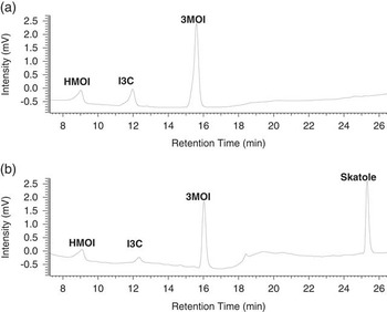
No metabolites were detected in S9 incubations in the absence of NADPH or skatole, and in the incubations without S9 fractions. In the S9 incubations with skatole and NADPH, three skatole metabolites were detected – 3-hydroxy-3-methyloxindole (HMOI), indole-3-carbinol (I3C) and 3-methyloxindole (3MOI). Skatole and skatole metabolites were detected by the UV detector at the following retention times: skatole 25 min; HMOI 9 min; I3C 12 min; and 3MOI 16 min (Figure 1). In addition, skatole and I3C were detected by the fluorescence detector. HMOI and 3MOI were quantified by UV absorption, and I3C was quantified using data from the fluorescence detector because of its higher sensitivity and accurate estimation of lower concentrations compared with the UV detector.
Typical HPLC chromatogram of skatole metabolites. (a) Standard solutions and (b) S9 incubations in the presence of 38 µM of skatole and β-nicotinamide adenine dinucleotide phosphate (NADPH).

The formation of HMOI and I3C was linear for 90 min of incubation time, and the formation of 3MOI for 60 min. The formation of all metabolites was linear up to at least 1.6 mg of S9 protein. Incubation time of 60 min and protein content of 0.5 mg were chosen as optimal for subsequent assays.
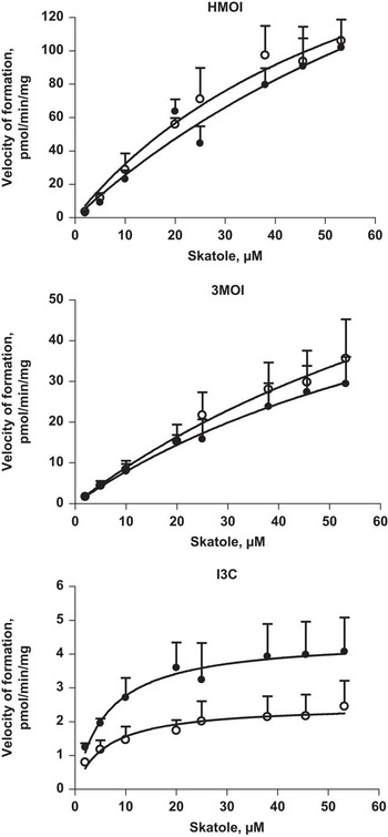
The effect of skatole concentration on the velocity of metabolite formation is presented in Figure 2. The most abundant metabolite of skatole was HMOI followed by 3MOI and I3C in both male and female S9 fractions. Concentrations of formed HMOI and 3MOI did not differ between the genders (P=0.124 for HMOI, and P=0.575 for 3MOI). Concentrations of formed I3C and Vmax were higher in S9 fractions from the female compared with male pigs (P=0.0001 for I3C concentration and P=0.008 for Vmax). Enzyme kinetic parameters were similar for both genders (P>0.05; Table 1).
Substrate–velocity plot for skatole metabolism in porcine liver S9 fractions. Data are presented as mean values and standard errors. Open dots represent metabolite formation by S9 fractions from male, and solid dots from female pigs.

Enzyme kinetic parameters for the formation of skatole metabolites by hepatic S9 fractions from male and female pigs

HMOI=3-hydroxy-3-methyloxindole; 3MOI=3-methyloxindole; IC3=indole-3-carbinol.
Kinetic parameters were calculated from data obtained separately for six male and four female pigs. Incubations were conducted using substrate (skatole) concentrations from 2 to 53 μM. Results are presented as best-fit values±standard errors from nonlinear regression analysis (GraphPad Prism program 4.0 kinetic software). Student’s t-test was used to compare K m and V max values in the incubations with male and female S9 fractions.
Inhibition study
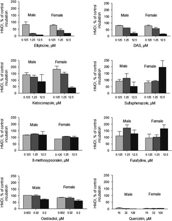
Formation of HMOI (Figure 3) was inhibited by ellipticine (CYP1A), DAS (CYP2E1) and quercetin (CYP1A, 2E1 and 3A). The highest degree of inhibition was observed at high concentration of the inhibitor (remaining activity, mean and standard deviation, 12.9±6.9% for ellipticine; 18.1±9.2% for DAS; and 2.8±2.0 for quercetin). Ketoconazole (CYP3A) at the concentration of 12.5 µM inhibited HMOI formation to a lesser degree, and this inhibition was more pronounced in female pigs (89.2±79.9% in male and 41.3±11.0% in female). Interestingly, at lower concentrations of ketoconazole, an increase in HMOI formation was observed in S9 fractions from female pigs. The rate of HMOI formation also increased in the presence of sulphaphenazole and furafylline.
Rate of HMOI (3-hydroxy-3-methyloxindole) formation in hepatic S9 fractions from six male and four female pigs in the presence of various cytochrome P450 (CYP450) inhibitors. Data are presented as mean values and standard errors. The activity was regarded as reduced if it differed from the control activity by at least 30%.

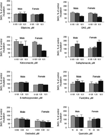
Formation of 3MOI (Figure 4) was lower in the presence of DAS (CYP2E1; 65.7±9.4% at 12.5 µM of DAS). In the S9 fractions from female pigs, 3MOI formation was inhibited by oestradiol (CYP2E1; 60.7±23.5% at 0.2 µM of oestradiol), although in the male pigs oestradiol did not affect 3MOI formation (89.8±14.0%). Formation of 3MOI increased in the presence of ketoconazole, sulphaphenazole, furafylline and quercetin.
Rate of 3MOI (3-methyloxindole) formation in hepatic S9 fractions from six male and four female pigs in the presence of various cytochrome P450 (CYP450) inhibitors. Data are presented as mean values and standard errors. The activity was regarded as reduced if it differed from the control activity by at least 30%.

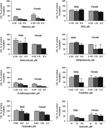
Formation of I3C (Figure 5) was inhibited by ellipticine (CYP1A1; 15.9±7.1% at 12.5 µM of ellipticine, 8-MOP (CYP2A; 15.1±6.2% at 12.5 µM of 8-MOP) and quercetin (CYP1A1, 2E1, 3A; 40.5±18.5% at 128 µM of quercetin). Sulphaphenazole (CYP2C) inhibited formation of I3C only in female pigs (54.3±60.9% at 12.5 µM of sulphaphenazole), but not in male pigs (93.4±21.9% at 12.5 µM of sulphaphenazole). A similar pattern was observed in the presence of ketoconazole (CYP3A): I3C formation decreased in female pigs (57.2±1.5% at 12.5 µM of ketoconazole), but not in male pigs (115.6±70.6% at 12.5 µM of ketoconazole).
Rate of I3C (indole-3-carbinol) formation in hepatic S9 fractions from six male and four female pigs in the presence of various cytochrome P450 (CYP450) inhibitors. Data are presented as mean values and standard errors. The activity was regarded as reduced if it differed from the control activity by at least 30%.

Discussion
The risk of boar taint in meat from intact male pigs is mainly associated with skatole and androstenone accumulation in the fat. High skatole levels in the fat might be due to insufficient skatole hepatic metabolism, which prevents appropriate elimination of skatole from the body. Several skatole metabolites were detected in pig plasma and urine (Bæk et al., Reference Bæk, Hansen-Møller, Friis, Cornett and Hansen1997). Major insights into skatole metabolism came from in vitro microsomal studies (Babol et al., Reference Babol, Squires and Lundström1998; Diaz et al., Reference Diaz, Skordos, Yost and Squires1999; Diaz and Squires, Reference Diaz and Squires2000). In these studies, CYP2A and CYP2E1 were identified as major isoforms involved in skatole metabolism. Further in vitro studies on primary cultured pig hepatocytes (Terner et al., Reference Terner, Gilmore, Lou and Squires2006), individual purified porcine enzymes (Matal et al., Reference Matal, Matuskova, Tunkova, Anzenbacherova and Anzenbacher2009) and individually expressed enzymes (Wiercinska et al., Reference Wiercinska, Lou and Squires2012) confirmed an involvement of CYP2A and CYP2E1, and revealed that other isoforms (CYP1A, CYP2C, CYP3A) are also involved in skatole metabolism. Moreover, gender-related differences in the rate of skatole metabolism are frequently suggested as a cause of the higher levels of skatole in male pigs compared with the female pigs. Indeed, the expression and activities of skatole-metabolising enzymes were higher in females compared with male pigs (Skaanild and Friis, Reference Skaanild and Friis1999; Zamaratskaia et al., Reference Zamaratskaia, Chen and Lundström2006; Rasmussen et al., Reference Rasmussen, Zamaratskaia and Ekstrand2011a). Interestingly, no data are available on the skatole metabolite formation in both male and female pigs.
The aim of the present study was to evaluate gender-related differences in skatole metabolism. We utilised S9 fractions, which contain a wide variety of Phase I and II metabolising enzymes, and more closely represent a more complete physiological environment compared with microsomes (Zhang et al., Reference Zhang, Luo, Ding and Lu2012). S9 fractions from both male and female pig livers were included. The following three skatole metabolites were identified: HMOI, I3C and 3MOI. Interestingly, in our pilot study performed on the porcine microsomes, two additional unidentified peaks that might be related to skatole metabolites were detected by fluorescence (unpublished data); however, these peaks were not detected when we used S9 fractions. We speculated that these two unidentified peaks produced by the microsomes were 6- and 5-OH-3-methylindole. In the S9 fractions, these metabolites may immediately undergo sulpho-conjugation by phase II enzymes and cannot be detected. Similarly, no 6- and 5-OH-3-methylindole formation was observed in the study with hepatocyte preparations (Terner et al., Reference Terner, Gilmore, Lou and Squires2006).
Surprisingly, the metabolic rates of production of the three skatole metabolites and kinetic parameters were similar in both male and female pigs. The activities of skatole-metabolising enzymes were previously measured in the liver samples from the same animals, and significantly higher activities of CYP1A and CYP2A in female pigs compared with male pigs were found (Rasmussen et al., Reference Rasmussen, Zamaratskaia and Ekstrand2011a). Obviously, these differences in the activities were not reflected in the rate of skatole metabolism. This might be due to the high substrate concentration used in the present in vitro study. Skatole concentrations in the range from 2 to 53 µM were used, which correspond to the high physiological concentrations of skatole in fat. It is likely that skatole levels in liver tissue are lower.
Ellipticine, DAS and quercetin inhibited HMOI formation, confirming the involvement of CYP1A1 and CYP2E1. In addition, HMOI formation in male pigs was inhibited by oestradiol. We have previously shown that oestradiol inhibits CYP2E1 activities in male pigs, but not in female pigs (Zamaratskaia et al., Reference Zamaratskaia, Gilmore, Lundström and Squires2007; Rasmussen et al., Reference Rasmussen, Zamaratskaia and Ekstrand2011b). This explains why HMOI formation was not decreased by oestradiol in female pigs in the present study, and confirms the involvement of CYP2E1 in HMOI formation. CYP2C (sulphaphenazole) and CYP1A2 (furafylline) were not involved in the formation of HMOI in either male or female pigs. On the contrary, involvement of CYP2C and CYP1A2 was demonstrated by previous in vitro studies (Wiercinska et al., Reference Wiercinska, Lou and Squires2012). The disagreement between these studies might be because of the different in vitro systems used. Interestingly, our study suggested that inhibition of CYP2C and CYP1A2 might stimulate other isoforms to produce more HMOI in female pigs. This, however, needs further investigation. CYP2A19 was not involved in HMOI formation (as supported by Wiercinska et al., Reference Wiercinska, Lou and Squires2012).
CYP2E1 was involved in the formation of 3MOI in both male and female pigs. CYP3A and CYP2C were not directly involved in the formation of 3MOI, but probably inhibition of CYP3A stimulated other isoforms to produce more 3MOI.
Our results indicated that CYP1A1 and CYP2A19 were involved in the formation of I3C in both male and female pigs. It is known that specificity of 8-MOP as CYP2A19 inhibitor is low; it inhibits both CYP2A19 and CYP2E1. However, if CYP2E1 is involved in the I3C formation, inhibition would also be expected in the presence of DAS. This was not true, suggesting a minor role, if any, of CYP2E1 in I3C formation. CYP2C and CYP3A were involved in I3C formation in female pigs only, as suggested by inhibition by sulphaphenazole and ketoconazole, respectively. This is a very essential finding supporting our hypothesis that gender-related differences in the rate of skatole metabolite formation might be because of the involvement of larger number of CYP450 isoforms in female pigs. It also explains why I3C formation in the present study was higher in females compared with male pigs. On the other hand, I3C is a minor skatole metabolite, and the physiological significance of CYP2C and CYP3A involvement in its formation in females but not in male pigs needs to be elucidated. CYP2E1, CYP3A and CYP1A2 were not involved in the formation of I3C in neither male nor female pigs.
It should be noted that our results should be interpreted with caution because of the low number of animals and possibility of breed and age effects on skatole metabolism. Thus, more research is needed to elucidate gender-related differences in skatole metabolism. In such research, larger number of animals of different breed and ages should be included.
In summary, skatole metabolites and CYP450 isoforms involved in skatole metabolism were for the first time investigated in hepatic S9 fractions from male and female pigs. Only minor differences in skatole metabolism between male and female pigs were observed, particularly the involvement of CYP2C and CYP3A in I3C formation in females but not in male pigs.
Acknowledgements
This work was supported by a research grant from C.F. Lundströms Stiftelse. The authors thank Dr Carl Brunius for the great work on the synthesis of 3-hydroxy-3-methyloxindole, Ms Ensieh Hajazimi for her technical expertise and Prof. Kerstin Lundström for her interest and support. The work of M.K.R. was supported by a personal grant from the Villum foundation, the Lundbeck foundation and Norma og Frode S. Jacobsens fond.